Review
. 2014 Mar 26;66(2):513-69.
doi: 10.1124/pr.112.007351. Print 2014.

Regulation of cellular communication by signaling microdomains in the blood vessel wall

Affiliations
Review

Regulation of cellular communication by signaling microdomains in the blood vessel wall

Marie Billaud et al. Pharmacol Rev. .

Abstract

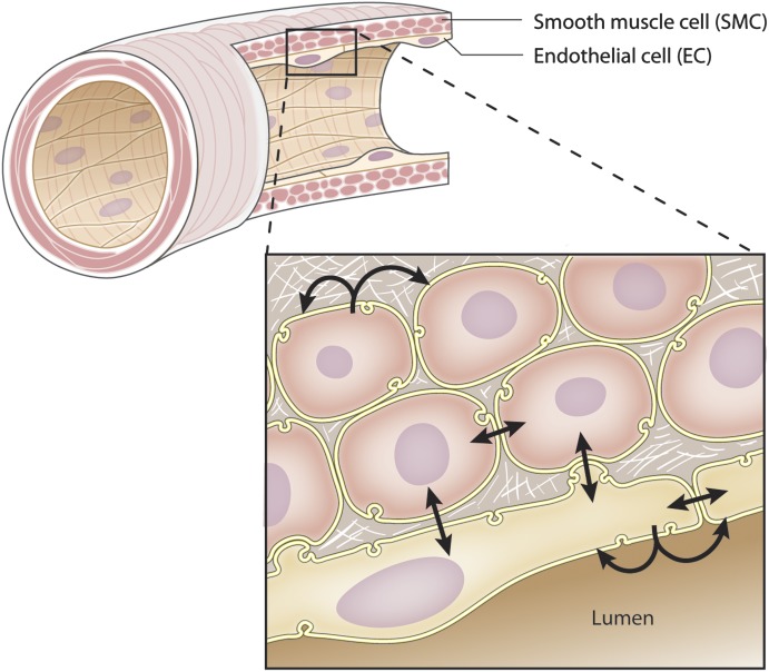
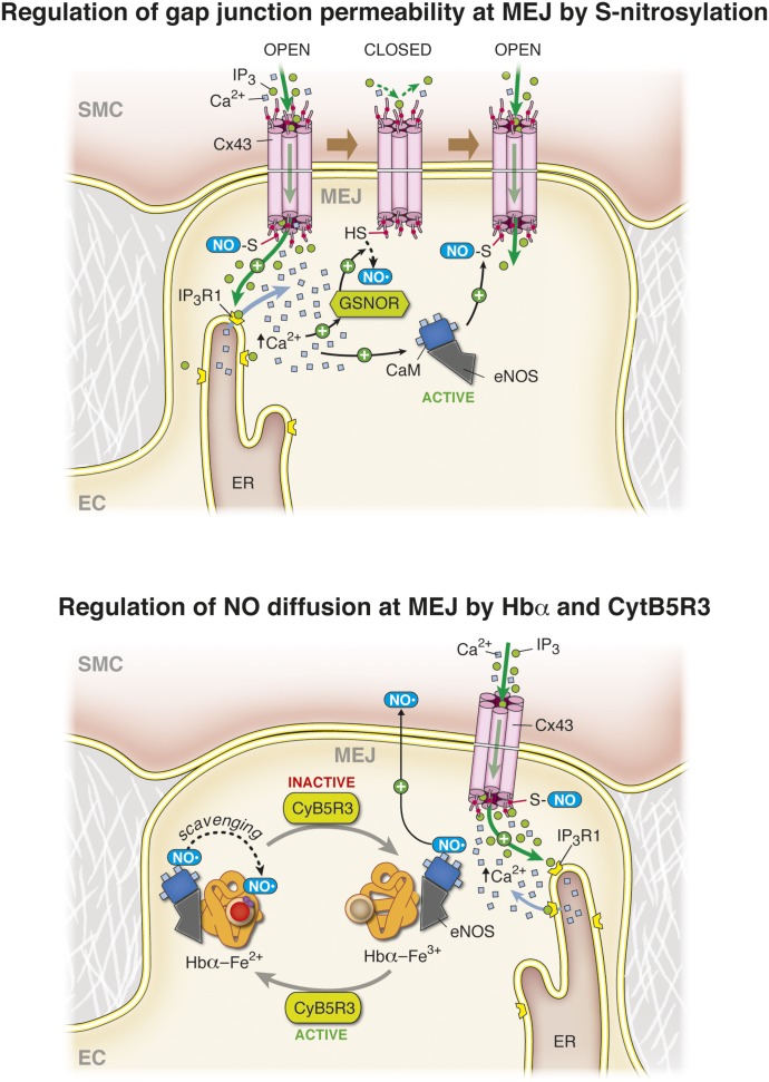
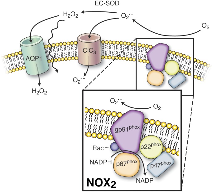
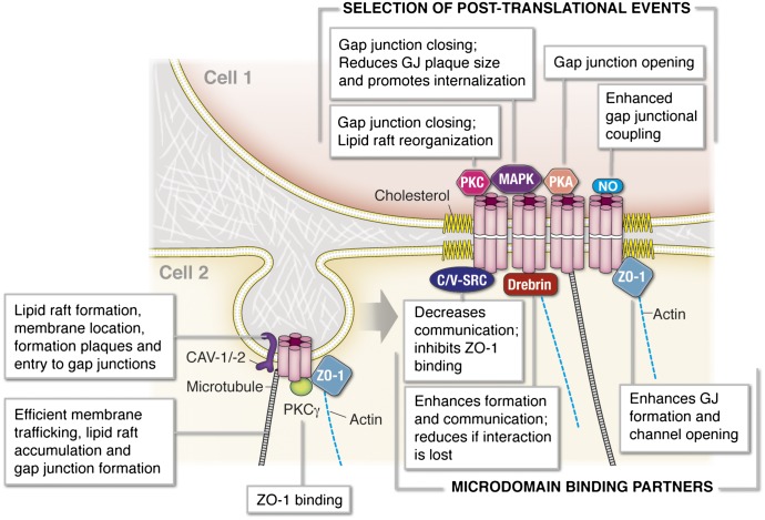
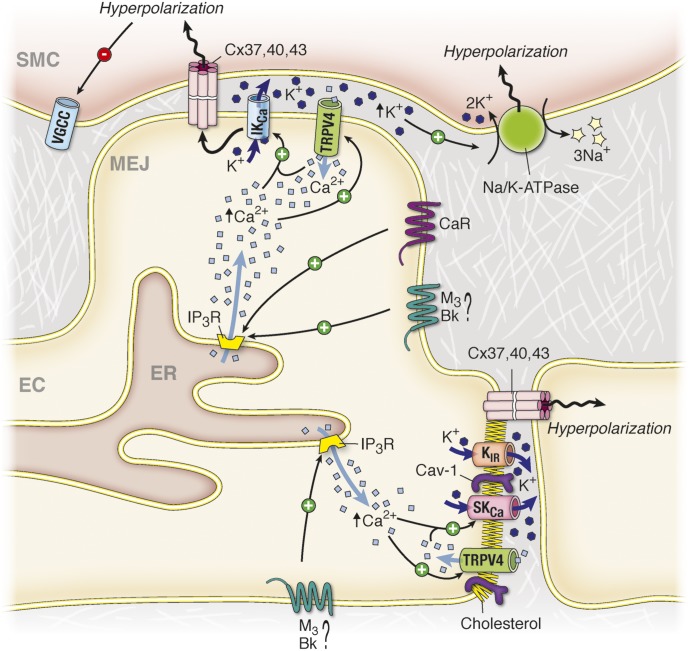
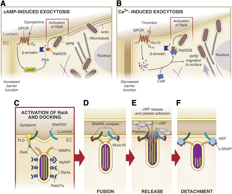
It has become increasingly clear that the accumulation of proteins in specific regions of the plasma membrane can facilitate cellular communication. These regions, termed signaling microdomains, are found throughout the blood vessel wall where cellular communication, both within and between cell types, must be tightly regulated to maintain proper vascular function. We will define a cellular signaling microdomain and apply this definition to the plethora of means by which cellular communication has been hypothesized to occur in the blood vessel wall. To that end, we make a case for three broad areas of cellular communication where signaling microdomains could play an important role: 1) paracrine release of free radicals and gaseous molecules such as nitric oxide and reactive oxygen species; 2) role of ion channels including gap junctions and potassium channels, especially those associated with the endothelium-derived hyperpolarization mediated signaling, and lastly, 3) mechanism of exocytosis that has considerable oversight by signaling microdomains, especially those associated with the release of von Willebrand factor. When summed, we believe that it is clear that the organization and regulation of signaling microdomains is an essential component to vessel wall function.


Figures

Fig. 1.
Fig. 1.
Schematic representation of intercellular communication in the arterial wall. The SMCs and ECs composing the vascular wall can communicate with each other either by releasing molecules to neighboring cells (paracrine communication) or directly via gap junction channels that link the cytoplasm of two adjacent cells. The different types of intercellular communications are represented by the arrows.
Fig. 2.
Fig. 2.
Schematic representation of calcium compartmentalization. (A) In VSMCs, calcium (Ca2+) can be stored in both the SR and in the mitochondria. Calcium release from both organelles is tightly coordinated to calcium influx at the PM, and this coordination is facilitated by the close proximity between the organelles and the PM. (B) In cerebral VSMCs, after increased intravascular pressure, there is a coordinated action of TRPV4, TRPM4, VGCC, and large conductance potassium channels (BKCa) at the plasma membrane and ryanodine receptors RyR at the SR membrane. In this configuration, increased pressure activates calcium influx in VSMCs via TRPV4, which stimulates calcium release from the SR through RyR, whereas opening of TRPM4 results in calcium influx through VGCC also activating calcium release from the SR through RyR. Calcium release from RyR (also termed calcium “sparks”) further activates potassium (K+) efflux via BKCa channels. The hyperpolarization resulting from potassium efflux reduces the activity of VGCC, making BKCa key in the autoregulation of calcium homeostasis in VSMCs. (C)Upon cerebral VSMC stimulation with ET-1, activation of IP3-R1 at the SR membrane activates calcium influx through TRPC3 independently of calcium release via IP3R but via a direct protein interaction between IP3-R and TRPC3. This IP3R/TRPC3 interaction is facilitated by the presence of Cav1. Calcium release via IP3R upon ET-1 stimulation further activates BKCa channels at the plasma membrane in a similar manner as RyR activates BKCa channels in (B). Activation of BKCa induces hyperpolarization of the plasma membrane, thus attenuating the activation of VGCC by cation influx through TRPC3. (D) After stimulation with ET-1, nicotinic acid adenine dinucleotide phosphate (NAADP) activates the release of calcium from intracellular lysosomes via the two pore calcium channel (TCP2). The calcium released from the lysosome further activates calcium release from the SR via RyRs. (E) The compartmentalization of VGCC, TRP channels, the NCX, and the SERCA are part of a signaling microdomain controlling calcium replenishment of the PM-SR junction. In this configuration, calcium and Na+ influxes via TRPC6 activate the adjacent VGCC and the NCX in reverse mode. Calcium influx via the VGCC and the NCX provide sources of calcium for ER/SR replenishment via the SERCA pump. Additionally, the STIM present at the SR membrane is capable of sensing decreased levels of calcium in the SR and activates calcium influx via Orai at the plasma membrane, again providing calcium for SR replenishment via the SERCA pumps. (F) Mitochondria also play a major role as a buffer and as a source of calcium for the SR. After stimulation of VSMCs, mitochondria take up the calcium released from IP3R via the VDAC on the outer mitochondrial membrane and the mitochondrial calcium uniporter (MCU) on the inner mitochondrial membrane. The buffering role of mitochondria is essential to prevent the formation of high local calcium concentrations surrounding the IP3-R, which would inhibit the IP3-R activity. The release of calcium from the mitochondria via the mitochondrial sodium/calcium exchanger (NLCX) present on the inner mitochondrial membrane and the permeability transition pore (PTP) on the outer mitochondrial membrane provides a source of calcium for SR replenishment by the SERCA pumps. Straight arrows with positive and negative signs indicate activation and inhibition by Ca2+, respectively. Wavy arrows with a positive or negative sign indicate activation by depolarization or an inhibition by hyperpolarization respectively.
Fig. 3.
Fig. 3.
eNOS: protein domains, phosphorylation sites, and higher order organization. (A) eNOS is composed of an N-terminal oxygenase domain containing binding sites for tetrahydrobiopterin (BH4), Zn2+, heme, and l-arginine and a C-terminal reductase domain containing NADPH, flavin adenine dinucleotide, and flavin mononucleotide binding sites. The oxygenase and reductase domains are separated by a linker region that harbors a regulatory CaM binding domain. Binding domains are indicated in italics. eNOS also harbors several serine, threonine, and tyrosine residues that are targeted for phosphorylation. The most extensively characterized phosphorylated residues are depicted with those that promote enzyme activation in green (Tyr81, Ser615, Ser633, and Ser1177; human sequence) and sites imparting inhibition in red (Ser114 and Thr495). (B) eNOS forms a homodimer coordinated by BH4 and Zn2+ binding in the N-terminal oxygenase domains of each monomer. Dimeric eNOS synthesizes NO from l-arginine and O2 through NADPH-dependent electron flux from the C-terminal reductase domain of one monomer to the heme moiety located on the oxygenase domain. Depletion of BH4 promotes eNOS uncoupling, leaving the enzyme in a monomeric form, resulting in the production of superoxide rather than NO.
Fig. 4.
Fig. 4.
Nitric oxide regulation at plasma membrane caveolae. (A) eNOS localizes to plasma membrane caveolae, where it directly binds to Cav1. This interaction inhibits basal eNOS activity and NO synthesis. Increases in Ca2+ facilitate activation of CaM, which is recruited to eNOS and promotes dissociation of the enzyme from Cav1. Binding of CaM to free eNOS increases its enzymatic activity, resulting in NO production from the substrates l-arginine, NADPH, and O2. (B) eNOS colocalizes with a number of membrane receptors in endothelial cell caveolae, including the angiotensin II type 1 receptor (AT1), the bradykinin B2 receptor (BKB2), the endothelin-1 type B receptor, the estrogen receptor (ERα), and the scavenger receptor (SR-B1). The GPCRs bind eNOS directly through an interaction with their fourth intracellular domain (ID4) and inhibit basal eNOS activity. Binding of GPCR ligands to their complement receptors promotes eNOS dissociation from these receptors, relieving the inhibitory clamp that is mediated through increases in intracellular Ca2+ and phosphorylation of eNOS and the GPCR, leading to NO production by eNOS. Activation of SR-B1 by high-density lipoprotein (HDL) or ERα by estradiol promotes activation of protein kinases, including Src, PI3K/Akt, and MAPKs, which promote eNOS phosphorylation and enzyme activation. (C) Activation of the AT1 receptor in caveolae signals eNOS activation as described in (B) as well as the recruitment and activation of NOX to caveolae. NOX activation produces superoxide anion (O2•−), which uncouples dimeric eNOS to its monomeric form, resulting in O2•− production. NO and O2•− generated at EC caveolae rapidly react to form the free radical peroxinitrite (ONOO). See Fig. 6 for NOX regulation. (D) eNOS enriched at EC caveolae colocalizes with and is regulated by the activity of the cationic amino acid transporter 1 (CAT1) and the plasma membrane Ca2+ ATPase (PMCA). In caveolae, PMCA functions to extrude Ca2+ from the local cytosolic compartment and the subsequent reduction in free intracellular Ca2+ prevents CaM recruitment and Cav1 dissociation from eNOS, inhibiting NO production. Conversely, CAT1 facilitates the cellular uptake of the eNOS substrate l-arginine in spatial proximity to the synthase, providing local enrichment in the precursor for NO synthesis.
Fig. 5.
Fig. 5.
Nitric oxide regulation at the MEJ. Top, Nitric oxide posttranslationally modifies the gap junction protein connexin 43 (Cx43) at the MEJ through S-nitrosylation. Under basal conditions, Cx43 is constitutively S-nitrosylated, which renders the channel in an open, permeable state. IP3 and Ca2+ from the smooth muscle cell layer diffuse through these open gap junctions and bind to IP3 receptor type 1 (IP3R1) on the EC endoplasmic reticulum that is poised at the MEJ, resulting in Ca2+ release from the internal store. This local increase in Ca2+ promotes the activation of the denitrosylase enzyme S-nitrosoglutathione reductase (GSNOR) whose activity leads to denitrosylation of Cx43. Reduction of the S-nitrosothiols on Cx43 closes the channel, preventing additional ion and metabolite diffusion into the ECs. After this event, the local rise in Ca2+ activates the eNOS localized at the MEJ, resulting in increased NO production and renitrosylation of Cx43 gap junctions, restoring gap junctional communication between ECs and SMCs in the arterial wall. The black dashed arrow indicates reduction of the nitrosothiol, and the red dots on Cx43 correspond to the cysteine residue on the carboxyl tail of Cx43 that is S-nitrosylated. Bottom, Hemoglobin α (Hbα) is synthesized by vascular ECs and is enriched at the MEJ, where it forms a complex with eNOS and the reductase CytB5R3. NO generated by eNOS at the MEJ is able to diffuse to the overlying smooth muscle cell layer when Hbα resides in the Fe3+ state (methemoglobin, maroon). Reduction of Hbα to the Fe2+ state (oxyhemoglobin, red) by the activity of CytB5R3 promotes NO scavenging by Hbα and prevents NO diffusion.
Fig. 6.
Fig. 6.
ROS microdomain at the plasma membrane. NADPH oxidase is the main source of O2•− at the plasma membrane where it produces O2•− in the extracellular space. The NADPH oxidase protein complex represented here corresponds to the Nox2 isoform (previously termed gp91phox). Upon stimulation, the regulators of gp91phox, including gp22phox, gp67phox, gp47phox, and Rac assemble with the gp91phox. The O2•− produced extracellularly can either be dismuted by the extracellular superoxide dismutase (EC-SOD) or traverse the chloride channel ClC3 at the plasma membrane. In the extracellular space, the dismutation of O2•− results in the production of H2O2, which can either cross the plasma membrane directly because of its lipophilic property or via the aquaporin channels (AQP1).
Fig. 7.
Fig. 7.
Connexins and gap junction signaling microdomains. The formation of gap junctional structures is regulated through organization of connexin hemichannels in caveolae that further assemble to a gap junction plaque through association with multiple protein partners. The C-terminal domain of connexins interacts with microtubule, PKC, and ZO-1, and these interactions promote integration of connexin hemichannels to the plasma membrane at lipid-enriched caveolae. Assembling of hemichannels in the lipid raft structures surrounding a gap junction plaque is enhanced through interactions with ZO-1/2 and drebrin. The different steps leading to the formation of gap junction assembly as well as gap junctional permeability are modulated through a dynamic process of posttranslational modifications at the C-terminal regions of connexins including phosphorylations by PKC and MAPK, which both reduce gap junction communication, and by PKA, which increases gap junctional signaling.
Fig. 8.
Fig. 8.
The endothelium-dependent hyperpolarization microdomain. The EDH response starts with the activation of IP3R at the ER in EC by a GqPCR [for example, muscarinic (M3), BK, or the calcium sensing receptor (CaR)]. The calcium released from the ER activates the calcium-sensitive potassium channels SKCa at the EC-EC junctions and IKCa at the MEJ, which induces hyperpolarization of the EC. In parallel, the calcium release from the ER activates the capacitive entry of calcium at both the EC-EC junctions and the MEJ via TRPV4 channels, which sustains the opening of the IKCa and SKCa channels. The activation of SKCa and IKCa at the plasma membrane of ECs activates the efflux of potassium and its accumulation in the extracellular space between the VSMCs and the ECs. This potassium accumulation further activates the sodium/potassium ATPase (Na/K/ATPase) at the plasma membrane of VSMC, producing hyperpolarization and relaxation of the VSMC by closing the voltage-gated calcium channels (VGCC). The hyperpolarization of EC can also be transferred to the adjacent ECs and VSMCs via gap junctions channels located at the MEJ and at the EC-EC junctions. Wavy arrows indicate hyperpolarization.
Fig. 9.
Fig. 9.
The exocytosis microdomain. (A) Epinephrine induces increased cAMP via Gs protein-coupled receptors, resulting in the activation of RalA by RalGDS. The exocytosis of the Weibel-Palade bodies present at the PM is activated, whereas the progression of the WPBs located in the perinuclear region is inhibited by the presence of a more prominent peripheral actin rim. This process is accompanied by an increase in barrier function. (B) Upon activation of GqPCR in ECs by agonists such as histamine or thrombin, there is an increase in [Ca2+]i, which associates with CaM. The Ca2+-CaM further binds to the amino terminus of Ral GDS, which leads to the dissociation from the inhibitory beta-arrestin and to activation of RalA. This whole process allows WPB migration to the surface and concurrently decreases the strength of the barrier function between ECs. (C) RalA promotes fusion of the membrane by increasing PLD activity. Rab27a helps to determine when exocytosis will occur via its ratio of fractional occupancy by MyRIP and Slp4a. (D) The V-SNARE VAMP3 and the t-SNAREs syntaxin4 and SNAP23 interact to pull the two membranes in close proximity for fusion to occur. Munc18 acts to inhibit the SNAREs from binding prematurely. (E) vWF is released into the lumen, where it can bind to and attract platelets in addition to exerting effects on neighboring ECs. (F) NSF/α-SNAP bind to SNAREs to facilitate their disassembly.

References

    1. Abo A, Pick E, Hall A, Totty N, Teahan CG, Segal AW. (1991) Activation of the NADPH oxidase involves the small GTP-binding protein p21rac1. Nature 353:668–670 - PubMed
    1. Absi M, Burnham MP, Weston AH, Harno E, Rogers M, Edwards G. (2007) Effects of methyl beta-cyclodextrin on EDHF responses in pig and rat arteries; association between SK(Ca) channels and caveolin-rich domains. Br J Pharmacol 151:332–340 - PMC - PubMed
    1. Abu-Soud HM, Stuehr DJ. (1993) Nitric oxide synthases reveal a role for calmodulin in controlling electron transfer. Proc Natl Acad Sci USA 90:10769–10772 - PMC - PubMed
    1. Adeagbo AS, Triggle CR. (1993) Varying extracellular [K+]: a functional approach to separating EDHF- and EDNO-related mechanisms in perfused rat mesenteric arterial bed. J Cardiovasc Pharmacol 21:423–429 - PubMed
    1. Adebiyi A, Narayanan D, Jaggar JH. (2011) Caveolin-1 assembles type 1 inositol 1,4,5-trisphosphate receptors and canonical transient receptor potential 3 channels into a functional signaling complex in arterial smooth muscle cells. J Biol Chem 286:4341–4348 - PMC - PubMed

Publication types

MeSH terms

Substances