. 2014 May 16;289(20):13838-50.
doi: 10.1074/jbc.M113.544288. Epub 2014 Mar 26.

Structural characterization of heparin-induced glyceraldehyde-3-phosphate dehydrogenase protofibrils preventing α-synuclein oligomeric species toxicity

Affiliations

Structural characterization of heparin-induced glyceraldehyde-3-phosphate dehydrogenase protofibrils preventing α-synuclein oligomeric species toxicity

César L Ávila et al. J Biol Chem. .

Abstract

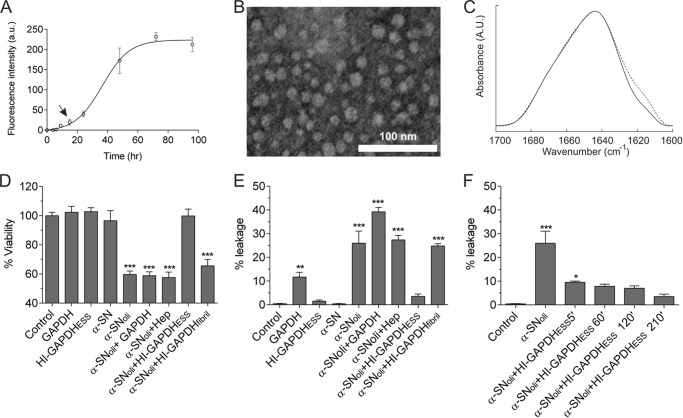
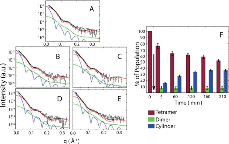
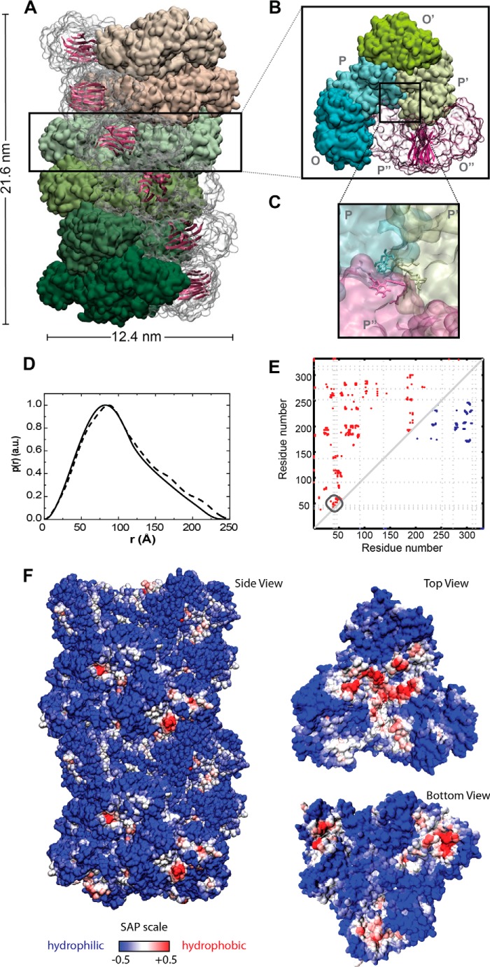
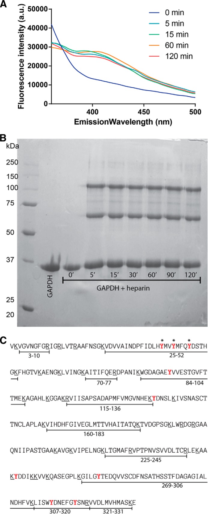
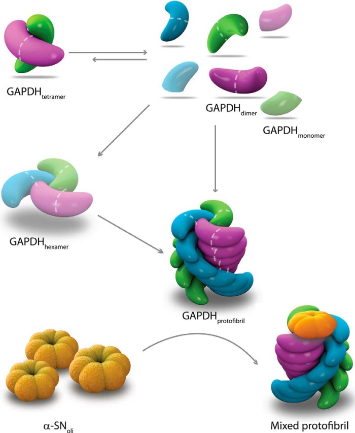
Glyceraldehyde-3-phosphate dehydrogenase (GAPDH) is a multifunctional enzyme that has been associated with neurodegenerative diseases. GAPDH colocalizes with α-synuclein in amyloid aggregates in post-mortem tissue of patients with sporadic Parkinson disease and promotes the formation of Lewy body-like inclusions in cell culture. In a previous work, we showed that glycosaminoglycan-induced GAPDH prefibrillar species accelerate the conversion of α-synuclein to fibrils. However, it remains to be determined whether the interplay among glycosaminoglycans, GAPDH, and α-synuclein has a role in pathological states. Here, we demonstrate that the toxic effect exerted by α-synuclein oligomers in dopaminergic cell culture is abolished in the presence of GAPDH prefibrillar species. Structural analysis of prefibrillar GAPDH performed by small angle x-ray scattering showed a particle compatible with a protofibril. This protofibril is shaped as a cylinder 22 nm long and a cross-section diameter of 12 nm. Using biocomputational techniques, we obtained the first all-atom model of the GAPDH protofibril, which was validated by cross-linking coupled to mass spectrometry experiments. Because GAPDH can be secreted outside the cell where glycosaminoglycans are present, it seems plausible that GAPDH protofibrils could be assembled in the extracellular space kidnapping α-synuclein toxic oligomers. Thus, the role of GAPDH protofibrils in neuronal proteostasis must be considered. The data reported here could open alternative ways in the development of therapeutic strategies against synucleinopathies like Parkinson disease.

Keywords: Cell Permeabilization; GAPDH; Molecular Modeling; Parkinson Disease; Protofibril Structure; X-ray Scattering; α-Synuclein.


Figures

FIGURE 1.
FIGURE 1.
HI-GAPDHESS sequesters α-SNoli abolishing its deleterious effects. A, kinetics of 140 μm α-SN aggregation monitored by ThT fluorescence. The arrow shows the point where α-SNoli is harvested. B, uranyl acetate-stained transmission electron microscopy images of α-SNoli. a.u., arbitrary units. C, FTIR spectra of the deconvoluted amide I′ region of monomeric α-SN (solid line) and α-SNoli (dashed line). Deconvolution was performed using a Lorentzian line shape of 18 cm−1 and a resolution enhancement factor of 1.75. D, cell viability of SH-SY5Y after the addition of 20 mm HEPES, pH 7.40 (control), or 50 μm GAPDH. For HI-GAPDHESS, the enzyme (50 μm) was preincubated with heparin (75 μg/ml) for 2 h at 37 °C. α-SN and α-SNoli corresponds to α-synuclein (140 μm) preincubated for 0 and 16 h at 37 °C with orbital agitation, respectively. For α-SNoli + GAPDH, α-SNoli was preincubated for 1 h at 37 °C with GAPDH (50 μm). α-SNoli + HI-GAPDHESS corresponds to α-SNoli preincubated for 1 h at 37 °C with HI-GAPDHESS. MTT test was used to estimate cell viability. E, changes in liposomal membrane permeability induced by distinct aggregated species. The fluorescence signal was normalized with the signal observed after Triton X-100 addition, which induced complete rupture of the vesicles. α-SN monomer was employed as leakage negative control. HI-GAPDHESS, α-SNoli, α-SNoli + GAPDH, α-SNoli, and α-SNoli + HI-GAPDHESS were prepared as described above. F, effect of heparin/GAPDH incubation time on the production of species with the ability to protect membranes against content leakage induced by α-SNoli. The α-SNoli was preincubated with HI-GAPDHESS harvested after 5, 60, 120, and 210 min of GAPDH/heparin mixture under orbital agitation at 37 °C. Results were evaluated by analysis of variance (p = 0.001), and asterisks indicate significant differences versus the control (***, p ≤ 0.001; **, p ≤ 0.01; *, p ≤ 0.05).
FIGURE 2.
FIGURE 2.
SAXS modeling of the heparin/GAPDH incubation mixture. Small angle x-ray scattering curves of GAPDH in the presence of heparin (open circles) at 5 min (A); 60 min (B); 120 min (C); 160 min (D); and 210 min (E). The solid black line represents the sum of three different models: GAPDH tetramer (red line), dimer (green line), and an effective cylinder (blue line). See text for details. F, relative population of GAPDH species present in the incubation mixture after the addition of heparin. The arrow indicates the moment when GAPDH and heparin were mixed. The bars represent the native-like tetramer (red vertical bar), the native-like dimer (green bar), and the protofibril (blue bar). a.u., arbitrary units.
FIGURE 3.
FIGURE 3.
All-atom model of the heparin-induced GAPDH protofibril. A, surface representation of the whole protofibril (side view), with each layer colored in a different shade of green or pink; B, surface representation for one of the repetitive layers along the elongation axis (top view). The dimer formed between subunits O and P is represented in a transparent surface shown in schematic representation the β-sheets forming the intersubunit interface and that might be involved in the cross-β structures in the mature fibril. C, surface representation of the contact between the dimeric building blocks at the core of the protofibril. Tyr residues 39, 42, and 46 are represented in Licorice. D, pair distance distribution functions, p(r), of the effective cylinder obtained from SAXS (solid line), along with the theoretical p(r) function of the model (dashed line). E, intersubunit contact map for the tetrameric (blue dots on upper right) versus putative protofibril (red dots on upper left) assembly of GAPDH. The dotted line represents the position of the tyrosine residues. F, spatial aggregation propensity at R = 10 mapped onto the protofibril solvent-accessible surface. The red regions indicate aggregation-prone sites with hydrophobic patches exposed.
FIGURE 4.
FIGURE 4.
Characterization of new protein-protein interfaces in GAPDH protofibril. A, fluorescence emission spectral changes of the photolyzed solution of GAPDH incubated in the presence of heparin; excitation at 275 nm reflects the formation of Tyr–Tyr bonds. A.U., arbitrary units. B, electrophoresis gel of the GAPDH alone or after incubation with heparin at different time intervals treated with PICUP. C, amino acid sequence of GAPDH. Putative trypsin cleavage sites (Arg and Lys residues) are underlined. The peptide fragments detected on the MS/MS analysis of the monomer band are indicated below the sequence. Tyr residues available for cross-linking are depicted in red, and those forming Tyr-Tyr bridges in the PICUP-stabilized dimer and trimer are marked with asterisks.
FIGURE 5.
FIGURE 5.
Schematic diagram of the heparin-induced GAPDH fibrillation pathway and its interaction with α-SNoli. Heparin interacts with GAPDH inducing dissociation of tetramer into dimers, which then reassemble into the growing protofibril. The existence of additional potential intermediates (shadowed monomer and hexamer) is also proposed. The model of interaction between GAPDH protofibrils and α-SNoli is based on the hydrophobic patch present at the edge of the protofibril allowing the recruitment α-SNoli into a mixed protofibril.

References

    1. Spillantini M. G., Crowther R. A., Jakes R., Hasegawa M., Goedert M. (1998) α-Synuclein in filamentous inclusions of Lewy bodies from Parkinson's disease and dementia with Lewy bodies. Proc. Natl. Acad. Sci. U.S.A. 95, 6469–6473 - PMC - PubMed
    1. Lee H. J., Patel S., Lee S. J. (2005) Intravesicular localization and exocytosis of α-synuclein and its aggregates. J. Neurosci. 25, 6016–6024 - PMC - PubMed
    1. Lee H. J., Suk J. E., Bae E. J., Lee S. J. (2008) Clearance and deposition of extracellular α-synuclein aggregates in microglia. Biochem. Biophys. Res. Commun. 372, 423–428 - PubMed
    1. Lee S. J. (2008) Origins and effects of extracellular α-synuclein: implications in Parkinson's disease. J. Mol. Neurosci. 34, 17–22 - PubMed
    1. Chai Y. J., Kim D., Park J., Zhao H., Lee S. J., Chang S. (2013) The secreted oligomeric form of α-synuclein affects multiple steps of membrane trafficking. FEBS Lett. 587, 452–459 - PubMed

Publication types

MeSH terms

Associated data