. 2014 May 23;289(21):14569-82.
doi: 10.1074/jbc.M114.563031. Epub 2014 Mar 26.

Lysine 27 ubiquitination of the mitochondrial transport protein Miro is dependent on serine 65 of the Parkin ubiquitin ligase

Affiliations

Lysine 27 ubiquitination of the mitochondrial transport protein Miro is dependent on serine 65 of the Parkin ubiquitin ligase

Nicol Birsa et al. J Biol Chem. .

Abstract

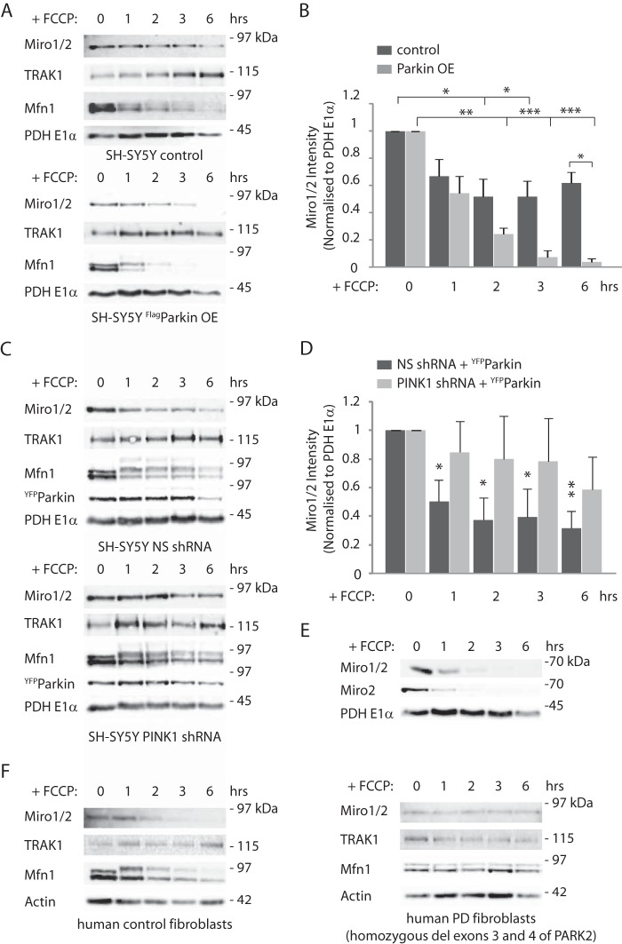
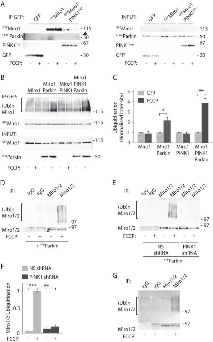
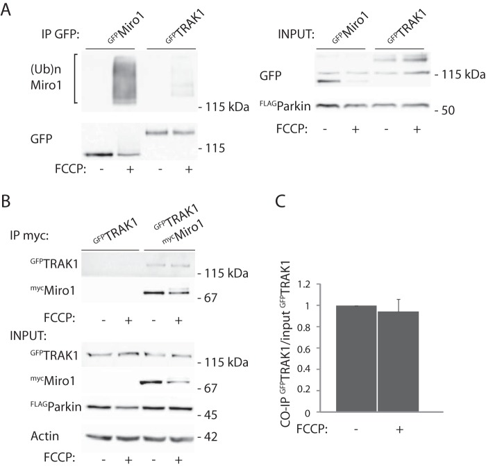
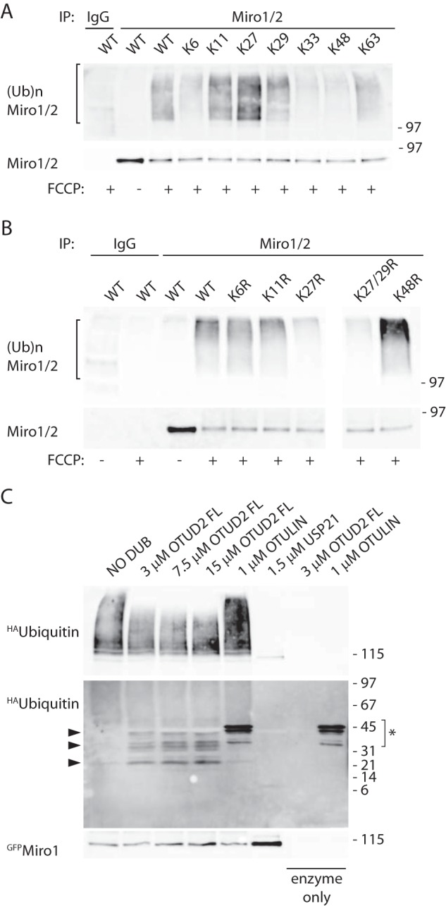
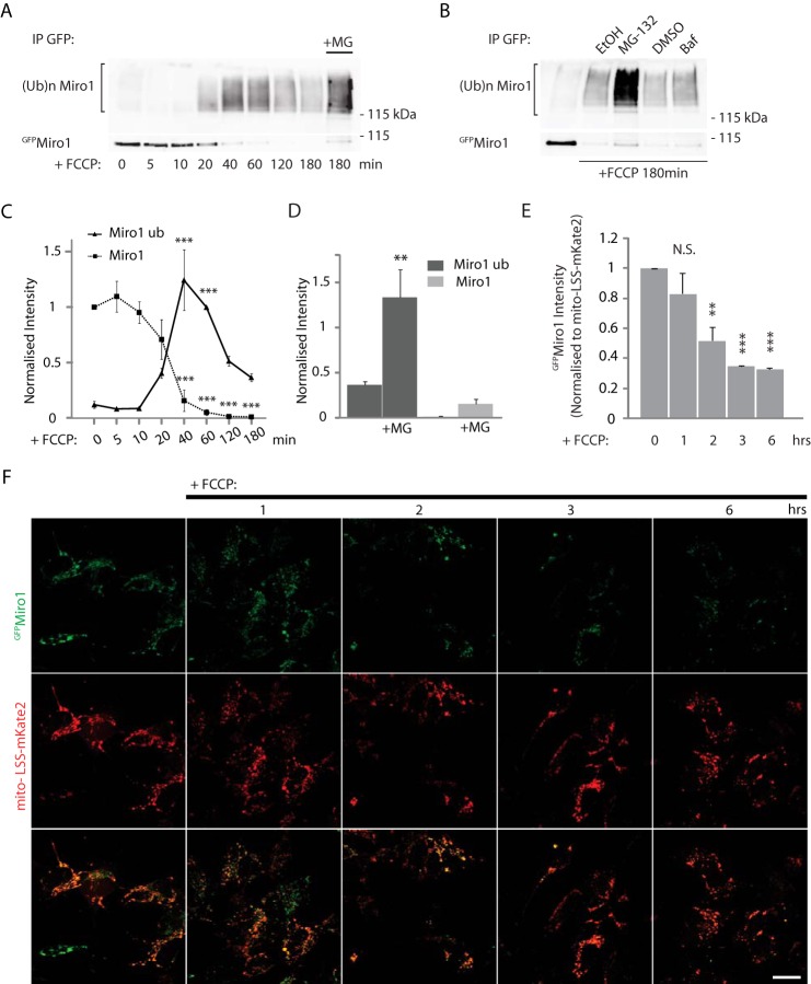
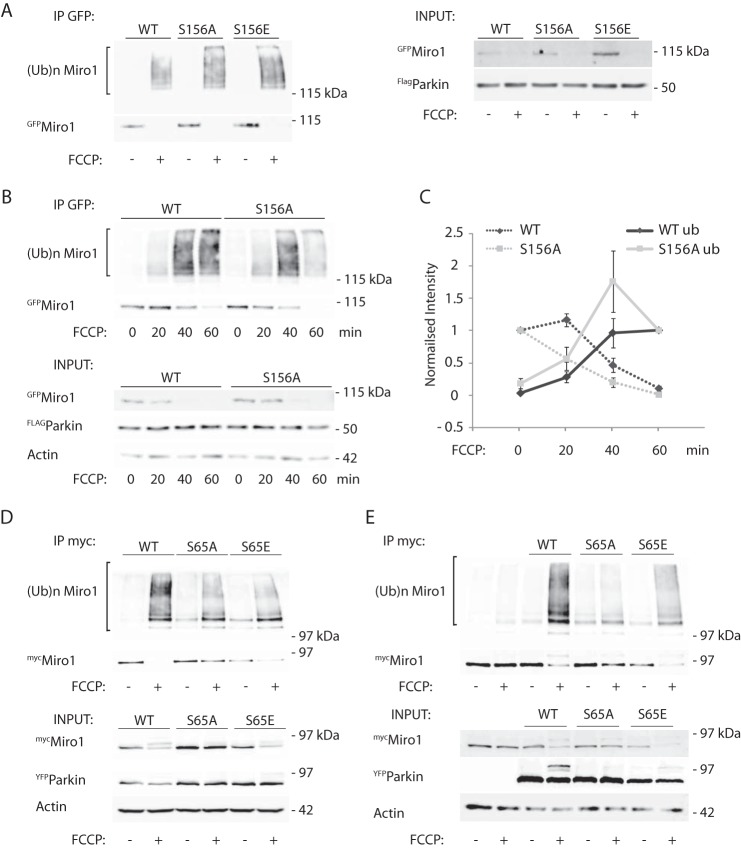
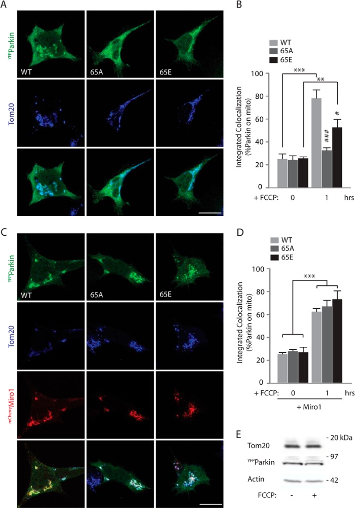
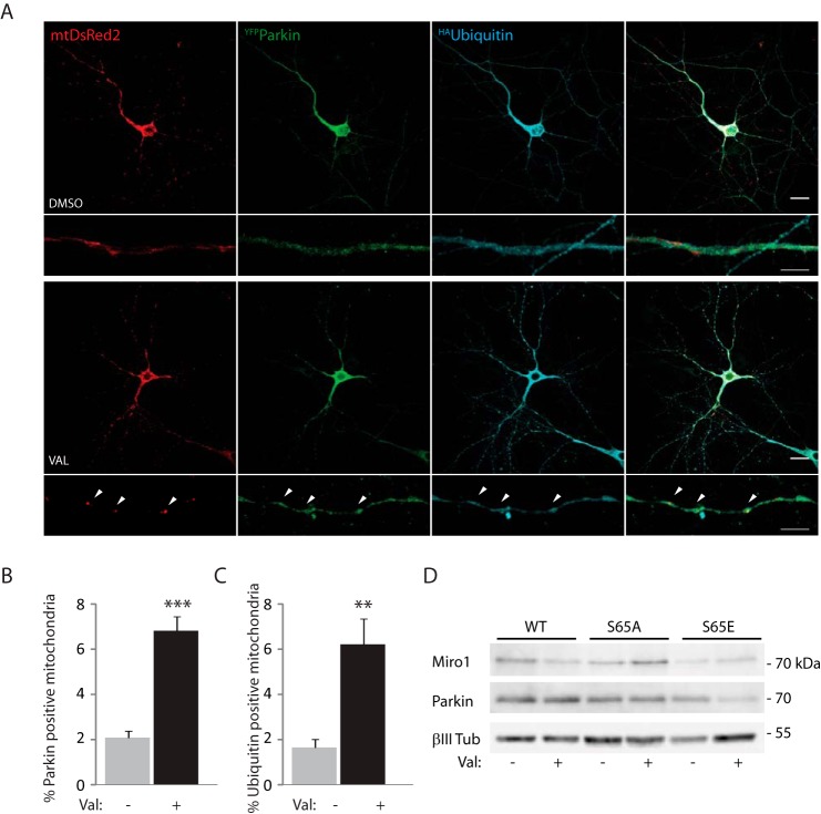
Mitochondrial transport plays an important role in matching mitochondrial distribution to localized energy production and calcium buffering requirements. Here, we demonstrate that Miro1, an outer mitochondrial membrane (OMM) protein crucial for the regulation of mitochondrial trafficking and distribution, is a substrate of the PINK1/Parkin mitochondrial quality control system in human dopaminergic neuroblastoma cells. Moreover, Miro1 turnover on damaged mitochondria is altered in Parkinson disease (PD) patient-derived fibroblasts containing a pathogenic mutation in the PARK2 gene (encoding Parkin). By analyzing the kinetics of Miro1 ubiquitination, we further demonstrate that mitochondrial damage triggers rapid (within minutes) and persistent Lys-27-type ubiquitination of Miro1 on the OMM, dependent on PINK1 and Parkin. Proteasomal degradation of Miro1 is then seen on a slower time scale, within 2-3 h of the onset of ubiquitination. We find Miro ubiquitination in dopaminergic neuroblastoma cells is independent of Miro1 phosphorylation at Ser-156 but is dependent on the recently identified Ser-65 residue within Parkin that is phosphorylated by PINK1. Interestingly, we find that Miro1 can stabilize phospho-mutant versions of Parkin on the OMM, suggesting that Miro is also part of a Parkin receptor complex. Moreover, we demonstrate that Ser-65 in Parkin is critical for regulating Miro levels upon mitochondrial damage in rodent cortical neurons. Our results provide new insights into the ubiquitination-dependent regulation of the Miro-mediated mitochondrial transport machinery by PINK1/Parkin and also suggest that disruption of this regulation may be implicated in Parkinson disease pathogenesis.

Keywords: Mitochondrial Transport; Mitophagy; Parkin; Parkinson Disease; Pink1; Ubiquitination.


Figures

FIGURE 1.
FIGURE 1.
Mitochondrial damage triggers PINK1/Parkin-dependent Miro1/2 degradation in a dopaminergic neuroblastoma cell line. A and B, control SH-SY5Y cells or stably overexpressing FLAGParkin SH-SY5Y (FLAGParkin OE) cells were incubated with FCCP (10 μm) for the indicated time. A, FCCP time course experiment indicating the loss of the outer membrane proteins Miro1/2 and Mfn1 but not of the trafficking adaptor TRAK1 or the mitochondrial matrix protein PDH E1α. Loss of Miro1/2 and Mfn1 is favored by Parkin overexpression. B, bar graph quantifying the FCCP-dependent loss of Miro1/2. Miro1/2 normalized mean intensity in control SH-SY5Y cells at 1 h FCCP 0.67 ± 0.12 S.E.; 2 h, 0.52 ± 0.13 S.E.; 3 h, 0.52 ± 0.12 S.E., and 6 h, 0.62 ± 0.08 S.E.; n = 6. Miro1/2 mean intensity in FLAGParkin OE SH-SY5Y cells at 1 h FCCP 0.54 ± 0.12 S.E.; 2 h, 0.24 ± 0.04 S.E.; 3 h, 0.07 ± 0.05; 6 h, 0.04 ± 0.02 S.E.; n = 3, *, p < 0.05; **, p < 0.01; ***, p < 0.001. C and D, SH-SY5Y cells stably expressing either nonsilencing (NS) shRNA or PINK1 shRNA and transfected with YFPParkin were treated with FCCP (10 μm) for the indicated time points. C, representative western blot of a FCCP time course. Miro1/2 loss is inhibited by the specific knockdown of PINK1. D, bar graph quantifying this effect. Miro1/2 normalized mean intensity in nonsilencing shRNAi at 1 h FCCP, 0.50 ± 0.15 S.E.; 2 h, FCCP 0.37 ± 0.16 S.E.; 3 h, FCCP 0.39 ± 0.20 S.E., and 6 h, FCCP 0.31 ± 0.12 S.E.; Miro1/2 mean intensity in PINK1 shRNAi expressing cells at 1 h FCCP 0.84 ± 0.22 S.E.; 2 h, FCCP 0.80 ± 0.30 S.E.; 3 h, FCCP 0.78 ± 0.30 S.E., and 6 h, FCCP 0.58 ± 0.23 S.E.; n = 4; significance is calculated comparing treated samples to control (0 h) *, p < 0.05; **, p < 0.01. E, SH-SY5Y cells stably overexpressing FLAGParkin were treated with FCCP (10 μm) for several time points. Miro1/2 and Miro2 show similar degradation kinetics, n = 3. F, FCCP treatment (10 μm) of human control fibroblasts triggers the loss of Miro1/2 as well as Mfn1 (left panel); however, FCCP-induced mitochondrial damage in human fibroblasts derived from a PD patient carrying a deletion of exons 3 and 4 of the PARK2 gene does not cause loss of Miro1/2 or Mfn1 (right panel).
FIGURE 2.
FIGURE 2.
GTPase Miro1 interacts with and is ubiquitinated by the PINK1-Parkin complex. A, coimmunoprecipitation experiments carried out in SH-SY5Y cells stably overexpressing FLAGParkin and transfected with GFP or GFPMiro1 and PINK1myc show that Miro1 interacts with PINK1, and mitochondrial depolarization (10 μm FCCP, 1 h) triggers the formation of a complex with Parkin. B and C, ubiquitination assays demonstrate that FCCP (10 μm, 1 h) triggers Parkin-dependent ubiquitination of exogenous GFPMiro1 in COS-7 cells, detected as a higher molecular weight smear of the immunopurified protein (amount of ubiquitinated Miro1 upon FCCP treatment in control cells 0.88 ± 0.23 S.E., in Parkin OE cells 2.17 ± 0.33 S.E., in PINK1 OE cells 0.88 ± 0.18 S.E., in PINK1 and Parkin OE cells 3.85 ± 0.48 S.E.; *, p = 0.027; **, p = 0.006, n = 4). D, FCCP triggers the ubiquitination of endogenous Miro1/2 in the neuroblastoma SH-SY5Y cell line, n = 3. E and F, this process is inhibited by the stable expression of a PINK1 RNAi (ubiquitinated Miro mean intensity in PINK1 shRNA cells 0.16 ± 0.07 S.E. compared with ubiquitinated Miro1 intensity in nonsilencing shRNA expressing cells 1.0; n = 3, **, p = 0.006; ***, p = 0.0008). G, ubiquitination of endogenous Miro1/2 can be detected with the endogenous ubiquitin antibody FK2, n = 3. IP, immunoprecipitation.
FIGURE 3.
FIGURE 3.
Effect of mitochondrial damage on TRAK1 ubiquitination and its interaction with Miro1. A, ubiquitination assays performed in SH-SY5Y cells stably overexpressing FLAGParkin and transfected with either GFPMiro1 or GFPTRAK1. FCCP treatment (10 μm, 1 h) triggers little ubiquitination of TRAK1 compared with Miro1; n = 3. B, coimmunopurification assay shows no change in the interaction between mycMiro1 and GFPTRAK1 upon mitochondrial damage. C, quantification of this effect (co-IP TRAK1 with FCCP 0.94 ± 0.11, compared to control 1); n = 3. IP, immunoprecipitation.
FIGURE 4.
FIGURE 4.
Analysis of Miro1 ubiquitin chains. A, ubiquitination assays performed in SH-SY5Y cells stably overexpressing FLAGParkin and transfected with either wild type HAubiquitin or several ubiquitin mutants that allow only specific chain types (Lys-6, Lys-11, Lys-27, Lys-29, Lys-33, Lys-48, and Lys-63) reveal that Miro1/2 is primarily ubiquitinated in a Lys-27-dependent manner; n = 3. B, same assay is carried out using HAubiquitin mutants that cannot mediate specific ubiquitin chain linkages (K6R, K11R, K27R, K27R/K29R, K48R; n = 3). C, immunopurified GFPMiro1 was incubated with increasing amounts of OTUD2 (that specifically cleaves Lys-11, Lys-27, Lys-29, and Lys-33, mediated ubiquitin linkages), Otulin (cleaves Met-linked chains) or USP21 (chain type aspecific DUB). Black arrowheads indicate HA-positive bands, likely to be ubiquitin trimers and dimers; asterisk indicates cross-reactivity between Otulin and the anti-HA antibody. IP, immunoprecipitation.
FIGURE 5.
FIGURE 5.
Temporal dynamics of Miro1 ubiquitination and degradation. A, FLAGParkin stably overexpressing SH-SY5Y cells were incubated with FCCP (10 μm) for several time points before ubiquitination assays were performed. GFPMiro1 ubiquitination can be detected after 20 min of incubation with the mitochondrial uncoupler (FCCP) and peaks after 40–60 min. Although the GFPMiro1 band disappears at 60 min, an actual decrease in the higher molecular weight species (ubiquitinated GFPMiro1) is detected only after 120 min. B, degradation is blocked by pretreatment with the proteasomal blocker MG-132 but not by the lysosomal inhibitor bafilomycin 1A. C, graph quantifying Miro1 bands (immunoprecipitated, dashed line) and ubiquitination smear intensity in A. Average intensities of Miro1 (immunoprecipitated (IP)) are normalized to Miro1 (IP) at 0 min, whereas Miro1 ubiquitination smear intensities are normalized to the smear intensity at 60 min. Mean intensity of ubiquitinated GFPMiro1 at 0 min, 0.12 ± 0.08; 5 min, 0.08 ± 0.03; 10 min, 0.09 ± 0.04; 20 min, 0.41 ± 0.09; 40 min 1.24 ± 0.23, 60 min 1; 120 min 0.52 ± 0.09; and 180 min 0.37 ± 0.08, n = 3, ***, p < 0.005. Data are shown as mean ± S.E. D, bar graph quantifying the effect of MG-132 in A, mean intensity of GFPMiro1 ubiquitination at 180 min with MG-132 1.33 ± 0.24 compared with 0.37 ± 0.08 at 180 min without MG-132, n = 3, **, p = 0.006. E and F, FLAGParkin overexpressing SH-SY5Y cells co-transfected with GFPMiro1 and the mitochondrial marker mito-LSS-mKate2. Confocal imaging of FCCP time course experiment shows a significant decrease of GFPMiro1 signal starting at 2 h, which is consistent with the biochemical data. E, quantification of GFPMiro1 intensity normalized to LSS-mito-mKate2 signal. Normalized GFPMiro1 intensity at 0 h, 1.0; 1 h, 0.83 ± 0.14; 2 h, 0.52 ± 0.09; 3 h, 0.35 ± 0.003; and at 6 h, 0.33 ± 0.01, n = 3, **, p < 0.01; ***, p < 0.005; scale bar, 20 μm. N.S. not significant. Data are shown as mean ± S.E.
FIGURE 6.
FIGURE 6.
Miro1 phosphorylation does not affect its ubiquitination dynamics, whereas Parkin phosphorylation does. A–C, GFPMiro1 phospho-null (S156A) or phospho-mimetic (S156E) mutants show no difference in ubiquitination dynamics compared with wild type Miro1 in Parkin OE SH-SY5Y cells; C, in ubiquitinated S156A Miro1 mean intensity at 0 min, 0.19 ± 0.08; at 20 min, 0.57 ± 0.17; at 40 min, 1.75 ± 0.47; at 60 min, 1.0, ubiquitinated wild type Miro1 mean intensity at 0 min, 0.04 ± 0.004; at 20 min, 0.29 ± 0.08; at 40 min, 0.96 ± 0.22; at 60 min, 1.0; n = 3. Data are shown as mean ± S.E. D, expression of the phospho-null YFPParkin S65A mutant in HeLa cells blocks FCCP-induced mycMiro1 ubiquitination and its subsequent loss. Phospho-mimetic S65E Parkin allows a residual Miro1 ubiquitination; n = 3. E, same effect is observed in SH-SY5Y dopaminergic cells; n = 1.
FIGURE 7.
FIGURE 7.
Miro1 facilitates the recruitment of Parkin S65A and S65E to damaged mitochondria. A, confocal images of HEK cells transfected with wild type YFPParkin or S65A or S65E Parkin mutants and treated with 10 μm FCCP for 1 h. Scale bar, 20 μm. B, Parkin recruitment to the mitochondria is analyzed as integrated colocalization of YFPParkin over the mitochondrial marker Tom20 and quantified in the bar graph. Percentage of wild type Parkin on mitochondria is 78.19 ± 4.86%; S65A Parkin is 33.27 ± 1.46%; S65E Parkin is 52.97 ± 4.68%; n = 9–14, #, p < 0.05; ###, p < 0.005. * is used to indicate significance between treated/untreated samples, whereas # is used to indicate significance within a treatment group, #, p < 0.05, **, p < 0.01; ###, ***p < 0.005. Data are shown as mean ± S.E. C, overexpression of mCherryMiro1 together with YFPParkin or the S65A and S65E mutants facilitates Parkin recruitment to the mitochondria after 1 h of FCCP treatment. Scale bar, 20 μm. D, quantification of this effect. Percentage of wild type Parkin on mitochondria at 1 h when Miro1 is overexpressed at 61.69 ± 2.55%; S65A Parkin is 63.55 ± 5.24%; S65E Parkin is 72.82 ± 4.85%; n = 12–14, ***, p < 0.005. Data are shown as mean ± S.E. E, western blot showing that the mitochondrial marker Tom20 levels are not altered after 1 h of FCCP treatment (10 μm).
FIGURE 8.
FIGURE 8.
Parkin is recruited to damaged mitochondria in neurons and determines Miro1 loss. A, confocal imaging of hippocampal neurons overexpressing YFPParkin, HAubiquitin, and the mitochondrial marker mtDsRed2 shows that valinomycin (Val) treatment (2 μm, 90 min) of the neuronal culture induces Parkin translocation to mitochondria and ubiquitination of the organelles (arrowheads; scale bar, 20 or 5 μm for the zoomed image). B and C, bar graphs quantifying, respectively, Parkin recruitment and ubiquitin-positive mitochondria (6.83 ± 0.63% of total mitochondria were Parkin-positive after 90 min of valinomycin compared with 2.08 ± 0.32% Parkin-positive mitochondria with DMSO control; ***, p < 0.001; 6.21 ± 1.14% of total mitochondria were ubiquitin-positive after 90 min of valinomycin treatment, compared with 1.65 ± 0.37% with DMSO control, **, p < 0.005; n = 3, 3 cells per n). Data are shown as mean ± S.E. D, mitochondrial damage (valinomycin, 2 μm, 90 min) triggers the loss of immunopurified mycMiro1 in cortical neurons overexpressing wild type Parkin but not S65A or S65E Parkin; n = 3.

References

    1. Macaskill A. F., Rinholm J. E., Twelvetrees A. E., Arancibia-Carcamo I. L., Muir J., Fransson A., Aspenstrom P., Attwell D., Kittler J. T. (2009) Miro1 is a calcium sensor for glutamate receptor-dependent localization of mitochondria at synapses. Neuron 61, 541–555 - PMC - PubMed
    1. Wang X., Schwarz T. L. (2009) The mechanism of Ca2+-dependent regulation of kinesin-mediated mitochondrial motility. Cell 136, 163–174 - PMC - PubMed
    1. Russo G. J., Louie K., Wellington A., Macleod G. T., Hu F., Panchumarthi S., Zinsmaier K. E. (2009) Drosophila Miro is required for both anterograde and retrograde axonal mitochondrial transport. J. Neurosci. 29, 5443–5455 - PMC - PubMed
    1. Saotome M., Safiulina D., Szabadkai G., Das S., Fransson A., Aspenstrom P., Rizzuto R., Hajnóczky G. (2008) Bidirectional Ca2+-dependent control of mitochondrial dynamics by the Miro GTPase. Proc. Natl. Acad. Sci. U.S.A. 105, 20728–20733 - PMC - PubMed
    1. Fransson A., Ruusala A., Aspenström P. (2003) Atypical Rho GTPases have roles in mitochondrial homeostasis and apoptosis. J. Biol. Chem. 278, 6495–6502 - PubMed

Publication types

MeSH terms

LinkOut - more resources