. 2014 May 9;289(19):13155-67.
doi: 10.1074/jbc.M113.530014. Epub 2014 Mar 26.

Hierarchical functional specificity of cytosolic heat shock protein 70 (Hsp70) nucleotide exchange factors in yeast

Affiliations

Hierarchical functional specificity of cytosolic heat shock protein 70 (Hsp70) nucleotide exchange factors in yeast

Jennifer L Abrams et al. J Biol Chem. .

Abstract

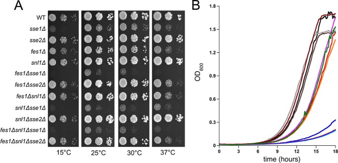
Heat shock protein 70 (Hsp70) molecular chaperones play critical roles in protein homeostasis. In the budding yeast Saccharomyces cerevisiae, cytosolic Hsp70 interacts with up to three types of nucleotide exchange factors (NEFs) homologous to human counterparts: Sse1/Sse2 (Heat shock protein 110 (Hsp110)), Fes1 (HspBP1), and Snl1 (Bag-1). All three NEFs stimulate ADP release; however, it is unclear why multiple distinct families have been maintained throughout eukaryotic evolution. In this study we investigate NEF roles in Hsp70 cell biology using an isogenic combinatorial collection of NEF deletion mutants. Utilizing well characterized model substrates, we find that Sse1 participates in most Hsp70-mediated processes and is of particular importance in protein biogenesis and degradation, whereas Fes1 contributes to a minimal extent. Surprisingly, disaggregation and resolubilization of thermally denatured firefly luciferase occurred independently of NEF activity. Simultaneous deletion of SSE1 and FES1 resulted in constitutive activation of heat shock protein expression mediated by the transcription factor Hsf1, suggesting that these two factors are important for modulating stress response. Fes1 was found to interact in vivo preferentially with the Ssa family of cytosolic Hsp70 and not the co-translational Ssb homolog, consistent with the lack of cold sensitivity and protein biogenesis phenotypes for fes1Δ cells. No significant consequence could be attributed to deletion of the minor Hsp110 SSE2 or the Bag homolog SNL1. Together, these lines of investigation provide a comparative analysis of NEF function in yeast that implies Hsp110 is the principal NEF for cytosolic Hsp70, making it an ideal candidate for therapeutic intervention in human protein folding disorders.

Keywords: Hsp70; Molecular Chaperone; Nucleotide Exchange Factor; Protein Degradation; Protein Folding; Protein Homeostasis; Protein Misfolding; Protein Stability; Saccharomyces cerevisiae; Yeast.


Figures

FIGURE 1.
FIGURE 1.
Growth analysis of wild type and nucleotide exchange factor deletion strains. A, serial dilutions of cells were plated onto rich (YPD) medium and incubated at the indicated temperatures. All other strains have the indicated genotypes. B, automated growth curves in liquid medium were generated as described under “Experimental Procedures.” WT, black; sse1Δ, blue; sse2Δ, gray; fes1Δ, red; snl1Δ, maroon; fes1Δ sse1Δ, yellow; fes1Δ sse2Δ, orange; fes1Δ snl1Δ, light purple; snl1Δ sse1Δ, light blue; snl1Δ sse2Δ, brown; fes1Δ snl1Δ sse1Δ, violet; fes1Δ snl1Δ sse2Δ, green.
FIGURE 2.
FIGURE 2.
Nucleotide exchange factor deletions differentially affect firefly luciferase GFP biogenesis. A, schematic of model folding construct firefly luciferase fused to GFP (FFL-GFP) and controlled by a methionine-repressible promoter. B, representative micrographs showing GFP only control (top panel) or steady state FFL-GFP fluorescence in log phase wild type or NEF single deletion strains (bottom panel). The FFL-GFP construct is grown in the presence of minimal methionine and is therefore not fully repressed, leading to low level expression. C, steady state FFL activity monitored in the same cells as B. D, de novo folding kinetics of wild type or NEF deletion strains monitored over 120 min. WT, black; sse1Δ, blue; sse2Δ, gray; fes1Δ, red; snl1Δ, maroon. Strains were shifted to methionine-free medium to fully induce FFL-GFP expression. E, Western blot of FFL-GFP protein levels from the same cells as in D. Monoclonal antibody against phosphoglycerate kinase (PGK) was used as a load control. RLU, relative light units.
FIGURE 3.
FIGURE 3.
Cytosolic nucleotide exchange factors are not required for luciferase refolding in vivo. A, schematic of refolding assay. B, representative micrographs showing GFP fluorescence for pre-heat shock cells and cells 0 (white bars in C) and 60 min (gray bars in C) after heat shock. GFP only controls are represented in the right panels, and they were visualized at 30 °C (before heat shock, pre-HS) or immediately after heat shock at 42 °C (after heat shock, post-HS). C, FFL activity from the same cells as in B. Refolding efficiency is calculated as a percentage of initial activity pre-heat shock. CHX, cycloheximide.
FIGURE 4.
FIGURE 4.
Sse1 and Fes1 contribute to regulation of the heat shock response through Hsf1. A, Hsf1 derepression in wild type, NEF single deletion strains, or the sse1Δfes1Δ strain monitored using an HSE-lacZ reporter. B, Western blot showing differential steady state expression of Hsf1 target proteins Cpr6, Hsp104, and Sti1, with PGK shown as a load control. C, growth analysis of wild type, sse1Δ, fes1Δ, and sse1Δfes1Δ strains in the presence or absence of proteotoxic stress caused by AZC.
FIGURE 5.
FIGURE 5.
Sse1 uniquely contributes to Hsp70-mediated protein degradation. A, Western blot of CPY-GFP degradation over a 2-h cycloheximide chase period. PGK was used as a load control. B, representative micrographs of wild type and NEF deletion cells from the time points sampled in A. C, quantitation of aggregate-containing fraction of the total population for each strain from B at 0 (light gray bars), 1 (dark gray bars), and 2 h (black bars) (n = ∼100 cells). D, Western blot analysis of CPY-GFP degradation in wild type and sse1Δ strains at control (30 °C) or heat shock (37 °C) temperatures.
FIGURE 6.
FIGURE 6.
Fes1 specifically interacts with Ssa chaperone in vivo. A, Coomassie Brilliant Blue (CBB, top panel) and Western blot (bottom panels) of in vitro immunoprecipitation (IP) of Hsp70 from wild type cell lysates added at concentrations of 0, 4.5, or 7.5 μg/μl with FLAG-Fes1-bound beads. Western analysis was done using anti-Ssa and anti-Ssb antibodies as indicated. B, Coomassie Brilliant Blue (top panel) and Western blot (bottom panels) of in vivo FLAG-Fes1 or FLAG-Sse1 immunoprecipitations in wild type cells. C, Coomassie Brilliant Blue (top panel) and Western blot (bottom panels) of in vivo FLAG-Fes1 immunoprecipitations from the indicated strains.
FIGURE 7.
FIGURE 7.
Model of nucleotide exchange factor roles in Hsp70-mediated protein biogenesis and quality control. See text for details. Translating ribosomes are depicted in orange, and the proteasome is depicted in blue and green.

Similar articles

Cited by

References

    1. Hartl F. U., Bracher A., Hayer-Hartl M. (2011) Molecular chaperones in protein folding and proteostasis. Nature 475, 324–332 - PubMed
    1. Soto C. (2003) Unfolding the role of protein misfolding in neurodegenerative diseases. Nat. Rev. Neurosci. 4, 49–60 - PubMed
    1. Mayer M. P. (2013) Hsp70 chaperone dynamics and molecular mechanism. Trends Biochem. Sci. 38, 507–514 - PubMed
    1. Swain J. F., Dinler G., Sivendran R., Montgomery D. L., Stotz M., Gierasch L. M. (2007) Hsp70 chaperone ligands control domain association via an allosteric mechanism mediated by the interdomain linker. Mol. Cell 26, 27–39 - PMC - PubMed
    1. Vogel M., Bukau B., Mayer M. P. (2006) Allosteric regulation of Hsp70 chaperones by a proline switch. Mol. Cell 21, 359–367 - PubMed

Publication types

MeSH terms