Review
. 2015:25:123-49.
doi: 10.1007/7854_2014_305.

Sleep and synaptic plasticity in the developing and adult brain

Affiliations
Review

Sleep and synaptic plasticity in the developing and adult brain

Marcos G Frank. Curr Top Behav Neurosci. 2015.

Abstract

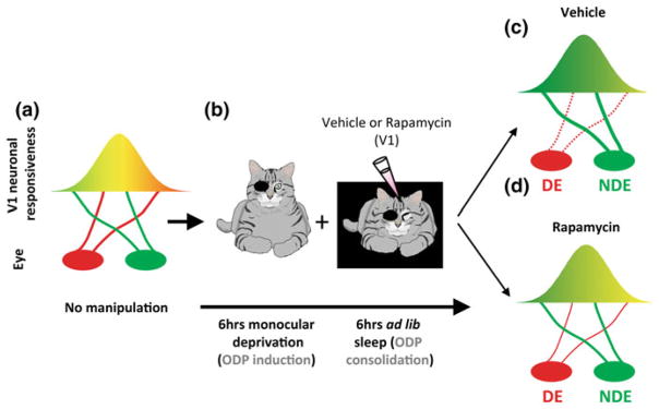
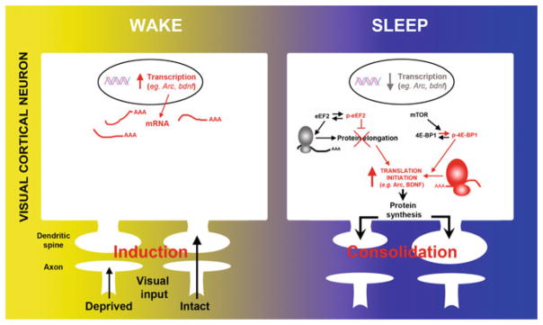
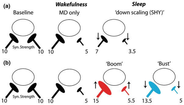
Sleep is hypothesized to play an integral role in brain plasticity. This has traditionally been investigated using behavioral assays. In the last 10-15 years, studies combining sleep measurements with in vitro and in vivo models of synaptic plasticity have provided exciting new insights into how sleep alters synaptic strength. In addition, new theories have been proposed that integrate older ideas about sleep function and recent discoveries in the field of synaptic plasticity. There remain, however, important challenges and unanswered questions. For example, sleep does not appear to have a single effect on synaptic strength. An unbiased review of the literature indicates that the effects of sleep vary widely depending on ontogenetic stage, the type of waking experience (or stimulation protocols) that precede sleep and the type of neuronal synapse under examination. In this review, I discuss these key findings in the context of current theories that posit different roles for sleep in synaptic plasticity.


Figures

Fig. 1
Fig. 1
Ocular dominance plasticity in the cat requires protein synthesis during sleep. a In developing cats with normal vision, most neurons in the primary visual cortex (V1) are binocular (i.e., equally responsive to inputs from either eye, represented as the yellow area). b When animals are deprived of patterned visual input in one eye (i.e., monocular deprivation) most neurons in V1 become responsive only to stimulation of the nondeprived eye (NDE). This canonical form of physiological plasticity is known as ocular dominance plasticity (ODP). It is induced very rapidly in awake cats (6 h) and is enhanced/consolidated by subsequent sleep (6 h). To test the role of protein synthesis in sleep-dependent ODP, visual cortices are infused with vehicle or the selective mammalian target of rapamycin (mTOR) inhibitor during the post-MD sleep period. c Sleep-dependent ODP is intact in the vehicle infused hemispheres and includes a maintenance of depression of the DE visual input (dotted red line) and potentiation of the NDE input (thick red line). d Inhibition of protein synthesis in V1 with rapamycin during post-MD sleep blocks sleep-dependent ODP. This essentially halts plastic changes at a stage induced by waking experience alone. Reproduced with permission from (Seibt and Frank 2012)
Fig. 2
Fig. 2
The transcription and translation of plasticity-related mRNAs are divided across wake and sleep. During wake, monocular deprivation triggers activity-dependent transcription of immediate-early and neurotrophin genes (e.g., arc, bdnf, c-fos) in V1. Subsequent sleep activates a cascade of translational events (increased translation initiation via 4E-BP1 phosphorylation and reduced global elongation via eEF2 phosphorylation) leading to a net increase in translation initiation of subsets of mRNA. Arc and bdnf are two examples of important plasticity-related genes where transcription is decreased and translation is increased during sleep. Reproduced with permission from (Seibt et al. 2012)
Fig. 3
Fig. 3
A “Boom and Bust” model of sleep-dependent plasticity explains the effects of sleep on ocular dominance plasticity. The initial effects of Monocular Deprivation (MD) in the cat are a weakening of responses to the deprived eye during wakefulness. After sleep, there is no further weakening in deprived eye circuits and instead responses to the nondeprived eye become stronger. a According to the Synaptic Homeostasis Hypothesis (SHY), sleep globally downscales synaptic strength in a manner proportionate to the strength at each synapse. This produces no net potentiation in the nondeprived circuits and increases depression in the deprived eye pathways. b According to the Boom and Bust model, sleep immediately after experience leads to synaptic potentiation (“Boom”). This is likely Hebbian, but may involve heterosynaptic changes due to synaptic tagging and capture of plasticity-related proteins in neighboring synapses (Redondo et al. 2010). As sleep progresses, global downscaling ensues, which reduces synaptic strength proportionately at each synapse (“Bust”). The net result is potentiation in the nondeprived eye pathways, and no further modifications in the deprived eye circuits, which fits empirical data. For illustration purposes, arbitrary units of synaptic strength are shown

References

    1. Aghajanian GK, Bloom FE. The formation of synaptic junctions in developing rat brain: a quantitative electron microscopic study. Brain Res. 1967;6:716–727. - PubMed
    1. Albensi BC, Oliver DR, Toupin J, Odero G. Electrical stimulation protocols for hippocampal synaptic plasticity and neuronal hyper-excitability: are they effective or relevant? Exp Neurol. 2007;204:1–13. - PubMed
    1. Alfoldi P, Tobler I, Borbely AA. Sleep regulation in rats during early development. Am J Physiol. 1990;258:R634–R644. - PubMed
    1. Arrigoni E, Lu J, Vetrivelan R, Saper CB. Long-term synaptic plasticity is impaired in rats with lesions of the ventrolateral preoptic nucleus. Eur J Neurosci. 2009;30:2112–2120. - PMC - PubMed
    1. Aton SJ, Broussard C, Dumoulin M, Seibt J, Watson A, Coleman T, Frank MG. Visual experience and subsequent sleep induce sequential plastic changes in putative inhibitory and excitatory cortical neurons. Proc Natl Acad Sci USA. 2013;110:3101–3106. - PMC - PubMed

LinkOut - more resources