. 2014 Jul;28(7):2840-51.
doi: 10.1096/fj.13-247254. Epub 2014 Mar 26.

Deimination of linker histones links neutrophil extracellular trap release with autoantibodies in systemic autoimmunity

Affiliations

Deimination of linker histones links neutrophil extracellular trap release with autoantibodies in systemic autoimmunity

Nishant Dwivedi et al. FASEB J. 2014 Jul.

Abstract

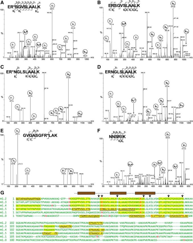
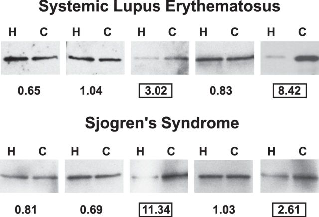
Autoantibodies to nuclear antigens arise in human autoimmune diseases, but a unifying pathogenetic mechanism remains elusive. Recently we reported that exposure of neutrophils to inflammatory conditions induces the citrullination of core histones by peptidylarginine deiminase 4 (PAD4) and that patients with autoimmune disorders produce autoantibodies that recognize such citrullinated histones. Here we identify histone H1 as an additional substrate of PAD4, localize H1 within neutrophil extracellular traps, and detect autoantibodies to citrullinated H1 in 6% of sera from patients with systemic lupus erythematosus and Sjögren's syndrome. No preference for deiminated H1 was observed in healthy control sera and sera from patients with scleroderma or rheumatoid arthritis. We map binding to the winged helix of H1 and determine that citrulline 53 represents a key determinant of the autoantibody epitope. In addition, we quantitate RNA for H1 histone subtypes in mature human neutrophils and identify citrulline residues by liquid chromatography and tandem mass spectrometry. Our results indicate that deimination of linker histones generates new autoantibody epitopes with enhanced potential for stimulating autoreactive human B cells.-Dwivedi, N., Neeli, I., Schall, N., Wan, H., Desiderio, D. M., Csernok, E., Thompson, P. R., Dali, H., Briand, J.-P., Muller, S., Radic, M. Deimination of linker histones links neutrophil extracellular trap release with autoantibodies in systemic autoimmunity.

Keywords: autoimmune disorders; chromatin; inflammation; peptidylarginine deiminase.


Figures

Figure 1.
Figure 1.
NETosis and histone deimination. A–D) NETosis was induced by treating purified neutrophils with calcium ionophore and chelerythrine for 1 h. Cells were fixed and bound by antibodies to deiminated histone H3 (dH3; A), to deiminated histone H4 (dH4; B), and to the linker histone H1 (C). Antibody binding was detected with fluorescence-conjugated secondary antibodies, (displayed in red), and DNA is shown in blue. The overlap between antibody binding and DNA staining thus yields violet. Control cells reacted poorly with antibody to dH3 (D). E, F) The extent of histone deimination in cells incubated in calcium buffer (Ca), ionophore (Io), or ionophore with chelerythrine (I/C) was detected with antibody to dH3 (E) or antibody to dH4 (F). Bands whose migration corresponds to dH3 and dH4 are marked by arrowheads. G) The total cell lysates (Tot. Prot.) from cells treated as in E and F were analyzed by SDS-PAGE, as were acid-extracted (Ac. Ex.) linker histones H1. Bands corresponding to H1 are indicated by a bracket. H) The same fractions as are shown in G were blotted to membrane, chemically modified, and probed with an antibody to modified citrulline. I) Only H1 histones from cells treated with ionophore and chelerythrine reacted with antibody to modified citrullines.
Figure 2.
Figure 2.
MS/MS analysis of H1 peptides from activated neutrophils A–F) MS/MS spectra of peptides yielding doubly charged precursor ions of H1 histone from Ca2+ ionophore-treated neutrophils. Fragmented ions (b series and y series) are marked on the peptide sequences or spectral peaks. Both peptide ER*SGVSLAALK (A) and ERSGVSLAALK (B) were observed, with a +1 Da shift resulting from deimination of arginine in evidence in b2 and b10 ions. The peptide ERSGVSLAALK is conserved between H1.2, H1.3, and H1.4 subtypes. Both ER*NGLSLAALK (C) and its nondeiminated form ERNGLSLAALK (D) derived from H1.5 were identified. Again, the mass shift of corresponding b ions is consistent with arginine deimination. Arginine-containing peptide GVGASGFR*LAK (E) from H1.0 was also identified, whereas peptide NNSRIK (F), conserved in subtypes H1.1–H1.5, was found only in its nondeiminated form. # indicates -NH3; @ indicates -H2O; ++ indicates a doubly charged ion. G) Summary of results obtained by MS is shown using sequences of 6 human H1 histones. Sequences of somatic linker histone H1 subtypes were aligned to maximize homology. Arginine residues are indicated in red. Peptides highlighted in yellow were identified by mass spectrometry, and H1 subtype-specific peptide sequences are underlined. The globular domain of H1 is indicated by a broken line above the sequences. Brown boxes represent helices that arrange into the H1 winged helix. Residues contributing to DNA binding (50) are indicated with arrows.
Figure 3.
Figure 3.
MS/MS analysis of in vitro deiminated recombinant H1.2. MS/MS spectra of doubly charged precursor ions of peptides obtained by LysC protease digestion of deiminated histone H1.2. Fragmented ions (b series and y series) are marked on respective peptide sequences and on the MS/MS spectra. A, B) Two of 3 arginines in H1.2, those contained in peptide ER*SGVSLAALK (A) and AGGTPR*K (B), showed mass consistent with deimination to citrulline. C) The peptide NNSRIK showed no evidence of deimination during the in vitro PAD4 reaction. # indicates -NH3; @ indicates -H2O; ++ indicates a doubly charged ion.
Figure 4.
Figure 4.
Preferential binding of autoimmune sera to deiminated H1.2. Equal amounts of nondeiminated (H) and deiminated (C) H1.2 were blotted on nitrocellulose and probed with sera from SLE and SS patients. Five representative blots from each set are shown. The ratio of binding to deiminated vs. nondeiminated H1.2 (C/H) was calculated, and values are listed below each blot. Two sera each of SLE and SS syndrome showed a >2.0 binding preference for deiminated H1.2.
Figure 5.
Figure 5.
Peptide inhibition of SLE6 binding to deiminated H1.2. A) The preferential binding of autoimmune sera to deiminated H1.2 was assessed after inhibition by either of 2 synthetic peptides. Equal amounts of deiminated (Cit H1.2) and nondeiminated (H1.2) linker histone were analyzed by SDS-PAGE (Coomassie) or blotted and probed with SLE6, SS63, and SS64 sera. Panels show results of Western blots performed following a preincubation in the presence of competitor A or B. Competitor B (R53Cit) was more effective at blocking preferential binding of SLE6 and SS63 to deiminated H1.2. B) The peptides were designed to match H1.2 with citrulline residues substituting for arginine 32 or 53.
Figure 6.
Figure 6.
Neutrophil activation increases SLE6 reactivity. A–C) SLE6 IgG binding (red) to unstimulated or LPS-treated neutrophils was compared to the binding of antideiminated H3 antibody (green) and the DNA binding dye, sytox orange (blue). A) Faint SLE6 binding is observed to the cytoplasm of untreated neutrophils. B, C) SLE6 binding is increased in LPS-treated neutrophils that have lost the integrity of nuclear membrane (B) and show increased reactivity with the antideiminated H3 antibody (C). D) SLE6 IgG binds to NETs that react with the antideiminated H3, but prominent cytoplasmic clusters of SLE6 binding were also observed. The micrographs are representative of SLE6 IgG binding to activated neutrophils. Lysates of unstimulated (U) and Ca2+ ionophore-treated (S) neutrophils were probed with SLE6 serum. Enhanced binding of IgG to a doublet of proteins migrating to a position near the 37kD marker is indicated (arrow). E) Equal amounts of nondeiminated (H) and deiminated (C) H1.2 and BSA were analyzed by Coomassie staining or probed with SLE6 serum. Preferential binding of SLE6 to deiminated H1.2 over nondeiminated H1.2 and no detectable binding to deiminated BSA indicate the contribution of flanking protein sequences to the specificity determinants of SLE6 IgG (B).
Figure 7.
Figure 7.
Analysis of linker histone transcripts from purified neutrophils. Relative mRNA levels of different H1 subtypes were determined from total mRNA extracted from neutrophils. cDNA was prepared by reverse transcription, and expression levels were measured on an Affymetrix Whole-Transcript Human Gene 1.0 ST Array. Relative fluorescence intensities were plotted for each H1 subtype.

References

    1. Muller S., Dieker J., Tincani A., Meroni P. L. (2008) Pathogenic anti-nucleosome antibodies. Lupus 17, 431–436 - PubMed
    1. Fauchais A. L., Martel C., Gondran G., Lambert M., Launay D., Jauberteau M. O., Hachulla E., Vidal E., Hatron P. Y. (2010) Immunological profile in primary Sjögren syndrome: clinical significance, prognosis and long-term evolution to other auto-immune disease. Autoimm. Rev. 9, 595–599 - PubMed
    1. El-Azhary R. A., Aponte C. C., Nelson A. M., Weaver A. L., Homburger H. A. (2006) Antihistone antibodies in linear scleroderma variants. Int. J. Dermatol. 45, 1296–1299 - PubMed
    1. Neeli I., Khan S. N., Radic M. (2008) Histone deimination as a response to inflammatory stimuli in neutrophils. J. Immunol. 180, 1895–1902 - PubMed
    1. Wang Y., Li M., Stadler S., Correll S., Li P., Wang D., Hayama R., Leonelli L., Han H., Grigoryev S. A., Allis C. D., Coonrod S. A. (2009) Histone hypercitrullination mediates chromatin decondensation and neutrophil extracellular trap formation. J. Cell Biol. 184, 205–213 - PMC - PubMed

Publication types