. 2014 Jul;28(7):2790-803.
doi: 10.1096/fj.13-244210. Epub 2014 Mar 26.

Inhibition of Janus kinase signaling during controlled mechanical ventilation prevents ventilation-induced diaphragm dysfunction

Affiliations

Inhibition of Janus kinase signaling during controlled mechanical ventilation prevents ventilation-induced diaphragm dysfunction

Ira J Smith et al. FASEB J. 2014 Jul.

Abstract

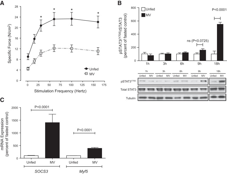
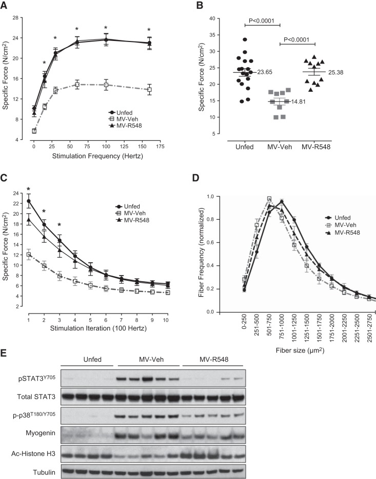
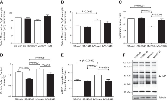
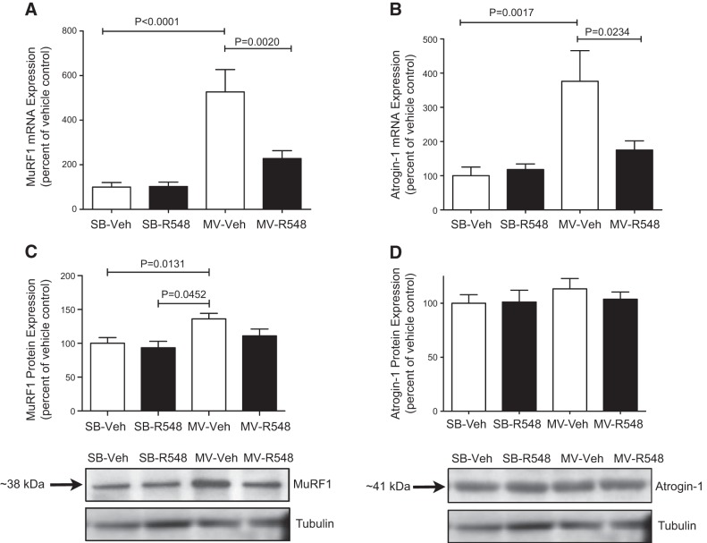
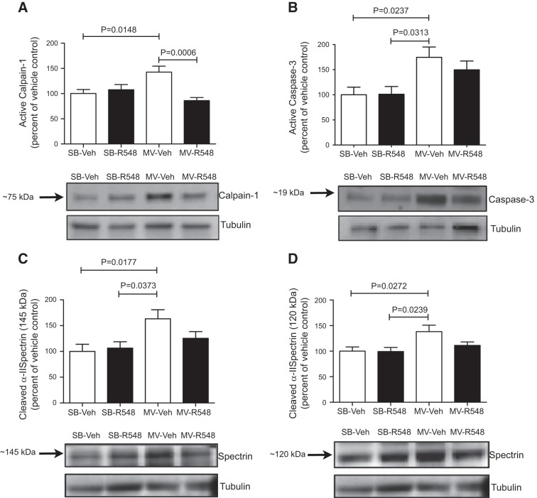
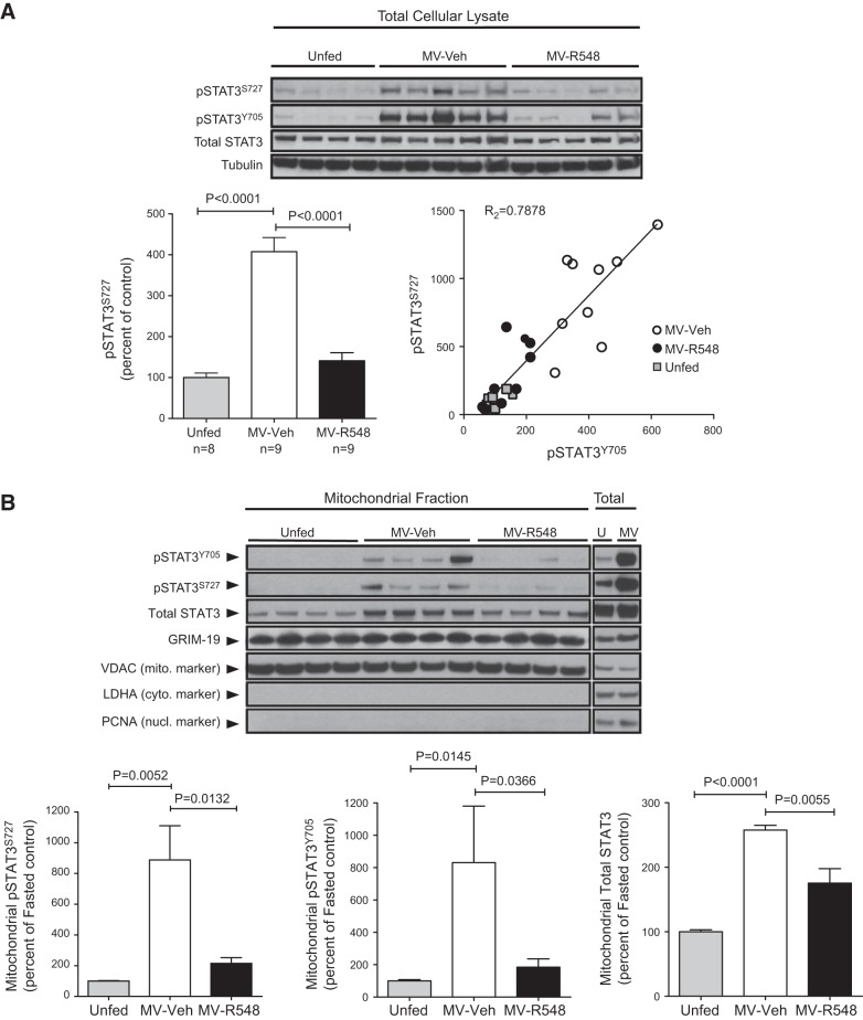
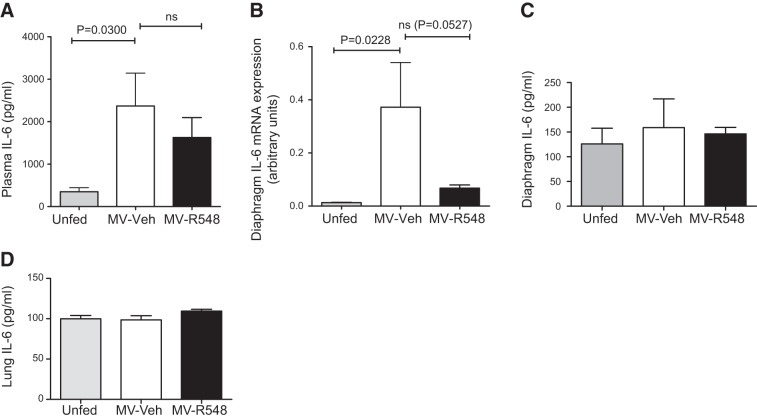
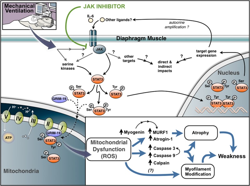
Controlled mechanical ventilation (CMV) is associated with the development of diaphragm atrophy and contractile dysfunction, and respiratory muscle weakness is thought to contribute significantly to delayed weaning of patients. Therefore, therapeutic strategies for preventing these processes may have clinical benefit. The aim of the current study was to investigate the role of the Janus kinase (JAK)/signal transducer and activator of transcription 3 (STAT3) signaling pathway in CMV-mediated diaphragm wasting and weakness in rats. CMV-induced diaphragm atrophy and contractile dysfunction coincided with marked increases in STAT3 phosphorylation on both tyrosine 705 (Tyr705) and serine 727 (Ser727). STAT3 activation was accompanied by its translocation into mitochondria within diaphragm muscle and mitochondrial dysfunction. Inhibition of JAK signaling during CMV prevented phosphorylation of both target sites on STAT3, eliminated the accumulation of phosphorylated STAT3 within the mitochondria, and reversed the pathologic alterations in mitochondrial function, reduced oxidative stress in the diaphragm, and maintained normal diaphragm contractility. In addition, JAK inhibition during CMV blunted the activation of key proteolytic pathways in the diaphragm, as well as diaphragm atrophy. These findings implicate JAK/STAT3 signaling in the development of diaphragm muscle atrophy and dysfunction during CMV and suggest that the delayed extubation times associated with CMV can be prevented by inhibition of Janus kinase signaling.-Smith, I. J., Godinez, G. L., Singh, B. K., McCaughey, K. M., Alcantara, R. R., Gururaja, T., Ho, M. S., Nguyen, H. N., Friera, A. M., White, K. A., McLaughlin, J. R., Hansen, D., Romero, J. M., Baltgalvis, K. A., Claypool, M. D., Li, W., Lang, W., Yam, G. C., Gelman, M. S., Ding, R., Yung, S. L., Creger, D. P., Chen, Y., Singh, R., Smuder, A. J., Wiggs, M. P., Kwon, O.-S., Sollanek, K. J., Powers, S. K., Masuda, E. S., Taylor, V. C., Payan, D. G., Kinoshita, T., Kinsella, T. M. Inhibition of Janus kinase signaling during controlled mechanical ventilation prevents ventilation-induced diaphragm dysfunction.

Keywords: STAT3; mitochondria; muscle wasting; oxidative stress.


Figures

Figure 1.
Figure 1.
CMV results in activated phosphorylation of STAT3 on Tyr705 and diaphragm contractile dysfunction. Diaphragms from mechanically ventilated (MV) rats and unfed controls were analyzed. A) Ex vivo force–frequency relationship from mechanically ventilated (n=13) rats and unfed controls (n=7). B) Western blots for total STAT3 and phospho-STAT3Y705 from a representative set of mechanically ventilated rats and unfed controls (1, 3, 6, and 9 h, n=5–6 rats/group; 18 h, n=6–8). C) Messenger RNA levels of the STAT3 downstream target genes SOCS3 and Myf5 from mechanically ventilated (n=9) rats and unfed controls (n=10). Mechanical ventilation period of 18 h. Results are means ± sem. *P < 0.05; Student's t test.
Figure 2.
Figure 2.
Treatment with a JAK1/JAK3 inhibitor prevents controlled mechanical VIDD and atrophy. Diaphragms from unfed control rats (n=17), MV-R548 rats (n=11), or MV-Veh rats (n=9) were analyzed. A) Diaphragm strip ex vivo force–frequency relationship. *P < 0.05, control and MV-R548 vs. MV-Veh; 2-way ANOVA. B, C) Specific force generation at 100 Hz (B) and (C) fatigue development after 18 h of CMV. *P < 0.05, control vs. MV-Veh; 2-way ANOVA. D) Muscle fiber CSA from a representative set of unfed control rats (n=9), MV-R548 rats (n=7), or MV-Veh rats (n=8). E) Representative Western blot from unfed control, MV-R548, and MV-Veh rats. Mechanical ventilation period of 18 h. Results are means ± sem.
Figure 3.
Figure 3.
Treatment with a JAK1/JAK3 inhibitor prevents CMV-induced mitochondrial dysfunction and oxidative damage. A–C) State 3 (A), State 4 (B), and RCR (C) in saponin-permeabilized diaphragm fibers of SB-Veh, SB-R548, MV-Veh, and MV-R548 rats. D, E) Protein carbonyl content (D) and 4-HNE-conjugated proteins (E) in total diaphragm homogenates. F) Representative blot for 4-HNE. CMV period of 12 h. Values are means ± sem (n=10/group). P values calculated by 1-way ANOVA with Tukey's post hoc analysis.
Figure 4.
Figure 4.
Treatment with a JAK1/JAK3 inhibitor prevents CMV-induced mRNA up-regulation of atrophic E3 ubiquitin ligases. Messenger RNA levels of E3 ubiquitin ligases MuRF1 (A) and atrogin-1 (B) mRNA levels and protein levels of MuRF1 (C) and atrogin-1 (D) in the diaphragms of SB-Veh, SB-R548, MV-Veh, and MV-R548 rats. CMV period of 12 h. Values are means ± sem (n=10/group). P values calculated by 1-way ANOVA with Tukey's post hoc analysis.
Figure 5.
Figure 5.
Treatment with a JAK1/JAK3 inhibitor attenuates activation of key proteases during CMV. Protein levels of active calpain-1 (A), active caspase-3 (B), calpain-cleaved 145 kDa α-II spectrin breakdown product (C), and 120 kDa caspase-3-cleaved α-II spectrin breakdown product (D) in the diaphragms of SB-Veh, SB-R548, MV-Veh, and MV-R548 rats. Mechanical ventilation period of 12 h. Values are means ± sem (n=10/group). P values calculated by 1-way ANOVA with Tukey's post hoc analysis.
Figure 6.
Figure 6.
CMV increases Ser705 phosphorylation and mitochondrial accumulation of phospho-STAT3 (Ser727 and/or Tyr705/Ser727) within diaphragm muscle. A) Diaphragms from unfed control rats (n=8), MV-R548 rats (n=9), or MV-Veh rats (n=9) were analyzed by Western blot of total muscle lysate (representative animals depicted in blot) and quantitation. Correlation between increases in phospho-STAT3S727 and phospho-STAT3Y705 were determined. B) Purified mitochondrial fractions (mito) and total lysates (total) were subjected to Western blot analysis for pSTAT3S727, pSTAT3Y705, total STAT3, GRIM-19, VDAC (mitochondrial marker), LDHA (cytoplasmic marker) and PCNA (nuclear marker). Unfed (U) controls, n = 10; MV-Veh (MV), n = 6; MV-R548, n = 7. Results are means ± sem. Mechanical ventilation period of 18 h. P values calculated by 1-way ANOVA with Tukey's post hoc analysis.
Figure 7.
Figure 7.
CMV increases plasma IL-6. A) Plasma IL-6 levels in unfed control (n=8) MV-Veh (n=6), and MV-R548 (n=10) rats. B) Messenger RNA levels of IL-6 in diaphragm of control (n=10), MV-Veh (n=9), and MV-R548 (n=10) rats. C, D) Protein levels of IL-6 in diaphragm (C) and lung (D) of control (n=8), MV-Veh (n=7), and MV-R548 (n=9) rats. CMV period of 18 h. Values are means ± sem. P values calculated by 1-way ANOVA with Tukey's post hoc analysis.
Figure 8.
Figure 8.
Proposed model for role of JAK signaling in VIDD. JAK signaling is activated by CMV and functions as a critical triggering mechanism upstream of STAT3 phosphorylation (Tyr705 and Ser705), mitochondrial dysfunction, ROS production, atrophy, and muscle weakness. Mechanical ventilation results in the mitochondrial accumulation of phospho-STAT3 (singly phosphorylated on Ser727 or doubly phosphorylated at both Ser727 and Tyr705). Import into mitochondria is facilitated through interaction of phospho-STAT3 with GRIM-19, a component of complex I of the ETC, and may directly affect mitochondrial function and ROS generation. Induction of various myogenic transcription factors and muscle-specific E3 ubiquitin ligases (MURF-1 and atrogin-1) and activation of calpain, caspase 9, and caspase 3 can contribute to muscle atrophy and proteolytic cleavage of myofilament proteins. Mitochondrial dysfunction may also lead to ROS-mediated modification of myofilament proteins in a manner that negatively affects contractile function.

References

    1. Wunsch H., Linde-Zwirble W. T., Angus D. C., Hartman M. E., Milbrandt E. B., Kahn J. M. (2010) The epidemiology of mechanical ventilation use in the United States. Crit. Care Med. 38, 1947–1953 - PubMed
    1. Esteban A., Anzueto A., Alia I., Gordo F., Apezteguia C., Palizas F., Cide D., Goldwaser R., Soto L., Bugedo G., Rodrigo C., Pimentel J., Raimondi G., Tobin M. J. (2000) How is mechanical ventilation employed in the intensive care unit? An international utilization review. Am. J. Respir. Crit. Care Med. 161, 1450–1458 - PubMed
    1. Esteban A., Anzueto A., Frutos F., Alia I., Brochard L., Stewart T. E., Benito S., Epstein S. K., Apezteguia C., Nightingale P., Arroliga A. C., Tobin M. J. (2002) Characteristics and outcomes in adult patients receiving mechanical ventilation: a 28-day international study. JAMA 287, 345–355 - PubMed
    1. Vassilakopoulos T., Petrof B. J. (2004) Ventilator-induced diaphragmatic dysfunction. Am. J. Respir. Crit. Care Med. 169, 336–341 - PubMed
    1. Levine S., Nguyen T., Taylor N., Friscia M. E., Budak M. T., Rothenberg P., Zhu J., Sachdeva R., Sonnad S., Kaiser L. R., Rubinstein N. A., Powers S. K., Shrager J. B. (2008) Rapid disuse atrophy of diaphragm fibers in mechanically ventilated humans. N. Engl. J. Med. 358, 1327–1335 - PubMed