. 2014 Mar 26;34(13):4509-18.
doi: 10.1523/JNEUROSCI.5071-13.2014.

A major external source of cholinergic innervation of the striatum and nucleus accumbens originates in the brainstem

Affiliations

A major external source of cholinergic innervation of the striatum and nucleus accumbens originates in the brainstem

Daniel Dautan et al. J Neurosci. .

Abstract

Cholinergic transmission in the striatal complex is critical for the modulation of the activity of local microcircuits and dopamine release. Release of acetylcholine has been considered to originate exclusively from a subtype of striatal interneuron that provides widespread innervation of the striatum. Cholinergic neurons of the pedunculopontine (PPN) and laterodorsal tegmental (LDT) nuclei indirectly influence the activity of the dorsal striatum and nucleus accumbens through their innervation of dopamine and thalamic neurons, which in turn converge at the same striatal levels. Here we show that cholinergic neurons in the brainstem also provide a direct innervation of the striatal complex. By the expression of fluorescent proteins in choline acetyltransferase (ChAT)::Cre(+) transgenic rats, we selectively labeled cholinergic neurons in the rostral PPN, caudal PPN, and LDT. We show that cholinergic neurons topographically innervate wide areas of the striatal complex: rostral PPN preferentially innervates the dorsolateral striatum, and LDT preferentially innervates the medial striatum and nucleus accumbens core in which they principally form asymmetric synapses. Retrograde labeling combined with immunohistochemistry in wild-type rats confirmed the topography and cholinergic nature of the projection. Furthermore, transynaptic gene activation and conventional double retrograde labeling suggest that LDT neurons that innervate the nucleus accumbens also send collaterals to the thalamus and the dopaminergic midbrain, thus providing both direct and indirect projections, to the striatal complex. The differential activity of cholinergic interneurons and cholinergic neurons of the brainstem during reward-related paradigms suggest that the two systems play different but complementary roles in the processing of information in the striatum.

Keywords: cholinergic; innervation; laterodorsal tegmental nucleus; pedunculopontine nucleus; striatum; topography.


Figures

Figure 1.
Figure 1.
Distribution of cholinergic axons in the striatal complex after viral vector injections into different groups of cholinergic neurons in the brainstem. Plots in the sagittal plane of the distribution of cholinergic axonal profiles from three representative animals that received viral vector injections in the rostral PPN (PPNr), the caudal PPN (PPNc), and the LDT. Injections in the rostral PPN gave rise to a dense innervation of the lateral striatum. Injections in the caudal PPN gave rise to a sparser innervation, with a tendency to innervate more medial regions of the striatum and parts of the NA. In contrast, injections into the LDT led to dense labeling in the most medial striatum and the NA core. Each injection led to labeling in the olfactory tubercle. Each red dot represents at least one immunopositive axonal profile in a bin of 40 μm2. The corresponding template (right) illustrates the ML levels (millimeters from the midline) and subdivisions of the striatum (DS, dorsal striatum), NA core (NA-core) and shell (NA-shell), and olfactory tubercle (OT) according to the stereotaxic rat brain atlas of Paxinos and Watson (1986). D, Dorsal; C, caudal. Scale bar, 2 mm.
Figure 2.
Figure 2.
Cholinergic axons arising in the rostral PPN preferentially innervate the lateral striatum. A, YFP-immunopositive axons arising from cholinergic neurons after the deposit of AAV2–EF1a–DIO–hChR2–YFP in the rostral PPN of a ChAT::Cre+ rat. The labeled cholinergic axons formed dense regions of innervation in the dorsolateral striatum. The green box in the outline represents the area in which the image was acquired. B, MOR immunolabeling to identify the striosomes. The distribution of the cholinergic axons was primarily confined to the MOR-negative matrix compartment. C, The cholinergic axons gave rise to a large number of varicosities, some of which were identified as VAChT immunopositive (arrowhead; see inset). D, Electron micrograph of a YFP-immunopositive cholinergic bouton (b) forming an asymmetric (Gray's type 1) synapse (arrow) with a dendritic shaft (d). E, Probability plot, calculated from all animals (n = 3) at three different ML levels (3.18, 2.10, and 1.13 mm from the midline, from top to bottom), of cholinergic axons arising in the rostral PPN. Cholinergic axons from this region of the PPN were more densely distributed in the lateral aspects of the dorsal striatum. Scale bars: A, 250 μm; B, 250 μm; C, 10 μm; D, 0.4 μm; E, 1000 μm.
Figure 3.
Figure 3.
Cholinergic axons arising in the caudal PPN project diffusely across the striatum and NA. A, YFP-immunopositive axons arising from cholinergic neurons in the caudal PPN form small patches mainly in the dorsal part of the striatum. The green box in the outline represents the area in which the image was acquired. B, Cholinergic axons from caudal PPN also avoided the striosomes. C, These cholinergic axons (top) gave rise to a smaller number of varicosities than those of the rostral PPN but were always immunopositive for VAChT (bottom). D, Electron micrograph of an immunopositive cholinergic (YFP-positive) bouton (b) establishing symmetric (Gray's type 2) synaptic contact (arrow) with the neck or base of a dendritic (d) spine (sp). E, Probability plot showing that cholinergic axons arising in the caudal PPN have a lower density distribution than those arising in the rostral PPN, with higher values in the lateral striatum, mainly in the more dorsal regions, and in the NA core and shell (n = 3; at three different ML levels as in Fig. 2). Scale bars: A, 250 μm; B, 250 μm; C, 10 μm; D, 0.2 μm; E, 1000 μm.
Figure 4.
Figure 4.
Cholinergic axons arising in the LDT preferentially innervate the medial striatum and the NA core. A, YFP-immunopositive axons from cholinergic neurons in the LDT form dense regions of innervation in the most medial levels of the striatum and NA core (depicted here). The green box in the outline represents the area in which the image was acquired. B, MOR immunolabeling in the NA shell revealed that cholinergic axons from the LDT tend to avoid striosomes. C, Cholinergic axons from the LDT formed large en passant varicosities that were immunopositive for VAChT (arrowheads). D, Electron micrograph of a cholinergic axon bouton (YFP-immunopositive; b) forming asymmetric (Gray's type 1) synapses (arrows) with a spine (sp). E, Probability plot showing that cholinergic axons arising in the LDT were more densely distributed in the medial striatum and the core and medial shell of the NA (n = 3; at three different ML levels as in Figs. 2, 3). Scale bars: A, 250 μm; B, 100 μm; C, 10 μm; D, 0.2 μm; E, 1000 μm.
Figure 5.
Figure 5.
Morphology of synapses differentiates brainstem cholinergic from cholinergic interneurons contacts. A–C, Electron micrographs of cholinergic boutons (b) arising from the rostral PPN (YFP-immunopositive) forming asymmetric (Gray's type 1) synapses with prominent (A) and moderate (B) postsynaptic densities onto a spine (sp) and dendritic shaft (d), respectively. C, A PPN cholinergic terminal forming a symmetrical (Gray's type 2) synapse with a dendritic shaft. D, YFP-immunopositive cell bodies from striatal cholinergic interneurons and their axons after the deposit of AAV2–EF1a–DIO–hChR2–YFP in the dorsolateral striatum of a ChAT::Cre+ rat. ChAT immunolabeling confirms their cholinergic nature. E, F, Electron micrographs of cholinergic boutons arising from striatal cholinergic interneurons (YFP-immunopositive) establishing asymmetrical synapses that possess prominent (E) and less prominent (F) postsynaptic densities onto a spine and dendritic shaft, respectively. G, A striatal cholinergic bouton forming a symmetrical (Gray's type 2) synapse with a spine. Note the unlabeled terminals (ut) forming synapses (black arrows) with unlabeled spines in A and G. H, Synapses from the rostral PPN are predominantly asymmetric, whereas the synapses from the cholinergic interneurons are predominantly symmetric. Scale bars: A, C, E–G, 0.2 μm; B, 0.5 μm; D, 50 μm.
Figure 6.
Figure 6.
Retrograde labeling from the striatum and NA shows a topographical distribution of cholinergic neurons in the brainstem. A, B, Confocal fluorescent images showing triple immunolabeling for ChAT, CTb, and FG in the rostral PPN (A; PPNr) and LDT (B). In A, CTb was injected in the medial striatum and FG was injected in the lateral striatum, whereas in B, CTb was injected in the NA core and FG was injected in the NA medial shell. In both cases, most of the retrogradely labeled neurons were immunopositive for ChAT. C, Plots of the location of retrogradely labeled neurons in the PPN and LDT (at 3 ML levels; millimeters from midline) after the injections of tracers in different regions of the striatal complex. Injections in the lateral aspects of the dorsal striatum (DS-lateral) led to retrograde labeling in the rostral and caudal PPN. Injections in the medial aspects of the dorsal striatum (DS-medial) led to retrograde labeling in the caudal PPN and the LDT. Injections in the lateral part of the NA core (NA-core) led to a similar pattern of labeling. Injections in the medial shell of the NA (NA-shell) produced retrograde labeling mainly in the LDT, whereas injections in the lateral shell produced labeling mainly in the rostral and caudal PPN. Scale bars: A, B, 50 μm; C, 500 μm.
Figure 7.
Figure 7.
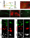
Labeling of axon collaterals from striatal-projecting brainstem neurons. A, Using a combination of two viral vectors, one of which possesses trans-neuronal retrograde capabilities, brainstem neurons that innervate striatal targets were selectively labeled, including their axon collaterals. B, Neurons in the LDT that retrogradely transported WGA–Cre from the NA core expressed the YFP after the local injection of a Cre-dependent virus. C–E, Axon collaterals expressing YFP were detected in the VTA (C), here defined by the border of the TH staining (D), and in the mediodorsal (MD) but not in the anteromedial (AM) thalamus (E), also in agreement with the study by Holmstrand and Sesack (2011). F, G, Fluorescent images showing triple immunolabeling for ChAT, CTb, and FG in the LDT after injections in the NA (FG) and the VTA (F; CTb) or the thalamus (G; CTb). Examples of neurons with triple labeling in the left panels of F and G (arrows) are shown at higher magnification in the right panels. Scale bars: B, 250 μm; (in E) C–E, 500 μm; F, G, 50 μm.

References

    1. Aosaki T, Tsubokawa H, Ishida A, Watanabe K, Graybiel AM, Kimura M. Responses of tonically active neurons in the primate's striatum undergo systematic changes during behavioral sensorimotor conditioning. J Neurosci. 1994;14:3969–3984. - PMC - PubMed
    1. Apicella P, Legallet E, Trouche E. Responses of tonically discharging neurons in the monkey striatum to primary rewards delivered during different behavioral states. Exp Brain Res. 1997;116:456–466. doi: 10.1007/PL00005773. - DOI - PubMed
    1. Barroso-Chinea P, Rico AJ, Conte-Perales L, Gómez-Bautista V, Luquin N, Sierra S, Roda E, Lanciego JL. Glutamatergic and cholinergic pedunculopontine neurons innervate the thalamic parafascicular nucleus in rats: changes following experimental parkinsonism. Brain Struct Funct. 2011;216:319–330. doi: 10.1007/s00429-011-0317-x. - DOI - PubMed
    1. Bennett BD, Wilson CJ. Spontaneous activity of neostriatal cholinergic interneurons in vitro. J Neurosci. 1999;19:5586–5596. - PMC - PubMed
    1. Bennett BD, Callaway JC, Wilson CJ. Intrinsic membrane properties underlying spontaneous tonic firing in neostriatal cholinergic interneurons. J Neurosci. 2000;20:8493–8503. - PMC - PubMed

Publication types

MeSH terms