. 2014 Mar 26;34(13):4627-33.
doi: 10.1523/JNEUROSCI.4887-13.2014.

Circadian perinatal photoperiod has enduring effects on retinal dopamine and visual function

Affiliations

Circadian perinatal photoperiod has enduring effects on retinal dopamine and visual function

Chad R Jackson et al. J Neurosci. .

Abstract

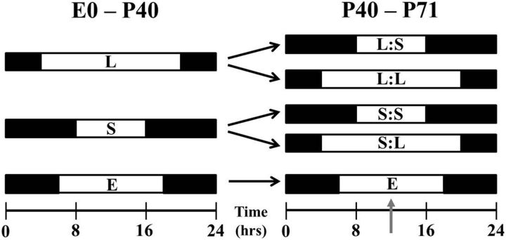
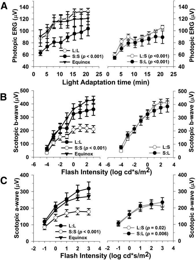
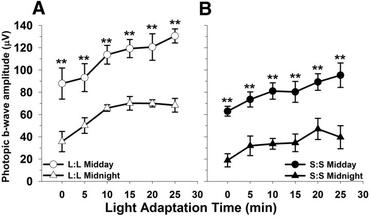
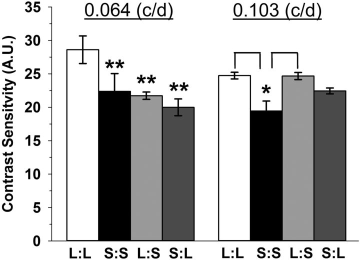
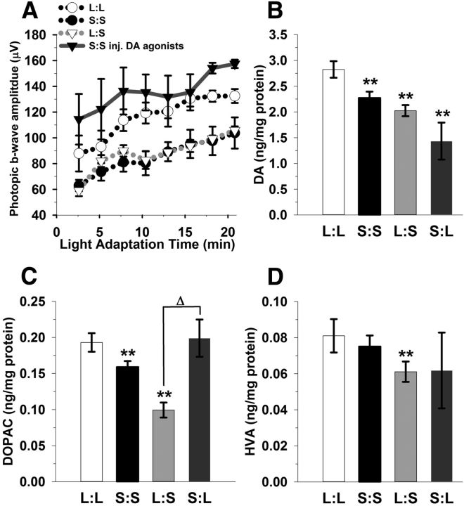
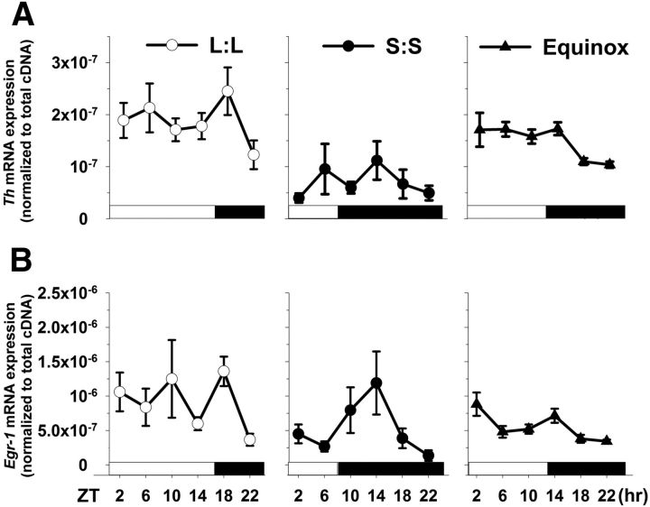
Visual system development depends on neural activity, driven by intrinsic and light-sensitive mechanisms. Here, we examined the effects on retinal function due to exposure to summer- and winter-like circadian light cycles during development and adulthood. Retinal light responses, visual behaviors, dopamine content, retinal morphology, and gene expression were assessed in mice reared in seasonal photoperiods consisting of light/dark cycles of 8:16, 16:8, and 12:12 h, respectively. Mice exposed to short, winter-like, light cycles showed enduring deficits in photopic retinal light responses and visual contrast sensitivity, but only transient changes were observed for scotopic measures. Dopamine levels were significantly lower in short photoperiod mice, and dopaminergic agonist treatment rescued the photopic light response deficits. Tyrosine hydroxylase and Early Growth Response factor-1 mRNA expression were reduced in short photoperiod retinas. Therefore, seasonal light cycles experienced during retinal development and maturation have lasting influence on retinal and visual function, likely through developmental programming of retinal dopamine.

Keywords: circadian; dopamine; electroretinogram; photoperiod; retina; vision.


Figures

Figure 1.
Figure 1.
Photoperiod paradigm. Experimental photoperiod for mice began at E0 with either long (L) or short (S) light exposure. Between P40 and P50, groups either remained in the initial photoperiod or entered the opposing photoperiod for ∼3 weeks. Other mice were exposed to an Equinox (E; 12:12 h) light/dark cycle for comparison. Gray arrow signifies midday test time point.
Figure 2.
Figure 2.
Photoperiod affects light- and dark-adapted retinal function. A, Light-adapted (photopic) b-wave amplitudes are significantly lower in mice exposed to S:S, L:S, and S:L photoperiods as compared with L:L (p < 0.001, all groups). B, Dark-adapted (scotopic) b-wave amplitudes are significantly reduced in the S:S mice compared with the L:L group (p < 0.001). In contrast, the L:S and S:L groups do not differ from L:L; however, they are significantly higher in amplitude compared with S:S mice (p < 0.05, both comparisons). C, Dark-adapted (scotopic) a-wave amplitudes are lower in the S:S, L:S, and S:L groups in relation to L:L mice (p = < 0.001, 0.022, 0.006, respectively). Also, the L:S and S:L groups significantly differ in amplitude compared with S:S mice (p = 0.018 and 0.024, respectively). All data points represent means ± SEM; n = 6–10.
Figure 3.
Figure 3.
Photoperiod does not affect photopic ERG rhythm phenotype, only the amplitude differs between groups. A, B, L:L and S:S animals display rhythmic responses to the photopic test (**p < 0.001, both groups: day vs night); however, the amplitude in the S:S group is significantly lower as compared with the L:L at the corresponding test time point (L:L vs S:S midday, p < 0.001; L:L vs S:S midnight, p < 0.001). All data points represent means ± SEM; n = 6.
Figure 4.
Figure 4.
Contrast sensitivity detection is impacted by perinatal photoperiod exposure. Contrast sensitivity is lower in S:S, L:S, and S:L groups at 0.064 cycles per degree (c/d) compared with L:L mice (**p < 001). Also, at 0.103 c/d, contrast sensitivity is significantly reduced in S:S mice compared with L:L and L:S exposed mice (*p = 0.004). All data represent means ± SEM; n = 4–6 mice. A.U., arbitrary units.
Figure 5.
Figure 5.
Stimulating dopaminergic signaling rescues the photopic ERG. A, Injection of SKF38393 and PD168077 (1 mg/kg, dopamine D1 and D4 receptor-selective agonists, respectively) significantly increases photopic ERG b-wave amplitudes in the S:S group (solid gray line with black triangles) compared with the untreated group (black dotted line; p = 0.018). B, Retinal dopamine is reduced in S:S, L:S, and S:L groups compared with L:L mice (**p < 0.05). C, Retinal DOPAC concentrations are lower in S:S and L:S mice in relation to L:L mice (**p < 0.05); however, S:L does not significantly differ. D, In contrast, only the L:S mice display a significant reduction in HVA compared with L:L mice (**p < 0.05). All data represent means ± SEM; n = 6–8 mice.
Figure 6.
Figure 6.
Tyrosine hydroxylase and Egr-1 mRNA expression levels are influenced by perinatal photoperiod. A, Th retinal mRNA levels, assayed over 24 h, differ among all photoperiods (p < 0.01); even so, mice exposed to the S:S photoperiod display the lowest Th mRNA levels compared with L:L and Equinox reared mice (p < 0.001). B, Egr-1 mRNA expression patterns substantially differ between the three groups (p = 0.015). S:S and Equinox levels are significantly lower compared with the L:L group (p < 0.01 and 0.001, respectively). Transcript expression peaks after the light to dark transition in each group. All data points represent means ± SEM; n = 6 mice per group per time point.

Similar articles

Cited by

References

    1. Cameron MA, Barnard AR, Hut RA, Bonnefont X, van der Horst GT, Hankins MW, Lucas RJ. Electroretinography of wild-type and Cry mutant mice reveals circadian tuning of photopic and mesopic retinal responses. J Biol Rhythms. 2008a;23:489–501. doi: 10.1177/0748730408325874. - DOI - PubMed
    1. Cameron MA, Barnard AR, Lucas RJ. The electroretinogram as a method for studying circadian rhythms in the mammalian retina. J Genet. 2008b;87:459–466. doi: 10.1007/s12041-008-0068-5. - DOI - PubMed
    1. Ciarleglio CM, Axley JC, Strauss BR, Gamble KL, McMahon DG. Perinatal photoperiod imprints the circadian clock. Nat Neurosci. 2011;14:25–27. doi: 10.1038/nn.2699. - DOI - PMC - PubMed
    1. Cransac H, Cottet-Emard JM, Pequignot JM, Peyrin L. Monoamines (norepinephrine, dopamine, serotonin) in the rat medial vestibular nucleus: endogenous levels and turnover. J Neural Transm. 1996;103:391–401. doi: 10.1007/BF01276416. - DOI - PubMed
    1. Doyle SE, Grace MS, McIvor W, Menaker M. Circadian rhythms of dopamine in mouse retina: the role of melatonin. Vis Neurosci. 2002;19:593–601. - PubMed

Publication types

MeSH terms

Substances

LinkOut - more resources