. 2014 Jun;88(11):6318-28.
doi: 10.1128/JVI.00381-14. Epub 2014 Mar 26.

NTPDASE4 gene products cooperate with the adenovirus E4orf4 protein through PP2A-dependent and -independent mechanisms and contribute to induction of cell death

Affiliations

NTPDASE4 gene products cooperate with the adenovirus E4orf4 protein through PP2A-dependent and -independent mechanisms and contribute to induction of cell death

Meirav Avital-Shacham et al. J Virol. 2014 Jun.

Abstract

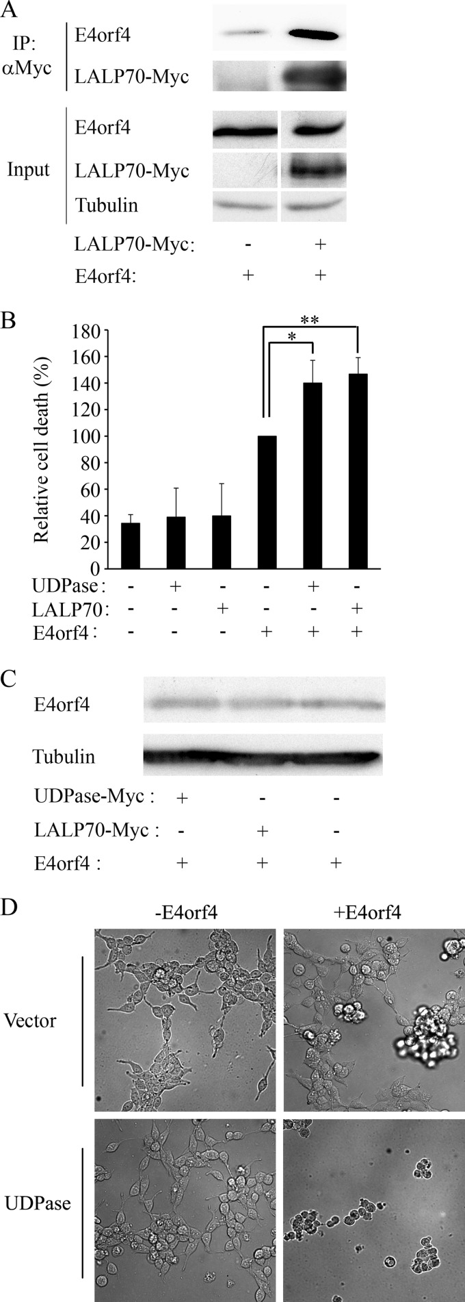
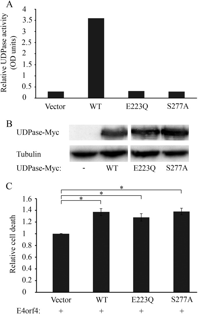
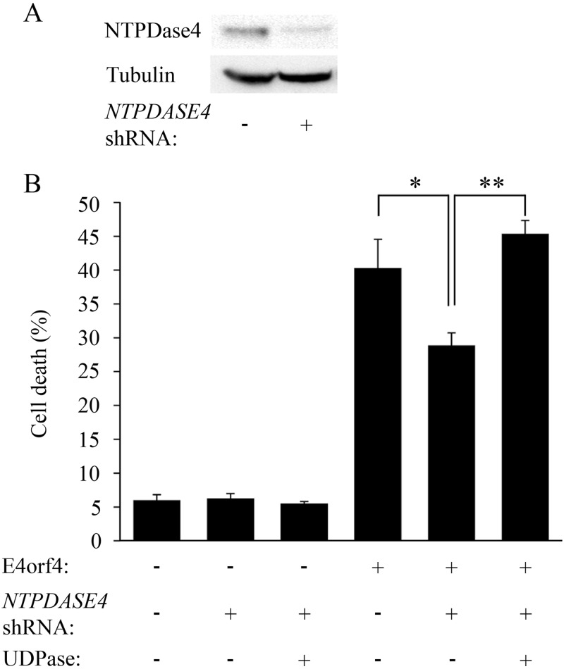
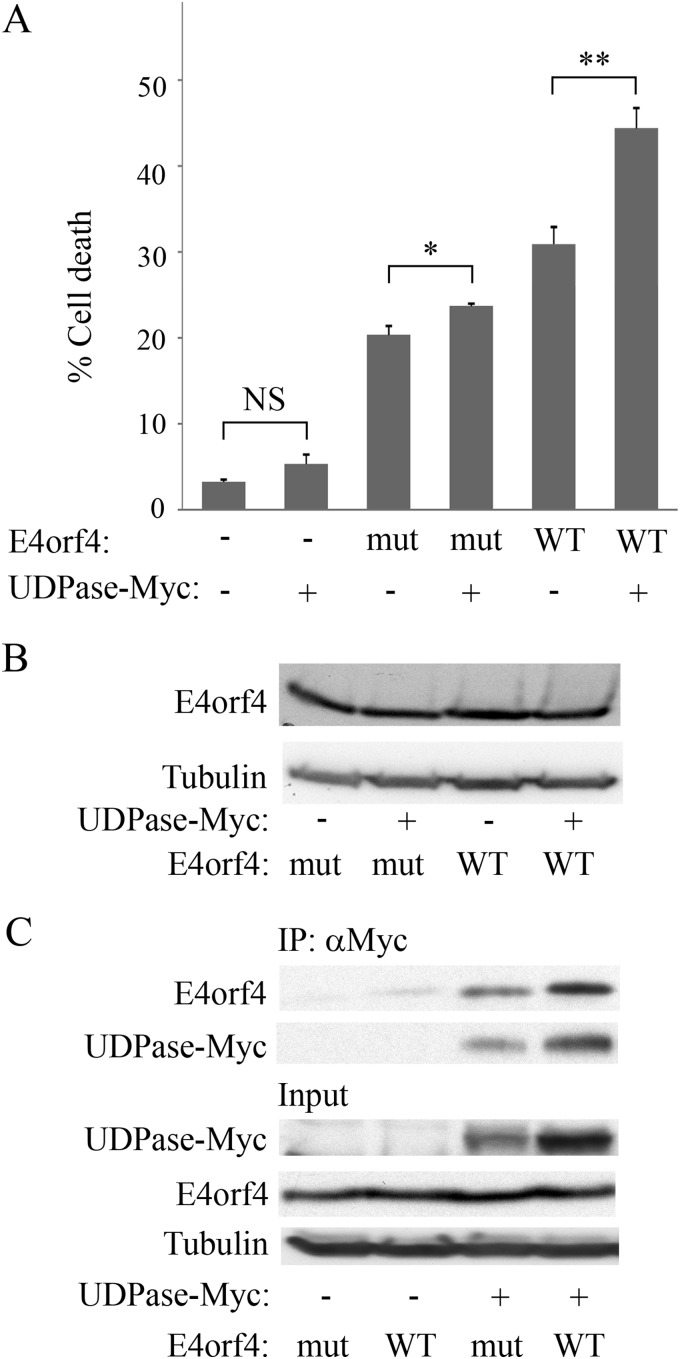
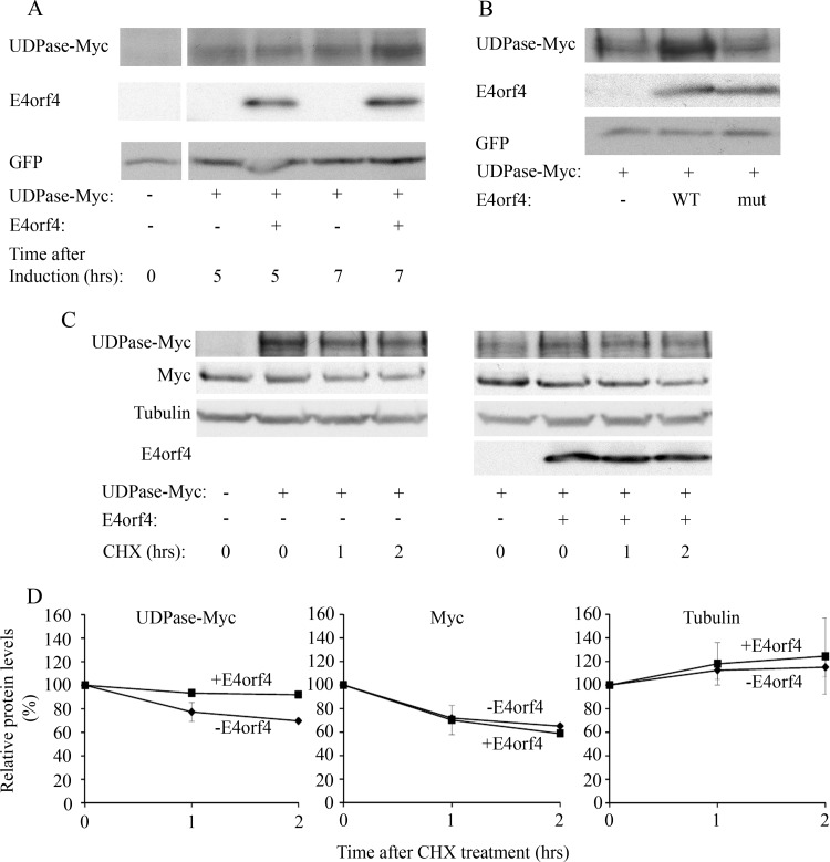
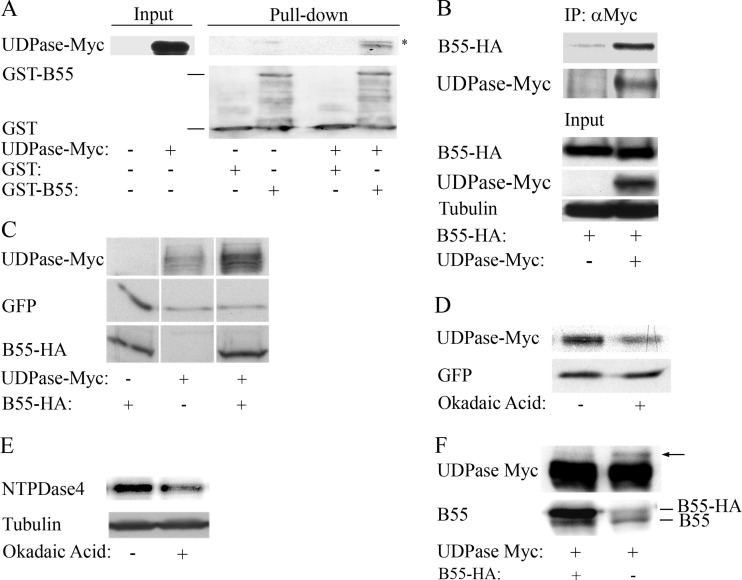
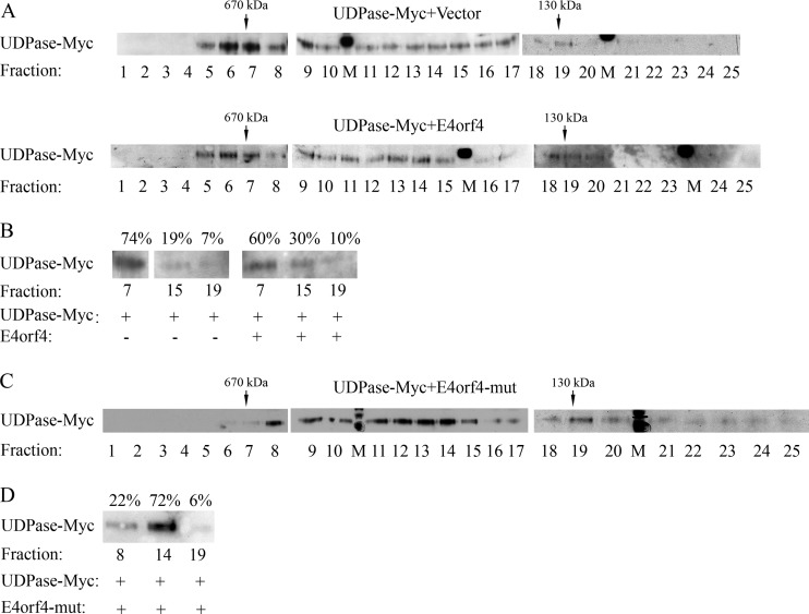
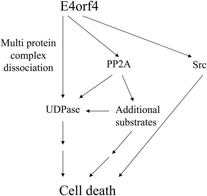
The adenovirus E4orf4 protein induces nonclassical apoptosis in mammalian cells through at least two complementing pathways regulated by the interactions of E4orf4 with protein phosphatase 2A (PP2A) and Src kinases. In Saccharomyces cerevisiae cells, which do not express Src, E4orf4 induces PP2A-dependent toxicity. The yeast Golgi apyrase Ynd1 was found to contribute to E4orf4-mediated toxicity and to interact with the PP2A-B55α regulatory subunit. In addition, a mammalian Ynd1 orthologue, the NTPDASE4 gene product Golgi UDPase, was shown to physically interact with E4orf4. Here we report that knockdown of NTPDASE4 suppressed E4orf4-induced cell death. Conversely, overexpression of the NTPDASE4 gene products Golgi UDPase and LALP70 enhanced E4orf4-induced cell killing. We found that similarly to results obtained in yeast, the apyrase activity of mammalian UDPase was not required for its contribution to E4orf4-induced toxicity. The interaction between E4orf4 and UDPase had two consequences: a PP2A-dependent one, resulting in increased UDPase levels, and a PP2A-independent outcome that led to dissociation of large UDPase-containing protein complexes. The present report extends our findings in yeast to E4orf4-mediated death of mammalian cells, and combined with previous results, it suggests that the E4orf4-NTPDase4 pathway, partly in association with PP2A, may provide an alternative mechanism for the E4orf4-Src pathway to contribute to the cytoplasmic death function of E4orf4.

Importance: The adenovirus E4orf4 protein contributes to regulation of the progression of virus infection from the early to the late phase, and when expressed alone, it induces a unique caspase-independent programmed cell death which is more efficient in cancer cells than in normal cells. The interactions of E4orf4 with cellular proteins that mediate its functions, such as PP2A and Src kinases, are highly conserved in evolution. The results presented here reveal that the NTPDASE4 gene product Golgi UDPase, first discovered to contribute to E4orf4 toxicity in Saccharomyces cerevisiae, associates with E4orf4 and plays a role in induction of cell death in mammalian cells. Details of the functional interaction between E4orf4, PP2A, and the UDPase are described. Identification of the evolutionarily conserved mechanisms underlying E4orf4 activity will increase our understanding of the interactions between the virus and the host cell and will contribute to our grasp of the unique mode of E4orf4-induced cell death.


Figures

FIG 1
FIG 1
Golgi UDPase and LALP70 enhance E4orf4-induced cell death. Plasmids expressing UDPase-Myc, LALP70-Myc, or an empty vector were transfected into 293T cells with a plasmid expressing E4orf4 or with the corresponding empty vector. (A) Cell extracts were subjected to immunoprecipitation (IP) with antibodies to the Myc tag, and the presence of E4orf4 and LALP70-Myc proteins in the immune complexes and in input lysates was determined by a Western blot. Alpha-tubulin served as a loading control. The amount of proteins in the input represents 10% of the amount of proteins used for immunoprecipitation. (B) Cells in duplicate plates were stained 24 h after transfection with antibodies to E4orf4 and the Myc tag and with 4′,6-diamidino-2-phenylindole (DAPI). Nuclei with apoptotic morphology were counted in the double-transfected cell population expressing both E4orf4 and UDPase-Myc and in single-transfected cells expressing either E4orf4 or UDPase-Myc, and the percentage of cell death was determined. Cell death induced by E4orf4 alone was defined as 100%, and relative cell death was calculated for the other samples. Two independent experiments, each with duplicates, were carried out. Error bars represent the pooled standard deviation, and statistical significance was determined using a one-tailed t test (*, P = 0.043; **, P = 0.04). (C) Proteins extracted from a parallel set of E4orf4-transfected plates, as described for panel B, were analyzed by Western blotting using antibodies to E4orf4 and to alpha-tubulin. (D) Representative photographs of cells containing the indicated plasmids were taken 24 h after transfection.
FIG 2
FIG 2
The UDPase catalytic activity is not required for E4orf4-induced cell death. (A) An empty vector and plasmids expressing WT UDPase-Myc and Myc tagged UDPase mutants (E223Q and S277A) were transfected into 293T cells. The cells were harvested 48 h later, and the UDPase catalytic activity was measured using an apyrase activity assay. (B) Protein extracts from the cells used for panel A were subjected to a Western blot analysis with antibodies to the Myc tag and alpha-tubulin. (C) Plasmids expressing WT UDPase-Myc and Myc tagged UDPase mutants (E223Q and S277A) or an empty vector were transfected into 293T cells together with a plasmid expressing E4orf4. The cells were fixed 24 h later and were stained with antibodies to E4orf4 and the Myc tag and with DAPI. The percentage of cell death in transfected cells was calculated as described in the legend to Fig. 1. Induction of cell death by E4orf4 in the presence of WT or mutant UDPase proteins was normalized to induction of cell death by E4orf4 in the absence of UDPase, defined as 1. Results of two independent experiments are shown. Error bars represent standard error. *, P < 0.002.
FIG 3
FIG 3
NTPDASE4 contributes to E4orf4-induced cell death. (A) 293T cells were transfected with an empty vector or a vector expressing both GFP and a shRNA sequence targeting NTPDASE4. Protein extracts were prepared 48 h later and subjected to Western blot analysis using antibodies to NTPDase4 and alpha-tubulin. NTPDase4 protein levels were determined by densitometry and normalized to tubulin levels, demonstrating a 3-fold reduction in NTPDase4 expression upon knockdown. (B) 293T cells were transfected with an empty vector or a plasmid expressing a NTPDASE4-specific shRNA. These cells were further transfected 2 days later with a plasmid expressing a shRNA-resistant UDPase-Myc or a vector control together with a plasmid expressing E4orf4 or its corresponding empty vector control. The cells were fixed 24 h after the second transfection and stained with antibodies to E4orf4 and the Myc tag and with DAPI. Cells transfected with the shRNA were identified by the presence of GFP expressed from the same plasmid. The percentage of cell death in transfected cells was calculated. Two independent experiments, each with duplicates, were carried out. Error bars represent the standard error. *, P = 0.017; **, P = 0.0001.
FIG 4
FIG 4
UDPase does not synergistically activate cell death induced by an E4orf4 mutant that binds Src but not PP2A. (A) The E4orf4 R81F84A mutant (mut) and WT E4orf4 were expressed in 293T cells alone or together with UDPase-Myc. Cells in duplicate plates were fixed 24 h after transfection and stained with antibodies to E4orf4 and the Myc tag and with DAPI. The percentage of cell death in transfected cells was calculated. Two independent experiments, each with duplicates, were carried out. Error bars represent standard errors. NS, not significant. *, P = 0.009; **, P = 0.002. (B) An additional set of cells that were transfected similarly was harvested 24 h after transfection, and a Western blot analysis was performed with E4orf4- and alpha-tubulin-specific antibodies. (C) Cell extracts as described for panel B were subjected to immunoprecipitation with antibodies to the Myc tag. A Western blot containing both immune complexes and input lysates was stained with antibodies to E4orf4 and the Myc tag. Alpha-tubulin served as a loading control. The blots were subjected to densitometry, and the results indicated that UDPase levels were 2-fold higher in the presence of WT E4orf4 than in the presence of the R81F84A mutant and the levels of WT and mutant E4orf4 proteins found in the immune complexes mirrored this difference (1.75:1).
FIG 5
FIG 5
E4orf4 increases UDPase protein levels. (A) UDPase-Myc was expressed in 293T cells together with inducibly expressed E4orf4 and with GFP. The transfection was carried out in duplicate. One day after transfection, E4orf4 expression was induced by the addition of 5 μg/ml doxycycline to the cells (+E4orf4), and a duplicate plate was treated with ethanol (-E4orf4). The cells were harvested 5 and 7 h later, and equal amounts of proteins were separated by SDS-PAGE. A Western blot was stained with the indicated antibodies. Densitometry of the blot and normalization to the levels of the GFP transfection control revealed that UDPase levels increased by 40% in the presence of E4orf4 after 7 h induction. (B) Plasmids expressing UDPase-Myc and GFP were transfected into 293T cells together with an empty vector or a vector expressing WT E4orf4 or the R81F84A mutant, which does not bind PP2A-B55α (mut). The cells were harvested 1 day after transfection, and protein levels of UDPase-Myc, E4orf4, and GFP were determined by Western blot analysis using specific antibodies. (C) UDPase-Myc was expressed in 293T cells with or without E4orf4. Cycloheximide (CHX; 50 μg/ml) or an equal volume of ethanol was added to the medium 1 day after the transfection, and cells were harvested at 0, 1, and 2 h after addition of the drug. The levels of UDPase-Myc, endogenous Myc, alpha-tubulin, and E4orf4 were visualized by Western blotting. (D) Protein levels were determined by densitometry of the Western blots. Protein levels at time zero were defined as 100%, and relative protein levels are shown in the graphs. Diamonds, samples with empty vector; squares, samples with E4orf4. Error bars represent the standard errors from two independent experiments. Errors smaller than 0.03 are not depicted.
FIG 6
FIG 6
PP2A-B55α interacts with UDPase and increases UDPase protein levels. (A) UDPase-Myc was expressed in 293T cells and protein extracts were prepared 24 h later. These extracts were incubated with bacterially produced PP2A-B55α fused to GST or with GST alone for 2 h at 4°C. A Western blot analysis was performed with Myc tag- and GST-specific antibodies. The amount of proteins in the input extracts from 293T cells represents 10% of the amount of proteins used for incubation with GST proteins. The asterisk marks a nonspecific band. (B) HA-tagged PP2A-B55α was expressed in 293T cells alone or together with UDPase-Myc. One day after transfection, the cells were harvested and protein extracts were subjected to immunoprecipitation (IP) with a Myc tag antibody. A Western blot shows the presence of PP2A-B55α–HA and UDPase-Myc in the immune complexes (IP) and in input lysates. Alpha-tubulin served as a loading control. The amount of proteins in the input represents 10% of the amount of proteins used for IP. Results of one representative experiment out of three are shown. (C) IRBα cells that constitutively express a PP2A-B55α-specific shRNA were transfected with a plasmid expressing UDPase-Myc and a plasmid expressing GFP (serving as a transfection efficiency control) with or without a plasmid expressing mutant PP2A-B55α–HA, which was resistant to the PP2A-B55α-specific shRNA. The cells were harvested 24 h later, and Western blot analysis was performed with antibodies to the HA and Myc tags and to GFP. (D) Two plates of IRBα cells were transfected with plasmids expressing UDPase-Myc, GFP, and shRNA-resistant PP2A-B55α–HA. After 24 h, 5 nM okadaic acid was added to one plate and ethanol was added to the other plate. The cells were harvested 4 h later, and protein extracts were subjected to Western blot analysis with antibodies to the Myc tag and GFP. Densitometry of the blot and normalization to the GFP control revealed that okadaic acid treatment led to a 2-fold reduction in UDPase levels. Similar results were obtained in a second independent experiment. (E) 293T cells were treated with 5 nM okadaic acid or with ethanol in three independent experiments and protein extracts were prepared 4 h later. Western blots were stained with antibodies to NTPDase4 proteins and alpha-tubulin. Densitometry of the blots and normalization to alpha-tubulin levels revealed an okadaic acid-induced drop of 33% to 75% in NTPDase4 protein levels in the three experiments. One representative blot is shown. (F) IRBα cells were transfected with a plasmid expressing UDPase-Myc and with a plasmid expressing PP2A-B55α–HA or an empty vector. The cells were harvested 1 day later, and protein extracts were prepared and loaded on a large SDS gel. Western blot analysis was performed with antibodies to the Myc tag and to PP2A-B55α. An arrow marks a slower-migrating UDPase-Myc isoform. The figure is representative of three independent experiments.
FIG 7
FIG 7
A high-molecular-weight UDPase-containing complex is partially dissociated in the presence of E4orf4, regardless of the ability of E4orf4 to bind PP2A. (A) Protein extracts from 293T cells expressing UDPase-Myc in the presence or absence of E4orf4 were separated independently on a Superose 6 column. Twenty-five fractions were collected from each run, and 10% of each fraction were loaded onto SDS gels and subjected to Western blot analysis with antibodies to the Myc tag. In addition, thyroglobulin and BSA dimers were chromatographed separately on the column as molecular mass markers (670 and 130 kDa, respectively). M denotes a marker lane. (B) Fractions 7, 15, and 19 from the two column runs whose results are shown in panel A were loaded onto one SDS gel, and a blot was stained with Myc tag-specific antibodies. The intensities of UDPase-Myc protein bands were quantified by densitometry and the sum of UDPase-Myc levels in the 3 fractions of each column run was defined as 100%. The percentage of UDPase-Myc in each fraction is shown above the blot. (C and D) A column run of protein extracts expressing UDPase-Myc in the presence of the R81F84A E4orf4 mutant (mut) was analyzed as described for panels A and B. The results are representative of 3 independent experiments.
FIG 8
FIG 8
A model of the interaction between UDPase, PP2A-B55α, and E4orf4, and their contribution to induction of cell death by E4orf4. E4orf4 affected UDPase in at least two ways. First, E4orf4 enhanced UDPase levels in a PP2A-dependent manner, and the increased UDPase levels enhanced E4orf4-induced cell death. Since high levels of UDPase alone did not cause cell death, we suggest that other substrates of PP2A were required for enhancing E4orf4 toxicity in parallel with UDPase or in cooperation with it. The finding that UDPase levels were not much influenced by an E4orf4 mutant that bound Src but not PP2A suggests that UDPase cooperated with the E4orf4-PP2A but not the E4orf4-Src pathway. Second, independently of PP2A, E4orf4 dissociated high-molecular-weight complexes that included UDPase. Because the Ynd1 cytoplasmic tail in yeast appears to act as a scaffold that binds E4orf4 and several proteins of the secretory pathway and mediates E4orf4 toxicity (29), we hypothesize that dissociation of a complex tethered to the UDPase cytosolic tail may contribute to induction of cell death. The Src- and UDPase-dependent pathways may provide partially overlapping contributions to E4orf4-induced cell death as they both interact, physically or functionally, with the protein trafficking machinery.

Similar articles

Cited by

References

    1. Ben-Israel H, Sharf R, Rechavi G, Kleinberger T. 2008. Adenovirus E4orf4 protein downregulates MYC expression through interaction with the PP2A-B55 subunit. J. Virol. 82:9381–9388. 10.1128/JVI.00791-08 - DOI - PMC - PubMed
    1. Bondesson M, Ohman K, Mannervik M, Fan S, Akusjarvi G. 1996. Adenovirus E4 open reading 4 protein autoregulates E4 transcription by inhibiting E1A transactivation of the E4 promoter. J. Virol. 70:3844–3851 - PMC - PubMed
    1. Estmer Nilsson C, Petersen-Mahrt S, Durot C, Shtrichman R, Krainer AR, Kleinberger T, Akusjarvi G. 2001. The adenovirus E4-ORF4 splicing enhancer protein interacts with a subset of phosphorylated SR proteins. EMBO J. 20:864–871. 10.1093/emboj/20.4.864 - DOI - PMC - PubMed
    1. Kanopka A, Muhlemann O, Petersen-Mahrt S, Estmer C, Ohrmalm C, Akusjarvi G. 1998. Regulation of adenovirus alternative RNA splicing by dephosphorylation of SR proteins, Nature 393:185–187 - PubMed
    1. Mannervik M, Fan S, Strom AC, Helin K, Akusjarvi G. 1999. Adenovirus E4 open reading frame 4-induced dephosphorylation inhibits E1A activation of the E2 promoter and E2F-1-mediated transactivation independently of the retinoblastoma tumor suppressor protein. Virology 256:313–321. 10.1006/viro.1999.9663 - DOI - PubMed

Publication types

MeSH terms