. 2014 Jun;88(11):6355-67.
doi: 10.1128/JVI.00219-14. Epub 2014 Mar 26.

Activation of Kaposi's sarcoma-associated herpesvirus (KSHV) by inhibitors of class III histone deacetylases: identification of sirtuin 1 as a regulator of the KSHV life cycle

Affiliations

Activation of Kaposi's sarcoma-associated herpesvirus (KSHV) by inhibitors of class III histone deacetylases: identification of sirtuin 1 as a regulator of the KSHV life cycle

Qiuhua Li et al. J Virol. 2014 Jun.

Abstract

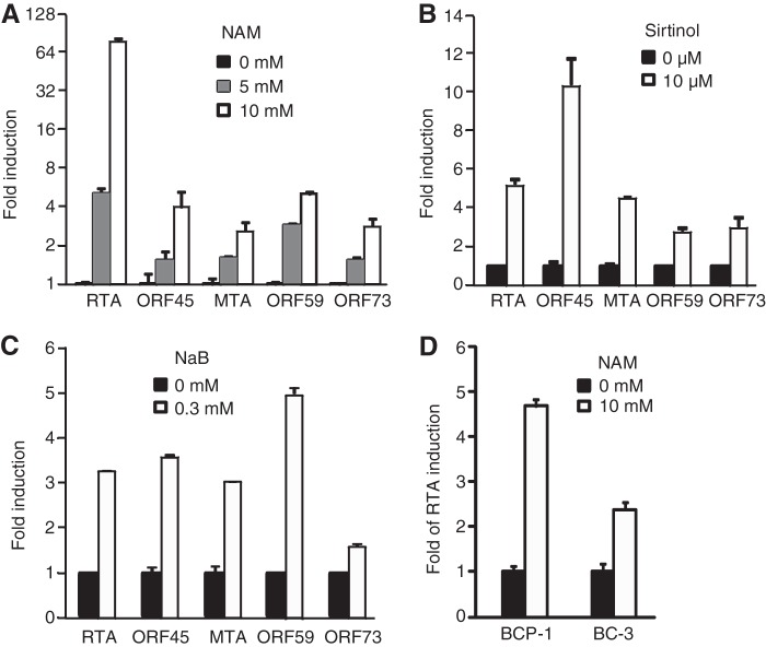
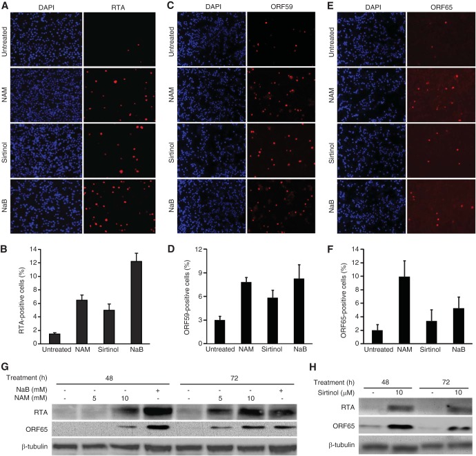
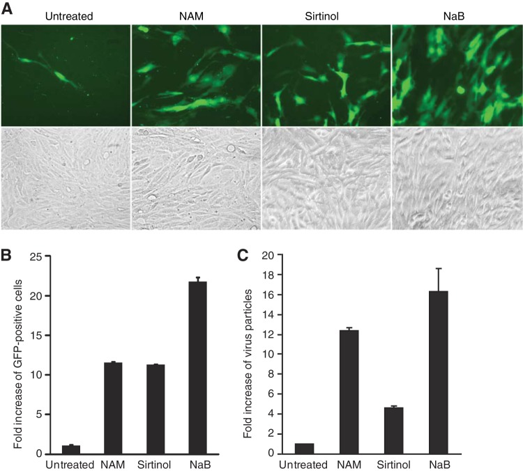
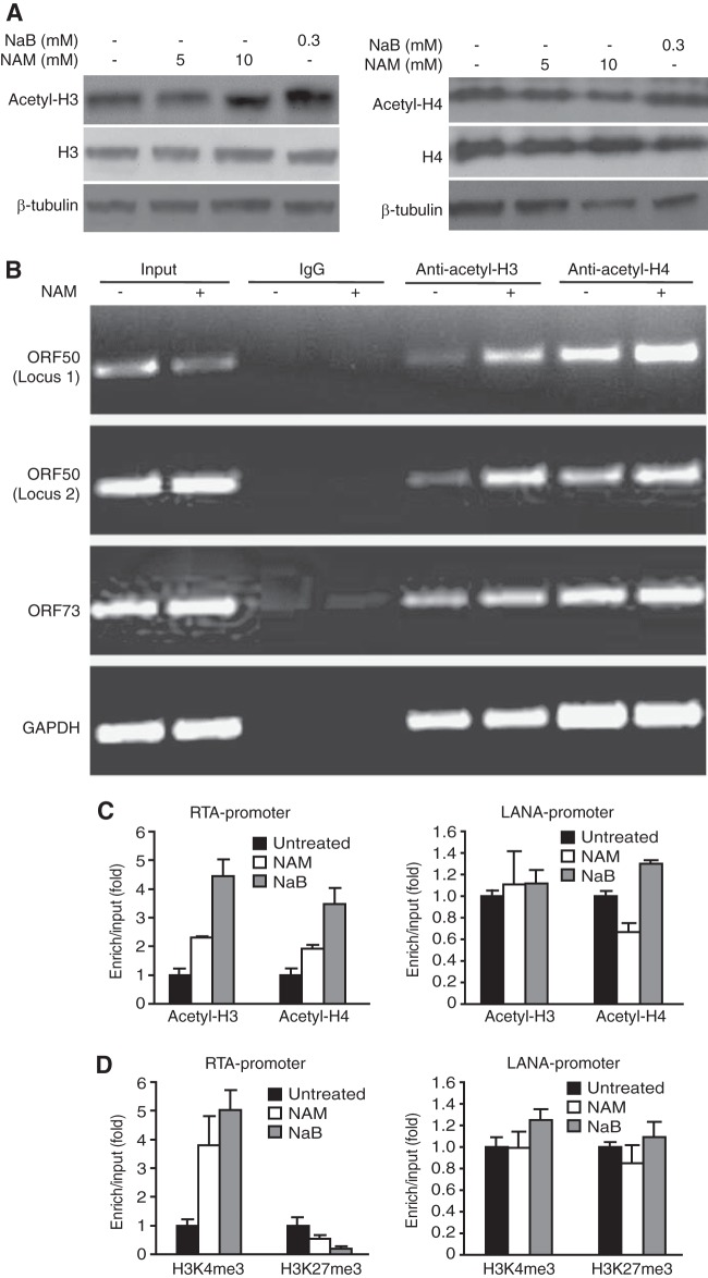
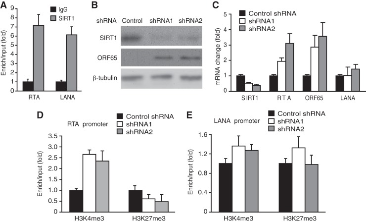
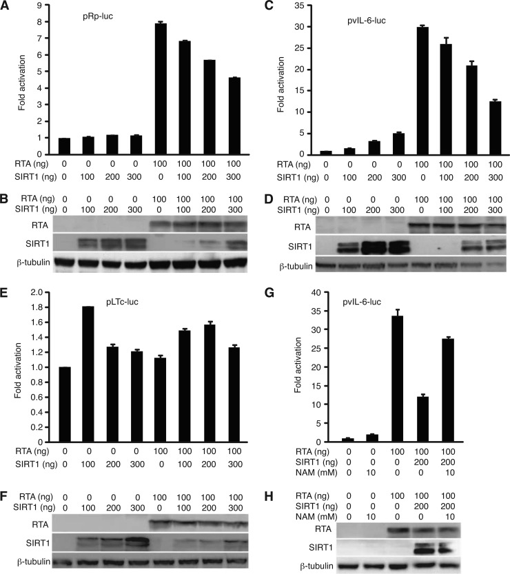
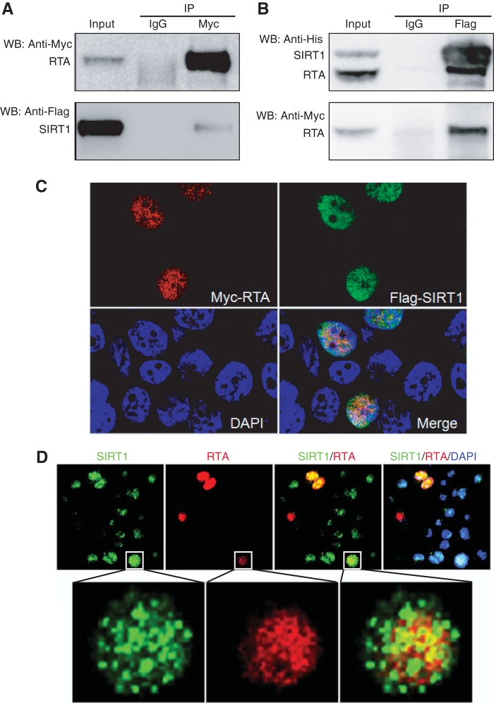
Kaposi's sarcoma-associated herpesvirus (KSHV) establishes persistent latent infection in immunocompetent hosts. Disruption of KSHV latency results in viral lytic replication, which promotes the development of KSHV-related malignancies in immunocompromised individuals. While inhibitors of classes I and II histone deacetylases (HDACs) potently reactivate KSHV from latency, the role of class III HDAC sirtuins (SIRTs) in KSHV latency remains unclear. Here, we examined the effects of inhibitors of SIRTs, nicotinamide (NAM) and sirtinol, on KSHV reactivation from latency. Treatment of latently KSHV-infected cells with NAM or sirtinol induced transcripts and proteins of the master lytic transactivator RTA (ORF50), early lytic genes ORF57 and ORF59, and late lytic gene ORF65 and increased the production of infectious virions. NAM increased the acetylation of histones H3 and H4 as well as the level of the active histone H3 trimethyl Lys4 (H3K4me3) mark but decreased the level of the repressive histone H3 trimethyl Lys27 (H3K27me3) mark in the RTA promoter. Consistent with these results, we detected SIRT1 binding to the RTA promoter. Importantly, knockdown of SIRT1 was sufficient to increase the expression of KSHV lytic genes. Accordingly, the level of the H3K4me3 mark in the RTA promoter was increased following SIRT1 knockdown, while that of the H3K27me3 mark was decreased. Furthermore, SIRT1 interacted with RTA and inhibited RTA transactivation of its own promoter and that of its downstream target, the viral interleukin-6 gene. These results indicate that SIRT1 regulates KSHV latency by inhibiting different stages of viral lytic replication and link the cellular metabolic state with the KSHV life cycle.

Importance: Kaposi's sarcoma-associated herpesvirus (KSHV) is the causal agent of several malignancies, including Kaposi's sarcoma, commonly found in immunocompromised patients. While latent infection is required for the development of KSHV-induced malignancies, viral lytic replication also promotes disease progression. However, the mechanism controlling KSHV latent versus lytic replication remains unclear. In this study, we found that class III histone deacetylases (HDACs), also known as SIRTs, whose activities are linked to the cellular metabolic state, mediate KSHV replication. Inhibitors of SIRTs can reactivate KSHV from latency. SIRTs mediate KSHV latency by epigenetically silencing a key KSHV lytic replication activator, RTA. We found that one of the SIRTs, SIRT1, binds to the RTA promoter to mediate KSHV latency. Knockdown of SIRT1 is sufficient to induce epigenetic remodeling and KSHV lytic replication. SIRT1 also interacts with RTA and inhibits RTA's transactivation function, preventing the expression of its downstream genes. Our results indicate that SIRTs regulate KSHV latency by inhibiting different stages of viral lytic replication and link the cellular metabolic state with the KSHV life cycle.


Figures

FIG 1
FIG 1
Treatment with NAM or sirtinol increases the expression of KSHV lytic transcripts. (A to C) Expression of KSHV transcripts in BCBL-1 cells following treatment with NAM (A), sirtinol (B), and NaB (C). (D) Expression of the RTA transcript in BCP-1 and BC-3 cells following treatment with NAM. Cells were treated with the indicated concentrations of the chemicals for 48 h. Viral transcripts were detected by RT-qPCR, and their levels of expression were normalized to the level of expression of cellular GAPDH. Experiments were repeated three times, and results are shown as averages and standard deviations, with the value for untreated cells being set equal to 1.
FIG 2
FIG 2
Treatment with NAM or sirtinol increases the expression of KSHV lytic proteins. (A to F) IFA detection of KSHV lytic proteins RTA (A and B), ORF59 (C and D), and ORF65 (E and F) following treatment with NAM, sirtinol, or NaB. BCBL-1 cells treated with 10 mM NAM, 10 μM sirtinol, or 0.3 mM NaB were subjected to IFA staining (A, C, and E) (DAPI, 4′,6-diamidino-2-phenylindole). The percentages of RTA-, ORF59-, and ORF65-positive cells are represented by the averages and standard deviations calculated from eight random fields (B, D, and F). (G and H) Western blot analysis of RTA and ORF65 proteins following treatment with NAM, sirtinol, or NaB. BCBL-1 cells treated with the indicated concentrations of NAM and 0.3 mM NaB (G) or 10 μM sirtinol (H) for the specified times were harvested and examined for the expression of RTA and ORF65 proteins using β-tubulin as a loading control.
FIG 3
FIG 3
Treatment with NAM or sirtinol induces the production of KSHV infectious virions. (A) Titration of KSHV infectious virions by inoculating MM cells with supernatants from BCBL-1–BAC36 KSHV-infected cells induced with NAM, sirtinol, or NaB. Cells were induced with 10 mM NAM, 10 μM sirtinol, or 0.3 mM NaB for 4 days. Cell-free supernatants were used to infect MM cells for 48 h. Images of GFP-positive cells illustrate the presence of infectious virions in the supernatants. (B) Relative virus titers were determined on the basis of the numbers of GFP-positive cells from five random fields and are shown as averages and standard deviations. (C) The relative amounts of virus particles produced by the induced cells were determined by qPCR for virion DNA. Supernatants of induced cells collected at the end of induction were filtered and subjected to differential centrifugation to obtain virus particles. The pellets were treated with DNase to eliminate nonspecifically attached nonvirion DNA. Viral DNA was then extracted and analyzed by qPCR.
FIG 4
FIG 4
Treatment with NAM induces the hyperacetylation of histones, increases the level of the active histone H3K4me3 mark, and decreases the level of the repressive histone H3K27me3 mark in the RTA promoter. (A) BCBL-1 cells were treated with the indicated concentrations of NAM or 0.3 mM NaB for 24 h and examined for the total and acetylated forms of histone H3 and histone H4 proteins by Western blotting. (B) Quantification of acetylated histones H3 and H4 in RTA and LANA promoters by ChIP-PCR assay. BCBL-1 cells treated with 10 mM NAM for 8 h were subjected to ChIP with an antibody to acetylated histone H3 (anti-acetyl-H3) or histone H4 (anti-acetyl-H4) or a control antibody. The immunoprecipitated DNAs were amplified by semiquantitative PCR for two loci in the RTA promoter, one locus in the LANA promoter, or one locus in the GAPDH intergenic region. (C) Quantification of acetylated histones H3 and H4 in the RTA and LANA promoters by ChIP-qPCR assays. Cells were treated as described in the legend to panel B, and the ChIP DNAs were quantified by qPCR. (D) Quantification of the H3K4me3 and H3K27me3 marks in RTA and LANA promoters by ChIP-qPCR assays. ChIP assays were performed with cells treated as described in the legend to B using an antibody to H3K4me3 or H3K27me3 or a control antibody. The ChIP DNAs were quantified by qPCR.
FIG 5
FIG 5
SIRT1 mediates KSHV latency. (A) SIRT1 binds to both RTA and LANA promoters. ChIP-qPCR was performed with BCBL-1 cells using an antibody to SIRT1 and a control antibody. (B) Short hairpin RNA knockdown of SIRT1 induces the expression of KSHV lytic protein ORF65. BCBL-1 cells infected with lentiviruses expressing shRNAs to SIRT1 or a scrambled control shRNA for 3 days were examined for the expression of SIRT1 and ORF65 proteins by Western blotting. (C) Expression of SIRT1, RTA, ORF65, and LANA transcripts in BCBL-1 cells following knockdown of SIRT1, as described in the legend to panel B. Viral transcripts were detected by RT-qPCR, and their levels were normalized to the level of cellular GAPDH. (D and E) Quantification of the H3K4me3 and H3K27me3 marks in RTA (D) and LANA (E) promoters by ChIP-qPCR assays following SIRT1 knockdown. Cells infected with lentiviruses expressing shRNAs to SIRT1 or a scrambled control shRNA as described in the legend to panel B were subjected to ChIP using an antibody to H3K4me3 or H3K27me3 or a control antibody. The ChIP DNAs were quantified by qPCR.
FIG 6
FIG 6
SIRT1 represses RTA transactivation in reporter assays. (A to F) SIRT1 represses RTA activation of its own promoter (A and B) and the vIL-6 promoter (C and D) but not LANA latent promoter LTc (E and F), as shown in reporter assays (A, C, and E) together with Western blots of the expressed RTA and SIRT1 proteins (B, D, and F). 293T cells were cotransfected with the luciferase reporter plasmid pRp-luc, pvIL-6-luc, or pLTc-luc together with RTA or without RTA in the presence of increased doses of a SIRT1 expression plasmid. A β-galactosidase expression construct was used to calibrate the transfection efficiency. The luciferase activity of the reporter without RTA and SIRT1 was given a value of 1. The protein extracts were also examined by Western blotting to detect the expression of the RTA and SIRT1 proteins. (G and H) NAM relieves SIRT1's inhibitory effect on RTA transactivation of the vIL-6 promoter. A reporter assay was carried out as described in the legend to panel A. NAM (10 mM) was added at 5 h posttransfection. The protein extracts were also examined by Western blotting to detect the expression of the RTA and SIRT1 proteins.
FIG 7
FIG 7
SIRT1 interacts with RTA. (A and B) Mutual coimmunoprecipitation of the SIRT1 and RTA proteins. (A) SIRT1 was coimmunoprecipitated by RTA; (B) RTA was coimmunoprecipitated by SIRT1. 293T cells were cotransfected with plasmids expressing Flag-His-SIRT1 and Myc-His-RTA. The whole-cell lysates were coimmunoprecipitated with agarose beads coated with an antibody to the Myc tag or Flag tag or control IgG. SIRT1 or RTA was detected by Western blotting (WB) with an antibody to the Myc tag, Flag tag, or His tag. IP, immunoprecipitation. (C) Detection of SIRT1 and RTA colocalization following transient coexpression in 293T cells. 293T cells cotransfected with plasmids expressing Flag-SIRT1 and Myc-RTA were stained with antibodies to the Flag tag and Myc tag and analyzed with a confocal microscope. Nuclei were counterstained with DAPI (4′,6-diamidino-2-phenylindole). (D) Detection of SIRT1 and RTA colocalization in KSHV-infected BCBL-1 cells. BCBL-1 cells were treated with 10 mM NAM, stained for SIRT1 and RTA proteins, and analyzed with a confocal microscope. Nuclei were counterstained with DAPI.

Similar articles

Cited by

References

    1. Ye FC, Lei XF, Gao SJ. 2011. Mechanisms of Kaposi's sarcoma-associated herpesvirus latency and reactivation. Adv. Virol. 2011:193860. 10.1155/2011/193860 - DOI - PMC - PubMed
    1. Lukac DM, Renne R, Kirshner JR, Ganem D. 1998. Reactivation of Kaposi's sarcoma-associated herpesvirus infection from latency by expression of the ORF 50 transactivator, a homolog of the EBV R protein. Virology 252:304–312. 10.1006/viro.1998.9486 - DOI - PubMed
    1. Sun R, Lin SF, Gradoville L, Yuan Y, Zhu F, Miller G. 1998. A viral gene that activates lytic cycle expression of Kaposi's sarcoma-associated herpesvirus. Proc. Natl. Acad. Sci. U. S. A. 95:10866–10871. 10.1073/pnas.95.18.10866 - DOI - PMC - PubMed
    1. Cattelan AM, Calabro ML, Gasperini P, Aversa SM, Zanchetta M, Meneghetti F, De Rossi A, Chieco-Bianchi L. 2001. Acquired immunodeficiency syndrome-related Kaposi's sarcoma regression after highly active antiretroviral therapy: biologic correlates of clinical outcome. J. Natl. Cancer Inst. Monogr. 28:44–49 http://jncimono.oxfordjournals.org/content/2000/28/44.long - PubMed
    1. Engels EA, Biggar RJ, Marshall VA, Walters MA, Gamache CJ, Whitby D, Goedert JJ. 2003. Detection and quantification of Kaposi's sarcoma-associated herpesvirus to predict AIDS-associated Kaposi's sarcoma. AIDS 17:1847–1851. 10.1097/00002030-200308150-00015 - DOI - PubMed

Publication types

MeSH terms