. 2014 Jun;88(11):6368-79.
doi: 10.1128/JVI.00332-14. Epub 2014 Mar 26.

Human metapneumovirus virus-like particles induce protective B and T cell responses in a mouse model

Affiliations

Human metapneumovirus virus-like particles induce protective B and T cell responses in a mouse model

Reagan G Cox et al. J Virol. 2014 Jun.

Abstract

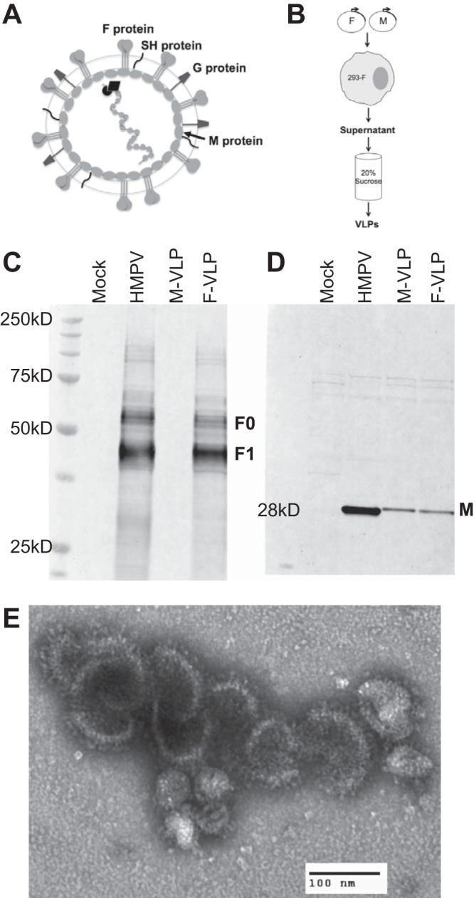
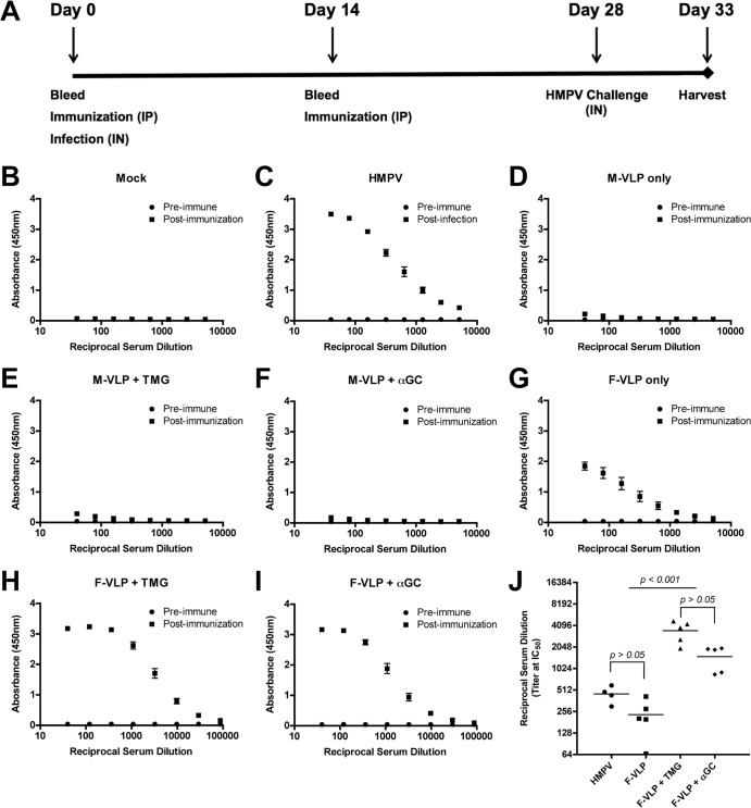
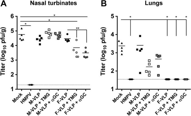
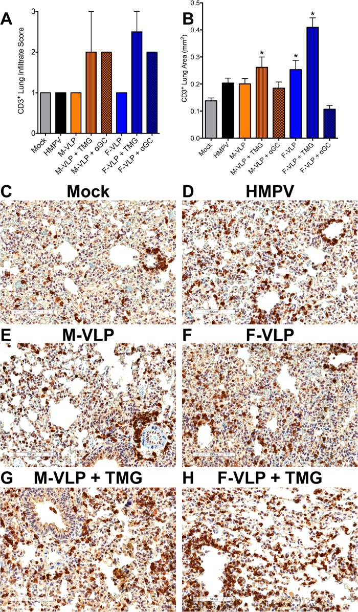
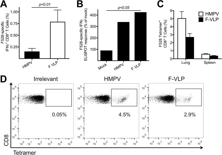
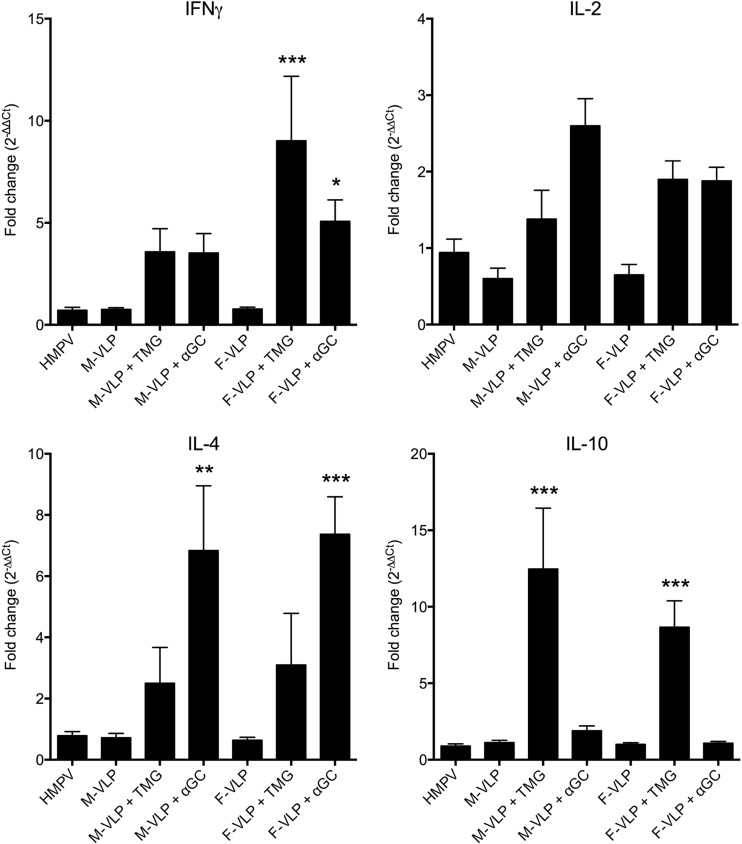
Human metapneumovirus (HMPV) is a leading cause of respiratory disease in infants, children, and the elderly worldwide, yet no licensed vaccines exist. Live-attenuated vaccines present safety challenges, and protein subunit vaccines induce primarily antibody responses. Virus-like particles (VLPs) are an attractive alternative vaccine approach because of reduced safety concerns compared with live vaccines. We generated HMPV VLPs by expressing viral proteins in suspension-adapted human embryonic kidney epithelial (293-F) cells and found that the viral matrix (M) and fusion (F) proteins were sufficient to form VLPs. We previously reported that the VLPs resemble virus morphology and incorporate fusion-competent F protein (R. G. Cox, S. B. Livesay, M. Johnson, M. D. Ohi, and J. V. Williams, J. Virol. 86:12148-12160, 2012), which we hypothesized would elicit F-specific antibody and T cell responses. In this study, we tested whether VLP immunization could induce protective immunity to HMPV by using a mouse model. C57BL/6 mice were injected twice intraperitoneally with VLPs alone or with adjuvant and subsequently challenged with HMPV. Mice were euthanized 5 days postinfection, and virus titers, levels of neutralizing antibodies, and numbers of CD3(+) T cells were quantified. Mice immunized with VLPs mounted an F-specific antibody response and generated CD8(+) T cells recognizing an F protein-derived epitope. VLP immunization induced a neutralizing-antibody response that was enhanced by the addition of either TiterMax Gold or α-galactosylceramide adjuvant, though adjuvant reduced cellular immune responses. Two doses of VLPs conferred complete protection from HMPV replication in the lungs of mice and were not associated with a Th2-skewed cytokine response. These results suggest that nonreplicating VLPs are a promising vaccine candidate for HMPV.

Importance: Human metapneumovirus (HMPV) is a leading cause of acute respiratory infection in infants, children, and the elderly worldwide, yet no licensed vaccines exist. Live-attenuated vaccines present safety challenges, and protein subunit vaccines induce primarily antibody responses. Virus-like particles (VLPs) are an attractive alternative vaccine approach. We generated HMPV VLPs by expressing the viral matrix (M) and fusion (F) proteins in mammalian cells. We found that mice immunized with VLPs mounted an F-specific antibody response and generated CD8(+) T cells recognizing an F protein-derived epitope. VLP immunization induced a neutralizing-antibody response that was enhanced by the addition of either TiterMax Gold or α-galactosylceramide adjuvant. Two doses of VLPs conferred complete protection against HMPV replication in the lungs of mice and were not associated with a Th2-skewed cytokine response. These results suggest that nonreplicating VLPs are a promising vaccine candidate for HMPV.


Figures

FIG 1
FIG 1
HMPV VLPs resemble virus in morphology and incorporate the F and M proteins. (A) Schematic representation of an HMPV virion. The F, G, and SH proteins are depicted at the virion surface. The M protein lines the inner leaflet of the virus membrane. Encapsidated within the viral envelope is the RNA genome that is associated with the nucleoprotein (N), viral polymerase (L), phosphoprotein (P), and matrix 2 (M2) proteins, which are not labeled. (B) Schematic showing the procedure used to generate VLPs. 293-F cells were transfected with HMPV M or M-plus-F protein expression plasmids. At 4 days posttransfection, cell supernatants were harvested and purified through 20% sucrose and VLP pellets were resuspended in PBS. (C, D) Particles released from untransfected (mock VLPs), HMPV-infected (HMPV), M-transfected (M-VLP), and M-plus-F-transfected (F-VLP) cells were harvested, purified, and analyzed by Western blot assay to verify their composition. The same amount (20 μg total protein of either VLP or virus) of each preparation was separated by denaturing, reducing SDS-PAGE and immunoblotted with anti-F MAb (C) or polyclonal anti-HMPV M antiserum (D). Molecular mass markers are shown to the left of the protein ladder. Bands corresponding to F0 (uncleaved F), F1 (cleaved F), and M are indicated. (E) F-VLPs released from cells transfected with HMPV M plus F were purified and analyzed by electron microcopy. An electron micrograph of a negatively stained sample showing a cluster of F-VLPs is shown. Magnification, ×28,000. F glycoprotein spikes are visible on the VLP surface.
FIG 2
FIG 2
Immunization with HMPV VLPs induces F-specific antibodies. (A) Schematic showing the immunization protocol. B6 mice (five per group) were immunized two times, 14 days apart, by i.p. injection with VLPs containing either HMPV M (M-VLP) or M plus F (F-VLP) protein. VLPs were administered alone, with TMG adjuvant, or with αGC adjuvant. Negative-control mice were injected with purified supernatant from untreated 293-F cells (mock VLPs). Positive-control mice were infected i.n. with HMPV on day 0. Blood was collected at the time points indicated and at euthanasia to determine the production of F-specific and neutralizing antibodies. (B to J) HMPV F-specific antibody levels in serum samples were measured by ELISA. Preimmune serum was collected on day 0. Postinfection and postimmunization serum samples were collected 5 days after an HMPV challenge. The absorbance resulting from serum antibody binding to plates coated with recombinant F protein is shown for each vaccination group as follows: panel B, mock VLPs; panel C, HMPV; panel D, M-VLP; panel E, M-VLP plus TMG; panel F, M-VLP plus αGC; panel G, F-VLP; panel H, F-VLP plus TMG; panel I, F-VLP plus αGC. Absorbance data are shown as the mean ± the SEM for five mice from three independent ELISAs. Nonlinear regression analyses were performed for groups with F-specific antibodies and used to calculate reciprocal serum titers at half-maximal absorbance (IC50), shown in panel J. Data points represent individual mice, and the bar depicts the mean IC50 titer of each immunization group. Comparisons of multiple groups were made by one-way ANOVA for the P values shown in panel J.
FIG 3
FIG 3
Immunization with HMPV VLPs protects mice against HMPV infection. The experimental protocol used is shown in Fig. 2A. Levels of replicating HMPV were quantified by plaque assay on day 33 (5 days after an HMPV challenge). (A) Nasal titers of HMPV. (B) Lung titers of HMPV. The dotted lines in panels A and B indicate the limits of detection (20 and 35 PFU/g, respectively); the bars represent the mean virus titers of groups of mice (n = 5). Comparisons of multiple groups were made by one-way ANOVA with Dunnett's posttest (*, P < 0.05). Comparisons of two groups were made with an unpaired Student t test (**, P < 0.05).
FIG 4
FIG 4
HMPV VLPs induce a CD3+ T cell infiltrate in the lungs of immunized animals after an HMPV challenge. At day 33 (5 days after an HMPV challenge), slices were collected from the left lung of each vaccinated mouse. Lung samples were fixed, paraffin embedded, sectioned at a 5-μm thickness, and stained with anti-CD3 antibody. (A) Lung samples were analyzed and scored for CD3+ T cell infiltration on a scale of 0 to 3. Infiltrate scores (mean ± SEM) of two mice from each group are shown. Comparisons of multiple groups were made by one-way ANOVA with Dunnett's posttest. No significant differences were noted. (B) Digital images of the CD3-stained lung sections were generated with an Aperio ScanScope CS2, and a color deconvolution algorithm was used to quantify CD3 staining. Ten random regions, covering the entire lung area, from two mice per group were chosen, and the CD3+ area of each region was calculated. Results are presented as the mean ± the SEM. Comparisons of multiple groups were made by one-way ANOVA with Dunnett's posttest (*, P < 0.05). (C to H) Representative images of lung sections from immunized or infected mice at 5 days after an HMPV challenge. Magnification, ×20. CD3+ T cells are stained dark brown. Scale bars are shown in the lower left corners.
FIG 5
FIG 5
HMPV VLPs generate HMPV-specific CD8+ T cells. (A) B6 mice were infected with HMPV or immunized with F-VLP without adjuvant, and splenocytes were isolated 7 days later. Lymphocytes were stimulated with either an HMPV-specific F528-536 peptide or an irrelevant influenza virus-specific NP366-374 peptide as described in Materials and Methods and analyzed for IFN-γ secretion. The two groups were compared by using an unpaired Student t test. (B) B6 mice were infected with HMPV on day 0 or immunized on days 0 and 14 with F-VLP without adjuvant, and then all of the groups were challenged on day 28 with HMPV. Splenocytes were collected on day 5 postchallenge and tested by ELISPOT assay by using stimulation with an HMPV-specific F528-536 peptide, an irrelevant peptide, or ConA. The average number of spots in the negative-control wells was subtracted from each experimental value, and the number of SFC per 106 lymphocytes was calculated and is expressed as a percentage of the response of mock VLP-immunized animals (set at 100%). Comparisons of multiple groups were made by one-way ANOVA with Dunnett's posttest (*, P < 0.05). (C) B6 mice were infected with HMPV or immunized with F-VLP plus TMG, and spleen and lung lymphocytes were isolated 10 days later. Lymphocytes were tetramer stained with either an HMPV-specific F528-536 tetramer or an irrelevant influenza virus-specific NP366-374 tetramer and anti-CD8 antibody as described in Materials and Methods. The percentage of tetramer+ F-specific lung TCD8 or spleen TCD8 cells was calculated. Data (mean ± SEM) represent an independent experiment with three to five mice per group. (D) Representative flow cytometry plots showing F528-536 tetramer staining on the gated CD8+ T cell population from the lungs of HMPV-infected or F-VLP-immunized mice. The values under the quadrants are the percentages of HMPV-specific F528-536 tetramer+ CD8+ T cells.
FIG 6
FIG 6
VLP immunization is associated with both Th1 and Th2 responses. Groups of mice were immunized with mock VLPs, M-VLP, M-VLP plus TMG, M-VLP plus αGC, F-VLP, F-VLP plus TMG, or F-VLP plus αGC on days 0 and 14 or infected with HMPV on day 0. All of the mice were then challenged with HMPV on day 28. Their lungs were collected and homogenized on day 5 postchallenge, and RNA was extracted and tested by real-time RT-PCR for multiple cytokines. All of the values were normalized to the housekeeping gene for HPRT and are reported as fold differences (determined by the ΔΔCT method) from mice that were mock VLP vaccinated. Comparisons of groups were made by one-way ANOVA with Dunnett's posttest (*, P < 0.01; **, P < 0.001; ***, P < 0.0001).

References

    1. Peiris JS, Tang WH, Chan KH, Khong PL, Guan Y, Lau YL, Chiu SS. 2003. Children with respiratory disease associated with metapneumovirus in Hong Kong. Emerg. Infect. Dis. 9:628–633. 10.3201/eid0906.030009 - DOI - PMC - PubMed
    1. Ebihara T, Endo R, Kikuta H, Ishiguro N, Ishiko H, Hara M, Takahashi Y, Kobayashi K. 2004. Human metapneumovirus infection in Japanese children. J. Clin. Microbiol. 42:126–132. 10.1128/JCM.42.1.126-132.2004 - DOI - PMC - PubMed
    1. Williams JV, Harris PA, Tollefson SJ, Halburnt-Rush LL, Pingsterhaus JM, Edwards KM, Wright PF, Crowe JE., Jr 2004. Human metapneumovirus and lower respiratory tract disease in otherwise healthy infants and children. N. Engl. J. Med. 350:443–450. 10.1056/NEJMoa025472 - DOI - PMC - PubMed
    1. Sloots TP, Mackay IM, Bialasiewicz S, Jacob KC, McQueen E, Harnett GB, Siebert DJ, Masters BI, Young PR, Nissen MD. 2006. Human metapneumovirus, Australia, 2001-2004. Emerg. Infect. Dis. 12:1263–1266. 10.3201/eid1708.051239 - DOI - PMC - PubMed
    1. Williams JV, Wang CK, Yang CF, Tollefson SJ, House FS, Heck JM, Chu M, Brown JB, Lintao LD, Quinto JD, Chu D, Spaete RR, Edwards KM, Wright PF, Crowe JE., Jr 2006. The role of human metapneumovirus in upper respiratory tract infections in children: a 20-year experience. J. Infect. Dis. 193:387–395. 10.1086/499274 - DOI - PMC - PubMed

Publication types

MeSH terms