. 2014 Jun;88(11):6380-93.
doi: 10.1128/JVI.03856-13. Epub 2014 Mar 26.

The Thr205 phosphorylation site within respiratory syncytial virus matrix (M) protein modulates M oligomerization and virus production

Affiliations

The Thr205 phosphorylation site within respiratory syncytial virus matrix (M) protein modulates M oligomerization and virus production

M Bajorek et al. J Virol. 2014 Jun.

Abstract

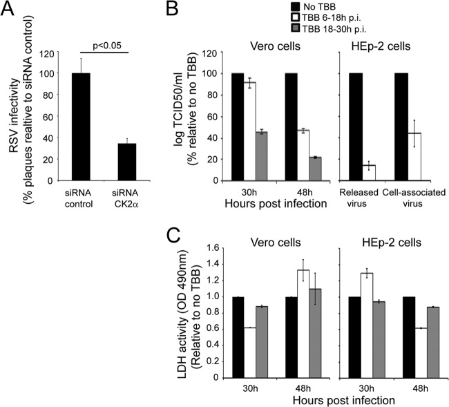
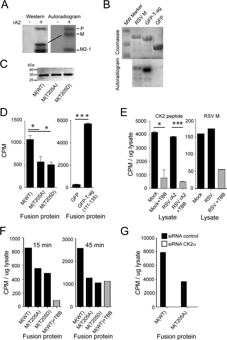
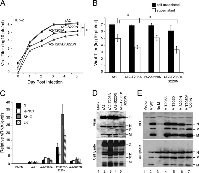
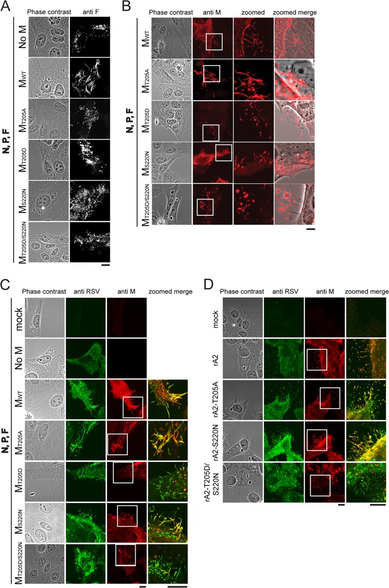
Human respiratory syncytial virus (RSV) is the most common cause of bronchiolitis and pneumonia in infants and the elderly worldwide; however, there is no licensed RSV vaccine or effective drug treatment available. The RSV matrix (M) protein plays key roles in virus assembly and budding, but the protein interactions that govern budding of infectious virus are not known. In this study, we focus on M protein and identify a key phosphorylation site (Thr205) in M that is critical for RSV infectious virus production. Recombinant virus with a nonphosphorylatable alanine (Ala) residue at the site was markedly attenuated, whereas virus with a phosphomimetic aspartate (Asp) resulted in a nonviable virus which could only be recovered with an additional mutation in M (serine to asparagine at position 220), strongly implying that Thr205 is critical for viral infectivity. Experiments in vitro showed that mutation of Thr205 does not affect M stability or the ability to form dimers but implicate an effect on higher-order oligomer assembly. In transfected and infected cells, Asp substitution of Thr205 appeared to impair M oligomerization; typical filamentous structures still formed at the plasma membrane, but M assembly during the ensuing elongation process seemed to be impaired, resulting in shorter and more branched filaments as observed using electron microscopy (EM). Our data thus imply for the first time that M oligomerization, regulated by a negative charge at Thr205, may be critical to production of infectious RSV.

Importance: We show here for the first time that RSV M's role in virus assembly/release is strongly dependent on threonine 205 (Thr205), a consensus site for CK2, which appears to play a key regulatory role in modulating M oligomerization and association with virus filaments. Our analysis indicates that T205 mutations do not impair M dimerization or viruslike filament formation per se but rather the ability of M to assemble in ordered fashion on the viral filaments themselves. This appears to impact in turn upon the infectivity of released virus rather than on virus production or release itself. Thus, M oligomerization would appear to be a target of interest for the development of anti-RSV agents; further, the recombinant T205-substituted mutant viruses described here would appear to be the first RSV mutants affected in viral maturation to our knowledge and hence of considerable interest for vaccine approaches in the future.


Figures

FIG 1
FIG 1
CK2 activity enhances RSV infectious virus production. (A) A549 cells were treated for 48 h with 20 nM siRNA specific for CK2α or control siRNA, followed by infection with RSV at an MOI of 1 for 24 h. Cells were harvested, and cell-associated virus was analyzed by plaque assay on Vero cells. Results shown are represented as the percentages of plaque compared to that of the negative control and are the means ± standard errors of the means (SEM) (n = 3 experiments); significant difference (Student's t test) is indicated by the P value. (B) Vero or HEp-2 cells were infected with RSV at an MOI of 1 and subsequently treated with the CK2-specific inhibitor TBB at 2.8 ng/ml (24, 25) for 12 h either 6 or 18 h postinfection (p.i.). In the case of Vero cells, cell-associated virus was harvested at 30 h and 48 h p.i. and titrated on Vero cells. In the case of HEp-2 cells, culture supernatants and cell-associated virus were collected at 48 h p.i. and infectious virus titers were determined on Vero cells. Results for log TCID50/ml (mean ± SEM, n = 3 experiments) are shown as percentages relative to the control (no TBB). (C) Vero and HEp-2 cells were infected with RSV A2 at an MOI of 1. Cells were treated without or with 2.8 ng/ml TBB for 12 h at 6 to 18 h p.i. or 18 to 30 h p.i. The medium was then replaced with fresh medium without TBB, and cells were cultured further up to 48 h p.i. Cell toxicity was determined by LDH release in culture supernatants as described in Materials and Methods. Data (means ± standard deviations [SD]) from two independent experiments are represented as LDH activity, determined in triplicate, relative to no treatment.
FIG 2
FIG 2
Thr205 within M protein is a key target for CK2. (A) RSV A2 (+)- or mock (−)-infected (MOI of 1) HEp-2 cells were metabolically labeled with 32Pi for 2 h at 18 h p.i. followed by lysis in RIPA buffer. RSV proteins were immunoprecipitated from the cleared lysates with goat anti-RSV antibody conjugated to protein G Sepharose beads (Merck Millipore), eluted by boiling in Laemmli buffer, and separated by SDS-PAGE. The gel was dried under vacuum and exposed to X-ray film. The P, M, and M2-1 proteins of RSV are indicated on the right. In parallel, samples were subjected to Western analysis using anti-RSV (Abcam) polyclonal antibody followed by donkey anti-goat HRP-conjugated secondary antibody (Santa Cruz Biotechnology). (B) M protein is phosphorylated by CK2 kinase in vitro. Recombinant His6-tagged M protein was expressed in E. coli and purified on Ni-NTA beads, along with His-tagged T-ag-GFP and GFP as controls. Purified proteins were incubated with 2 ng purified CK2 (Millipore) in the presence of γ32P[ATP] for 30 min at 30°C, and the reaction was stopped by boiling in Laemmli buffer. Reaction products were separated by SDS-PAGE and visualized by Coomassie blue staining (top), and the gel was dried and processed for autoradiography using the Typhoon Trio (GE Healthcare) (bottom). (C) M proteins used for phosphorylation experiments. Recombinant His6-tagged M protein derivatives were expressed in E. coli and purified on Ni-NTA beads. Proteins were run on SDS-PAGE and visualized by Coomassie blue staining. (D) M protein can be phosphorylated by CK2 at Thr205 in vitro. Purified proteins from panel C, along with His-tagged T-ag-GFP (positive control; see Materials and Methods) and GFP (negative control) were incubated with 2 ng purified CK2 (Millipore) in the presence of γ32P[ATP] for 120 min at 30°C, and the reaction was stopped by spotting onto P-81 paper/washing in phosphoric acid. Counts incorporated were determined by scintillation counting. Results represent the means ± SD (n = 2 experiments). (E) M protein can be phosphorylated in a TBB-sensitive manner by lysates prepared from RSV-infected cells in vitro. Phosphorylation was carried out for 15 min in the presence of ca. 24 μg cell lysate prepared as described in Materials and Methods from mock- or RSV-infected cells treated without or with TBB (2.8 ng/ml), prior to analysis of incorporated counts as described for panel D. Positive control (left) used a peptide CK2 substrate. Results represent the means ± SD (n = 2 experiments). (F) Thr205 within M is a key site for phosphorylation by cell lysates. M protein derivatives were incubated with cell lysates as described for panel E, without or with TBB (25 μM) treatment. Analysis was as described for panels D and E. (G) M protein phosphorylation by cell lysates is dependent on CK2. A549 cells were transfected with siRNA (scrambled or CK2α specific) at 20 nM, and then lysates were prepared 48 h later. Phosphorylation of recombinant purified M was carried out for 15 min in the presence of ca. 24 μg cell lysate prepared as described in Materials and Methods, prior to analysis of incorporated Pi as described for panel E; results are from a single typical experiment from a series of 2 experiments. Significant differences are denoted: *, P < 0.05; ***, P < 0.0001.
FIG 3
FIG 3
M protein Thr205 is required for optimal RSV production. (A) Monolayers of HEp-2 cells were infected by WT rA2 or the indicated M mutant rRSVs at an MOI of 0.01, and the supernatant was sampled every 24 h for 5 days prior to determination of infectious virus, performed by standard plaque assay on Vero cells as described previously (26). Results represent the means ± SEM (n = 3 experiments). *, P < 0.05. (B) HEp-2 cells were infected with the indicated viruses at an MOI of 3; samples were collected 24 h later, and titers of cell-associated and released virus were determined on HEp-2 cells using an immune plaque assay as described in Materials and Methods. Results represent the means ± SD (n = 3 experiments). *, P < 0.05. (C) Results of qRT-PCR analysis performed on viral RNA from 106 PFU of each virus (supernatant fraction) using primers specific for the N, leader-NS1, SH-G, and L-trailer regions of the genome. Results are for the means ± SEM (n = 3 experiments). (D) HEp-2 cells were infected without (mock) or with WT rA2 or the indicated M mutant rRSVs at an MOI of 3. Twenty-four hours p.i., virus was isolated from the supernatant after centrifugation (1,300 × g, 10 min, 4°C) to remove cell debris and pelleted through a sucrose cushion. Cell lysates were generated using RIPA buffer. Virus (top) and cell lysate (bottom) were then subjected to Western analysis using anti-RSV (Abcam) polyclonal antibody followed by donkey anti-goat HRP-conjugated secondary antibody (Santa Cruz Biotechnology). (E) HEp-2 cells were cotransfected with pcDNA3.1 vector plasmid (lane 1), with pcDNA3.1 plasmids encoding RSV P, N, and F proteins plus pcDNA3.1 RSV M WT (lane 2, positive control), with pcDNA3.1 plasmids encoding RSV P, N, and F proteins plus empty pcDNA3.1 vector (lane 3, negative control), or with the indicated RSV M mutant constructs (lanes 4 to 7). Forty-eight hours posttransfection, VLPs (top) were isolated from the supernatant after centrifugation (1,300 × g, 10 min, 4°C) to get rid of cell debris, and the clean supernatant was pelleted through a sucrose cushion. Cell lysates (bottom) were generated using RIPA buffer. VLPs and cell lysates were then subjected to Western analysis using anti-RSV (Abcam) polyclonal antibody followed by donkey anti-goat HRP-conjugated secondary antibody (Santa Cruz Biotechnology).
FIG 4
FIG 4
Mutations of T205 do not abolish M's ability to form stable dimers. (A) Purification of bacterially expressed His6-tagged WT M and the T205A, T205D, and T205D/S220N mutant derivative proteins using nickel affinity chromatography. Purified proteins were concentrated to 3 mg/ml and pelleted to separate higher oligomers (P) from the soluble fraction (S), run on an SDS-PAGE gel, and stained with Coomassie. (B) Analytical S200 gel filtration chromatography was performed on the soluble fraction of WT M and the T205A, T205D, and T205D/S220N mutants. (C) Molecular masses were estimated by comparing the gel-phase distribution coefficient (Kav) of the M protein peaks with the values obtained for known calibration standards (GE Healthcare). Mw of M peaks was estimated by fitting to the calibration curve (plot Kav versus log Mr); the single peak was estimated to be a dimer (estimated Mw, 65 kDa) and is indicated by an arrow. (D) Fractions 8 to 18 were run on an SDS-PAGE gel and stained with Coomassie, identifying the peak as purified M protein.
FIG 5
FIG 5
The T205D mutation alters M distribution on viruslike filaments. (A) HEp-2 cells were cotransfected with pcDNA3.1 plasmids encoding RSV P, N, and F proteins plus empty pcDNA3.1 vector (top row; negative control), pcDNA3.1 RSV M WT (second row; positive control), or the indicated RSV M mutant constructs (rows 3 to 6). Cells were fixed, permeabilized 24 h p.t., immunostained with anti-F (Millipore) monoclonal antibody and Alexa Fluor 568-coupled donkey anti-mouse secondary antibody (Invitrogen), and analyzed by confocal microscopy. (B) HEp-2 cells were transfected as described for panel A, fixed, permeabilized 24 h later, immunostained using monoclonal anti-M primary and Alexa Fluor 568-coupled donkey anti-mouse (Invitrogen) secondary antibodies, and analyzed by confocal microscopy. (C) HEp-2 cells were transfected as described for panel A, fixed, permeabilized 24 h later, immunostained using monoclonal anti-M and with polyclonal anti-RSV (Abcam) primary and Alexa Fluor 568-coupled donkey anti-mouse (Invitrogen) and Alexa Fluor 488-coupled rabbit anti-goat (Invitrogen) secondary antibodies, and analyzed by confocal microscopy. (D) HEp-2 cells were infected with the indicated recombinant viruses at an MOI of 3 and fixed and permeabilized 24 h later prior to immunostaining and confocal microscopy as described for panel C. Areas that are blown up in the details (zoomed column) are marked with white squares in the original panel. Scale bars represent 10 μm.
FIG 6
FIG 6
(A) HEp-2 cells were infected with rA2 (left) or rA2–T205D/S220N (right) recombinant viruses at an MOI of 3. Cells were fixed 24 h later, ultrathin sections were cut parallel to the dish, and cells were stained and processed for transmission electron microscopy as described in Materials and Methods. Different magnifications shown are denoted by the scale bars; in the higher magnifications, fv marks filamentous and sv marks spherical virus particles, with the M layer and glycoprotein spikes indicated. (B) For each virus strain, 150 viral filaments were randomly selected and analyzed for filament dimensions, with the results shown as a percentage of the total. (C) A model diagram of viral filament is shown based on reference , with the indicated M layer below the membrane and glycoprotein spikes on the outside.
FIG 7
FIG 7
Structural conservation among different Mononegavirales matrix proteins. (A) Structure-based sequence (single-letter amino acid code) alignment (adapted from reference 23) around the loop from RSV M containing Thr205 to Ser220 (Thr205 and Ser220 marked), with residues proposed to play a key role in higher-order oligomerization based on structural alignment to NDV M highlighted in green, and the residues within the respective proteins comprising the loop highlighted in yellow based on alignment to RSV M. CK2 consensus site in RSV M indicated in red. Residues in each protein are numbered. (B) Structure-based alignment of known M structures, shown as a ribbon diagram; RSV M (pink) (41), NDV M (blue) (23), BDV M (cyan) (42), and EBV M (orange) (43), with the RSV M Thr205-Ser220 loop or comparable loops shown in yellow and oligomerization residues in green.

Similar articles

Cited by

References

    1. Collins PL, Graham BS. 2008. Viral and host factors in human respiratory syncytial virus pathogenesis. J. Virol. 82:2040–2055. 10.1128/JVI.01625-07 - DOI - PMC - PubMed
    1. Huang YT, Collins PL, Wertz GW. 1985. Characterization of the 10 proteins of human respiratory syncytial virus: identification of a fourth envelope-associated protein. Virus Res. 2:157–173. 10.1016/0168-1702(85)90246-1 - DOI - PubMed
    1. Fearns R, Collins PL. 1999. Role of the M2–1 transcription antitermination protein of respiratory syncytial virus in sequential transcription. J. Virol. 73:5852–5864 - PMC - PubMed
    1. Collins PL, Crowe JE. 2007. Respiratory syncytial virus and human metapneumovirus, p 1601–1646 In Knipe H, Griffin DE, Lamb RA, Martin MA, Roizman B, Straus SE. (ed), Fields virology, 5th ed, vol 2 Lippincott, Williams, and Wilkins, Baltimore, MD
    1. Garcia J, Garcia-Barreno B, Vivo A, Melero JA. 1993. Cytoplasmic inclusions of respiratory syncytial virus-infected cells: formation of inclusion bodies in transfected cells that coexpress the nucleoprotein, the phosphoprotein, and the 22K protein. Virology 195:243–247. 10.1006/viro.1993.1366 - DOI - PubMed

Publication types

MeSH terms