. 2014 Jun;88(11):6470-82.
doi: 10.1128/JVI.00465-14. Epub 2014 Mar 26.

Substitution of herpes simplex virus 1 entry glycoproteins with those of saimiriine herpesvirus 1 reveals a gD-gH/gL functional interaction and a region within the gD profusion domain that is critical for fusion

Affiliations

Substitution of herpes simplex virus 1 entry glycoproteins with those of saimiriine herpesvirus 1 reveals a gD-gH/gL functional interaction and a region within the gD profusion domain that is critical for fusion

Qing Fan et al. J Virol. 2014 Jun.

Abstract

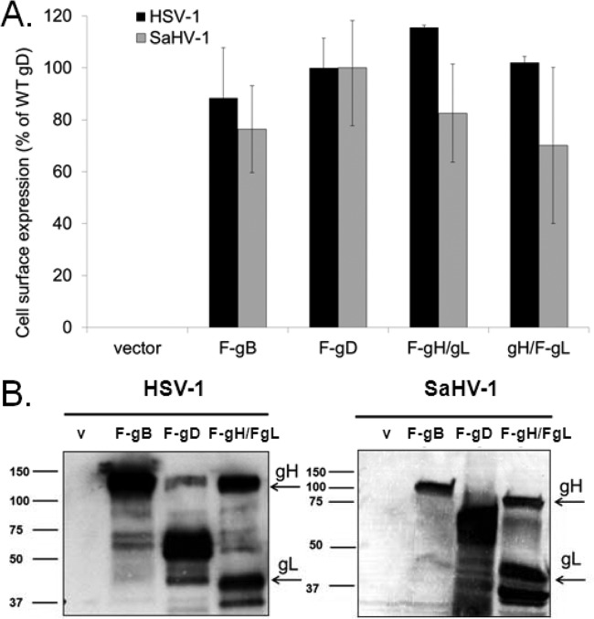
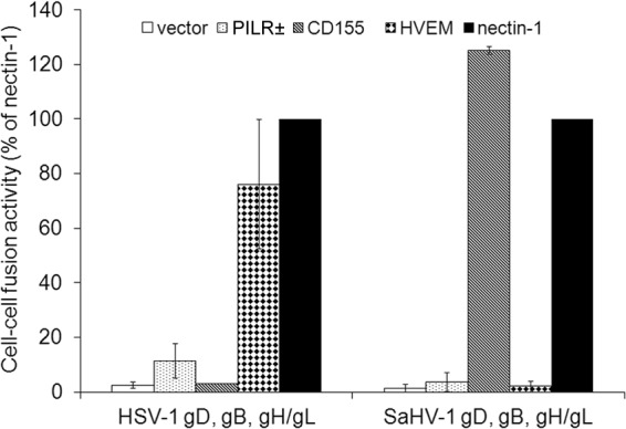
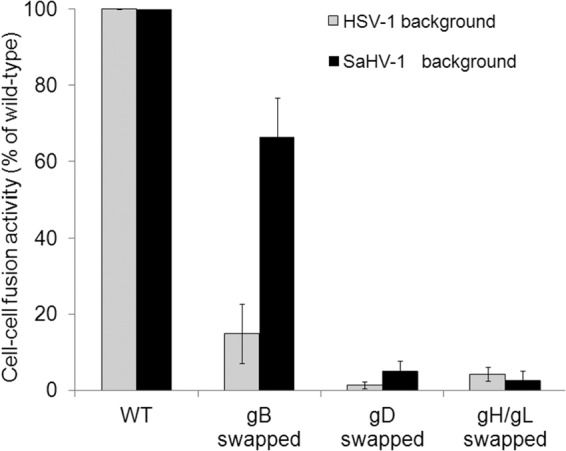
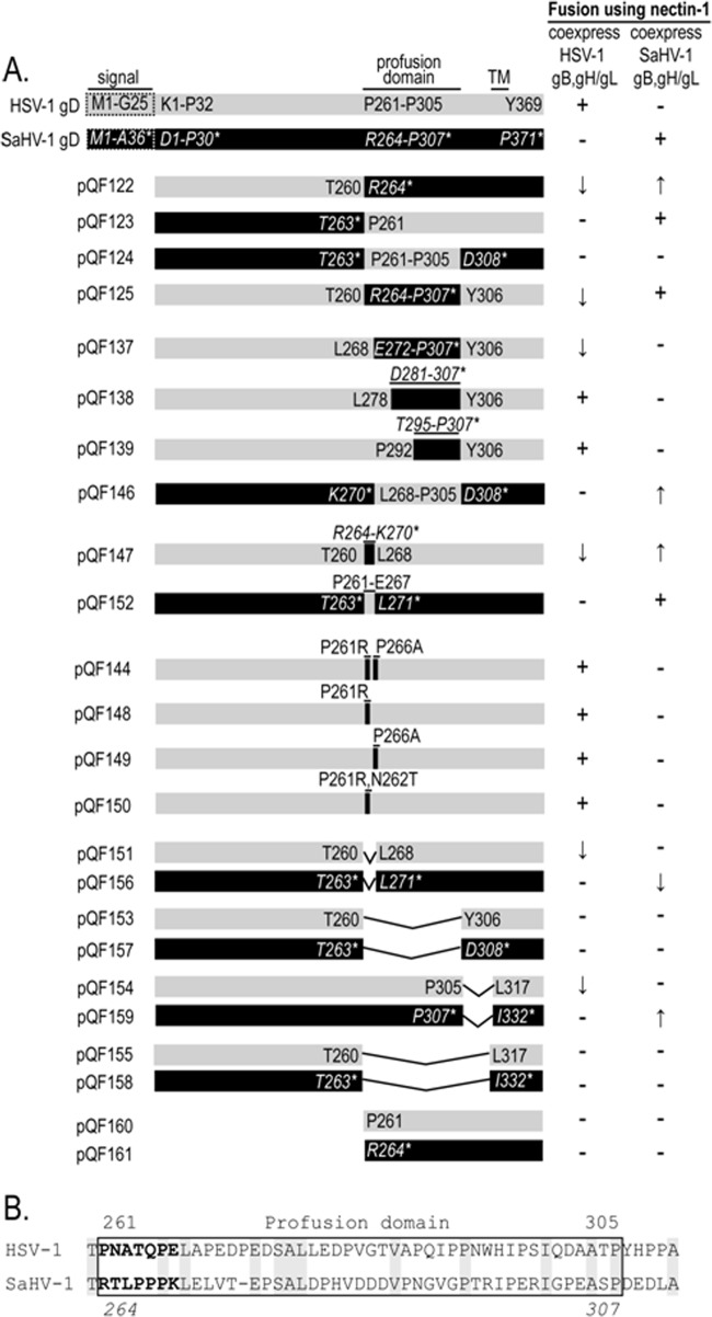
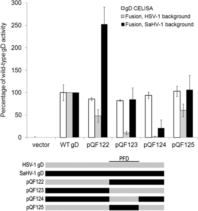
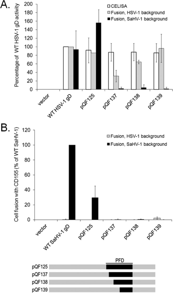
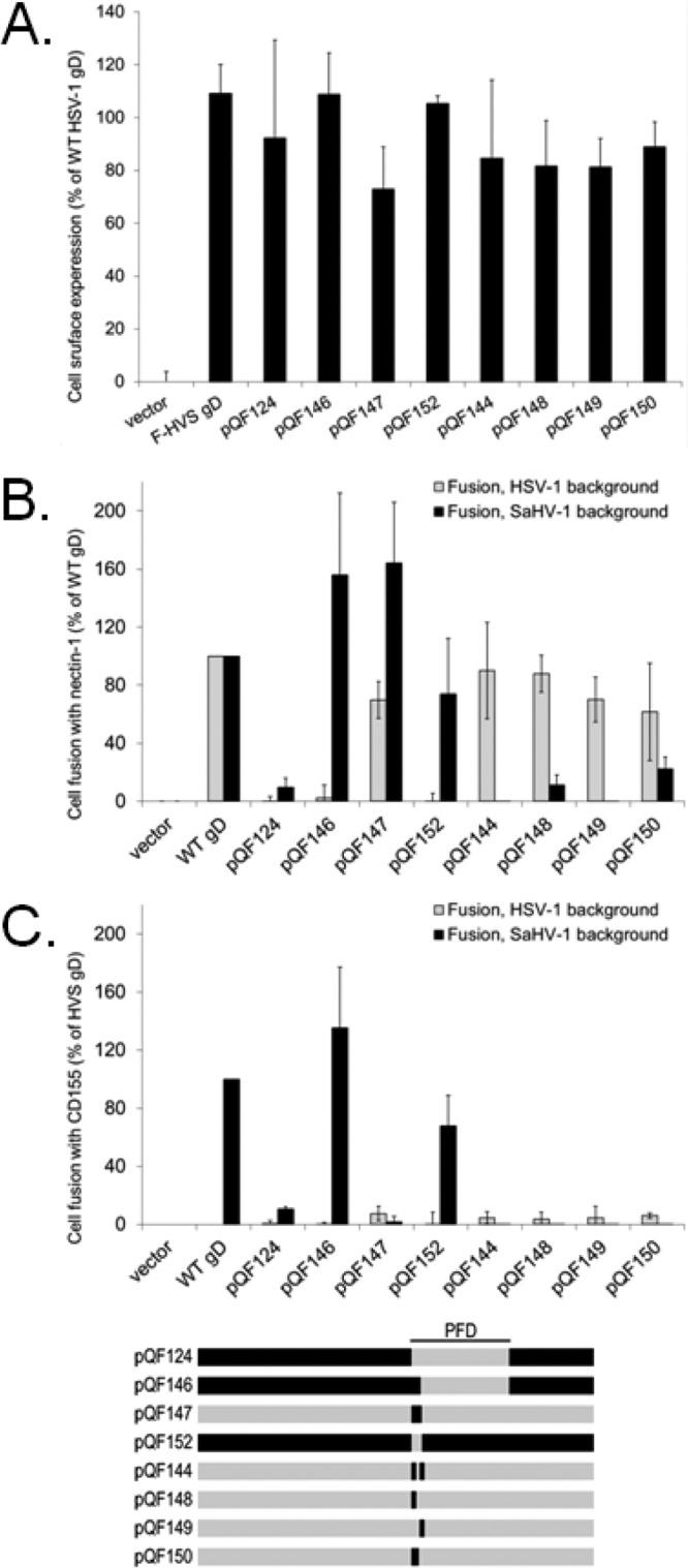
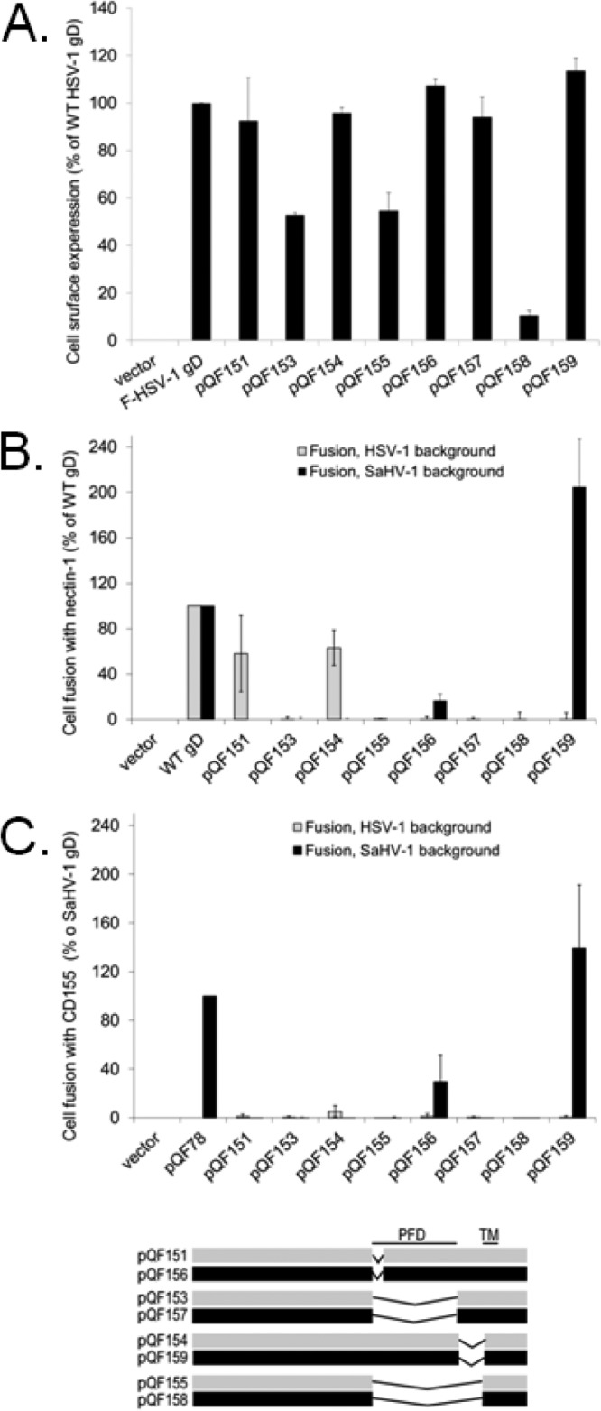
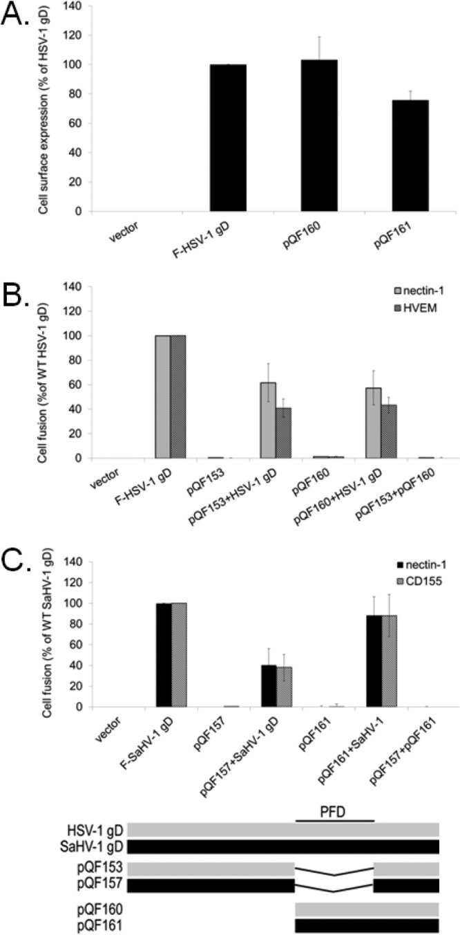
To gain insight into the mechanism of herpesvirus entry into cells, the four glycoproteins that are necessary for herpes simplex virus (HSV) fusion were cloned from the saimiriine herpesvirus 1 (SaHV-1) genome, a primate member of the alphaherpesvirus family. Cell-cell fusion assays indicate that SaHV-1 entry glycoproteins function with the previously identified alphaherpesvirus entry receptors nectin-1 and CD155 but not with herpesvirus entry mediator (HVEM) or paired immunoglobulin-like type 2 receptor alpha (PILRα). Replacement of HSV-1 gD with the SaHV-1 gD homolog resulted in a complete loss of fusion function when coexpressed with HSV-1 gB and gH/gL. HSV-1 gD was also unable to substitute for SaHV-1 gD when coexpressed with SaHV-1 gB and gH/gL. Similarly, the gH/gL heterodimers from HSV-1 and SaHV-1 were not interchangeable. In contrast, both the HSV-1 and SaHV-1 gB homologs retained function in a heterotypic context. These results suggest that an essential interaction between homotypic gD and gH/gL occurs during both HSV-1 and SaHV-1 entry. To map the site of this homotypic interaction, we created a series of gD chimeras, focusing on the "profusion domain" (PFD) that consists of HSV-1 gD residues 261 to 305 or SaHV-1 gD residues 264 to 307. We identified a seven-amino-acid stretch (264 RTLPPPK 270) at the N terminus of the SaHV-1 gD PFD that contributes to homotypic fusion. Finally, we found that the gD receptor-binding region and PFD cannot function independently but that both can inhibit the function of wild-type gD.

Importance: The herpesvirus entry machinery requires the concerted action of at least four glycoproteins; however, details of the interactions among these glycoproteins are not well understood. Like HSV-1, SaHV-1 belongs to the alphaherpesvirus subfamily. Using cell-cell fusion experiments, we found that SaHV-1 uses the entry receptors nectin-1 and CD155 but not HVEM or PILRα. By swapping the entry glycoproteins between HSV-1 and SaHV-1, we revealed a functional interaction between gD and gH/gL. To examine the homotypic interaction site on gD, we evaluated the function of a panel of HSV-1/SaHV-1 gD chimeras and identified a small region in the SaHV-1 gD profusion domain that is critical for SaHV-1 fusion. This study contributes to our understanding of the molecular mechanisms of herpesvirus entry and membrane fusion.


Figures

FIG 1
FIG 1
Expression of entry glycoproteins from HSV-1 and SaHV-1. (A) Cell surface expression measured by CELISA. CHO cells in a 96-well plate were transfected overnight with plasmids encoding FLAG-tagged gB, FLAG-tagged gD, gL plus FLAG-tagged gH, gH plus FLAG-tagged gL, or empty vector. The “F-” indicates the constructs that were FLAG tagged. The cells were washed and incubated with an anti-FLAG M2 antibody. After extensive washing, cells were fixed and incubated with an anti-mouse secondary antibody for detection. Each bar shows the mean and standard deviation of three independent determinations. Background signals detected after transfection with the vector alone were subtracted from the values. Data for each set of glycoproteins were normalized to the expression level of HSV-1 F-gD or SaHV-1 F-gD. (B) Total protein expression measured by Western blot of cell lysates. CHO cells expressing the constructs above were lysed, and proteins were resolved by SDS-PAGE. FLAG-tagged gH was coexpressed with FLAG-tagged gL. Proteins were transferred to nitrocellulose and probed with rabbit anti-FLAG antibody followed by goat anti-rabbit IgG. gB, gD, gH, and gL migrated to their expected molecular weights (shown [in thousands] at left). V, empty vector.
FIG 2
FIG 2
Receptor usage for HSV-1 and SaHV-1 entry glycoproteins. Target CHO cells were transfected with a reporter plasmid encoding luciferase under the control of the T7 promoter along with plasmids encoding PILRα, CD155, HVEM, nectin-1, or empty vector. The transfected cells were replated with effector CHO cells that had been transfected with plasmids encoding T7 polymerase and the complete set of either WT HSV-1 or FLAG-tagged SaHV-1 entry glycoproteins (gD, gB, gH, gL). After coincubation overnight, luciferase activity was measured as an indication of cell-cell fusion. For each set of viral glycoproteins, data were normalized to the fusion activity mediated by nectin-1, which was set at 100%. The relative light units after fusion with nectin-1 expressing cells averaged 110,000 for SaHV-1 and 187,000 for HSV-1. Each bar shows the mean and standard deviation of three independent determinations.
FIG 3
FIG 3
Heterotypic fusion activity of the HSV-1 and SaHV-1 entry glycoproteins. Target CHO cells were transfected with a reporter plasmid encoding luciferase under the control of the T7 promoter along with plasmids encoding nectin-1 or empty vector. Effector CHO cells were transfected with a plasmid encoding T7 polymerase along with a combination of plasmids encoding HSV-1 or SaHV-1 entry glycoproteins. Glycoproteins were swapped for heterotypic counterparts, as indicated. Target and effector cells were coincubated overnight, and luciferase activity was measured as an indication of cell-cell fusion. Data were normalized to the fusion activity measured when a homotypic set of glycoproteins was coexpressed (either all HSV-1 or all SaHV-1 glycoproteins). Each bar shows the mean and standard deviation of three independent determinations.
FIG 4
FIG 4
Mutant gD constructs examining the profusion domain (PFD). (A) Schematic representation of the HSV-1 and SaHV-1 gD constructs. Chimeras, point mutants, and deletion mutants were generated. Gray bars represent HSV-1 gD sequence, whereas black bars represent SaHV-1 sequence. The construct names begin with “QF.” For all constructs, the native signal sequence (dashed box) was replaced by an exogenous signal sequence and an N-terminal FLAG tag. Amino acids for each construct are numbered. Regular type indicates HSV-1 gD numbering, whereas italicized type with an asterisk indicates SaHV-1 gD numbering. The HSV-1 gD PFD residues are denoted P261-P305, and the SaHV-1 gD PFD residues are denoted R264-P307. The table on the right summarizes the ability of the constructs to mediate fusion with nectin-1-expressing cells when coexpressed with gB, gH, and gL from either HSV-1 or SaHV-1. A minus sign indicates that the construct failed to mediate fusion, and a plus sign indicates that it mediated fusion at near wild-type levels. Arrows indicate that the construct mediated fusion at levels higher (up arrow) or lower (down arrow) than that of wild-type gD. (B) Sequence alignment of the C termini of the HSV-1 and SaHV-1 gD ectodomains. The upper numbers refer to HSV-1 gD, and the lower numbers refer to SaHV-1 gD. The PFD is enclosed in a box. Conserved residues are highlighted in gray. The N-terminal seven residues of the PFD are in bold.
FIG 5
FIG 5
Cell surface expression and fusion activities of the HSV-1 and SaHV-1 gD chimeras. Target cells were transfected with a plasmid carrying the luciferase gene under the control of the T7 promoter and either nectin-1 or empty vector. Effector CHO cells were transfected with plasmids encoding T7 polymerase, gB, gH, and gL from either SaHV-1 or HSV-1, and either wild-type gD or a chimeric gD. “HSV-1 background” refers to HSV-1 gB, gH, and gL coexpression, whereas “SaHV-1 background” refers to SaHV-1 gB, gH, and gL coexpression. One set of effector cells was used for CELISA, and the rest were coincubated with target cells for the cell-cell fusion assay. CELISA data are presented as a percentage of wild-type HSV-1 gD expression. The fusion results are expressed as a percentage of wild-type HSV-1 gD or wild-type SaHV-1 gD activity after subtraction of background values (luciferase activity after coincubation with target cells transfected with vector). Means and standard deviations of results of three independent experiments are shown. For clarity, a schematic representation of the constructs is included, with HSV-1 sequence in gray and SaHV-1 sequence in black. Refer to Fig. 4 for a more detailed representation.
FIG 6
FIG 6
Defining a region in the SaHV-1 gD PFD that is important for fusion. (A) Cell surface expression and nectin-1 usage by pQF125 and its derivatives. Target cells were transfected with a plasmid carrying luciferase under the control of the T7 promoter along with nectin-1 or empty vector. Effector CHO cells were transfected with plasmids encoding T7 polymerase, gB, gH, and gL from either SaHV-1 or HSV-1, and either wild-type gD or a chimeric gD. One set of effector cells was used for CELISA, and the rest were coincubated with target cells for the cell-cell fusion assay. The fusion results are expressed as a percentage of wild-type HSV-1 gD or wild-type SaHV-1 gD activity after subtraction of background values (luciferase activity after coincubation with target cells transfected with vector). Means and standard deviations of results of three independent experiments are shown. (B) CD155 usage by pQF125 and its derivatives. The procedures from Fig. 6A were repeated, except that the target cells were transfected with CD155 instead of nectin-1. For clarity, a schematic representation of the constructs is included, with HSV-1 sequence in gray and SaHV-1 sequence in black. Refer to Fig. 4 for a more detailed representation.
FIG 7
FIG 7
The seven N-terminal amino acids of the SaHV-1 gD PFD are critical for SaHV-1 fusion. (A) Cell surface expression of gD chimeras and HSV-1 gD point mutants. The level of surface expression of gD mutants was determined by CELISA with effector cells used in the fusion assays below. (B and C) Cell fusion activity of gD mutants. Target cells were transfected with a plasmid carrying luciferase under the control of the T7 promoter along with nectin-1 (B), CD155 (C), or empty vector. Effector CHO cells were transfected with plasmids encoding T7 polymerase, gB, gH, and gL from either SaHV-1 or HSV-1, and either wild-type gD or a chimeric gD. Effector and target cells were coincubated, and luciferase activity was determined as an indication of fusion activity. The results are expressed as a percentage of wild-type HSV-1 gD or wild-type SaHV-1 gD activity, after subtraction of background (vector). Means and standard deviations of results of three independent experiments are shown. For clarity, a schematic representation of the constructs is included, with HSV-1 sequence in gray and SaHV-1 sequence in black. Refer to Fig. 4 for a more detailed representation.
FIG 8
FIG 8
Deletion of the PFD abrogates fusion. (A) Cell surface expression of gD deletion mutants. The level of surface expression of gD mutants was determined by CELISA with effector cells used in the fusion assays below. (B and C) Cell fusion activity of gD deletion mutants. Target cells were transfected with a plasmid carrying luciferase under the control of the T7 promoter along with nectin-1 (B), CD155 (C), or empty vector. Effector CHO cells were transfected with plasmids encoding T7 polymerase, gB, gH, and gL from either SaHV-1 or HSV-1, and either wild-type gD or a chimeric gD. Effector and target cells were coincubated, and luciferase activity was determined as an indication of fusion activity. The results are expressed as a percentage of wild-type HSV-1 gD or wild-type SaHV-1 gD activity, after subtraction of background (vector). Means and standard deviations of results of three independent experiments are shown. For clarity, a schematic representation of the constructs is included, with HSV-1 sequence in gray and SaHV-1 sequence in black. Refer to Fig. 4 for a more detailed representation. TM, transmembrane domain.
FIG 9
FIG 9
The gD PFD inhibits fusion activity of wild-type gD. (A) Cell surface expression of the PFD-only constructs. The level of surface expression of the HSV-1 and SaHV-1 gD PFD-only mutants was determined by CELISA with effector cells used in the fusion assays below. (B) Target cells were transfected with a plasmid carrying luciferase under the control of the T7 promoter along with nectin-1, HVEM, or empty vector. Effector cells were transfected with plasmids encoding luciferase, gB, gH, and gL from HSV-1, and combinations of wild-type HSV-1 gD, HSV-1 gD with a deletion of the PFD (pQF153), and HSV-1 gD lacking a receptor-binding domain (pQF160). After coincubation of target and effector cells, luciferase activity was measured as an indication of cell-cell fusion activity. (C) Target cells were transfected with a plasmid carrying luciferase under the control of the T7 promoter along with nectin-1, CD155, or empty vector. Effector cells were transfected with plasmids encoding luciferase, gB, gH, and gL from SaHV-1, and combinations of wild-type SaHV-1 gD, SaHV-1 gD with a deletion of the PFD (pQF157), or SaHV-1 gD lacking a receptor-binding domain (pQF161). After coincubation of target and effector cells, luciferase activity was measured as an indication of cell-cell fusion activity. Means and standard deviations of results of three independent experiments are shown for all panels. For clarity, a schematic representation of the constructs is included, with HSV-1 sequence in gray and SaHV-1 sequence in black. Refer to Fig. 4 for a more detailed representation.
FIG 10
FIG 10
(A) Phylogenetic tree of gD from seven alphaherpesviruses, generated using Phylogeny.fr (http://www.phylogeny.fr). (B) Amino acid alignment of gD from seven alphaherpesviruses, generated using ClustalW2 (http://www.ebi.ac.uk/Tools/msa/clustalw2/). Proteins are ordered by relatedness to HSV-1 gD. Minor variations in the HSV-1 and SaHV-1 alignments compared to Fig. 4 are a consequence of performing a multiple alignment versus a pairwise alignment. The HSV-1 gD V-like Ig fold (K1 to R184), profusion domain (P261 to P305), and transmembrane domain (L317 to M339) are in bold italics, as are the corresponding homologous regions in SaHV-1 gD. The N-terminal loop of HSV-1 gD that binds to HVEM is underlined, and the PFDs are boxed based on to HSV-1 gD sequence. HSV-1 numbering begins after the signal sequence. Cysteines involved in disulfide bonds are highlighted in gray.

Similar articles

Cited by

References

    1. Daniel MD, Karpas A, Melendez LV, King NW, Hunt RD. 1967. Isolation of herpes-T virus from a spontaneous disease in squirrel monkeys (Saimiri sciureus). Arch. Gesamte Virusforsch. 22:324–331. 10.1007/BF01242953 - DOI - PubMed
    1. King NW, Hunt RD, Daniel MD, Melendez LV. 1967. Overt herpes-T infection in squirrel monkeys (Saimiri sciureus). Lab. Anim. Care 17:413–423 - PubMed
    1. Tyler S, Severini A, Black D, Walker M, Eberle R. 2011. Structure and sequence of the saimiriine herpesvirus 1 genome. Virology 410:181–191. 10.1016/j.virol.2010.11.008 - DOI - PMC - PubMed
    1. Connolly SA, Jackson JO, Jardetzky TS, Longnecker R. 2011. Fusing structure and function: a structural view of the herpesvirus entry machinery. Nature Rev. Microbiol. 9:369–381. 10.1038/nrmicro2548 - DOI - PMC - PubMed
    1. Eisenberg RJ, Atanasiu D, Cairns TM, Gallagher JR, Krummenacher C, Cohen GH. 2012. Herpes virus fusion and entry: a story with many characters. Viruses 4:800–832. 10.3390/v4050800 - DOI - PMC - PubMed

Publication types

LinkOut - more resources