. 2014 Jun;88(12):6586-98.
doi: 10.1128/JVI.00208-14. Epub 2014 Mar 26.

A complex comprising phosphatidylinositol 4-kinase IIIβ, ACBD3, and Aichi virus proteins enhances phosphatidylinositol 4-phosphate synthesis and is critical for formation of the viral replication complex

Affiliations

A complex comprising phosphatidylinositol 4-kinase IIIβ, ACBD3, and Aichi virus proteins enhances phosphatidylinositol 4-phosphate synthesis and is critical for formation of the viral replication complex

Kumiko Ishikawa-Sasaki et al. J Virol. 2014 Jun.

Abstract

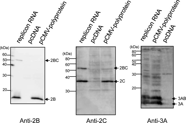
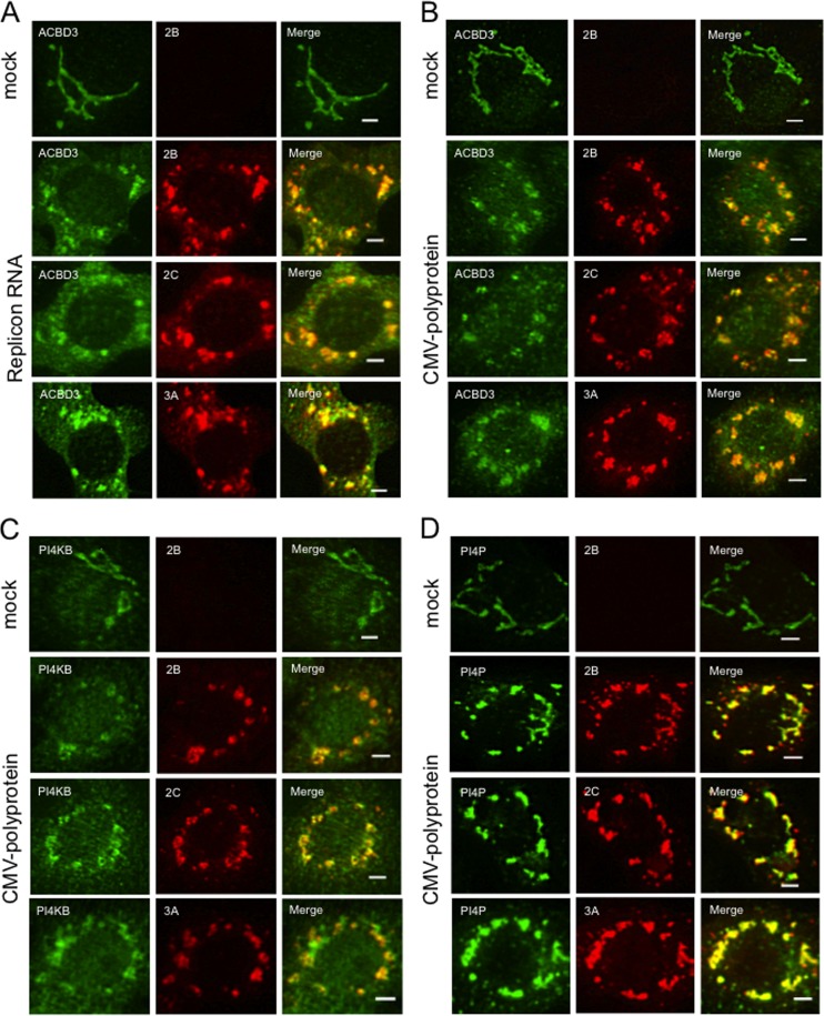
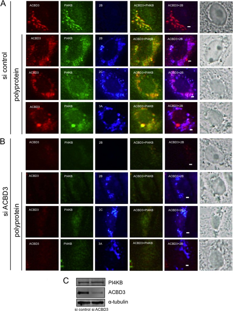
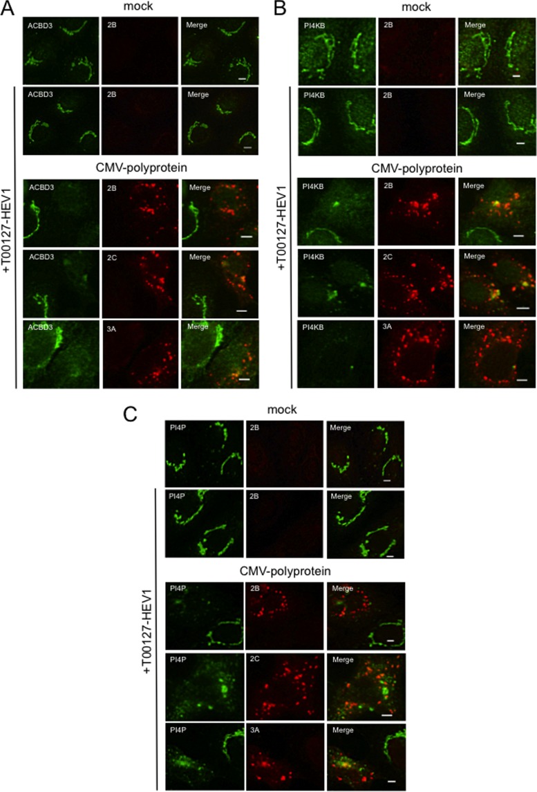
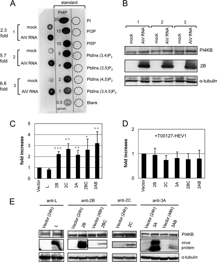
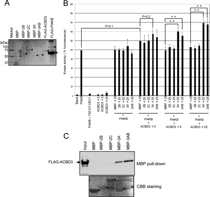
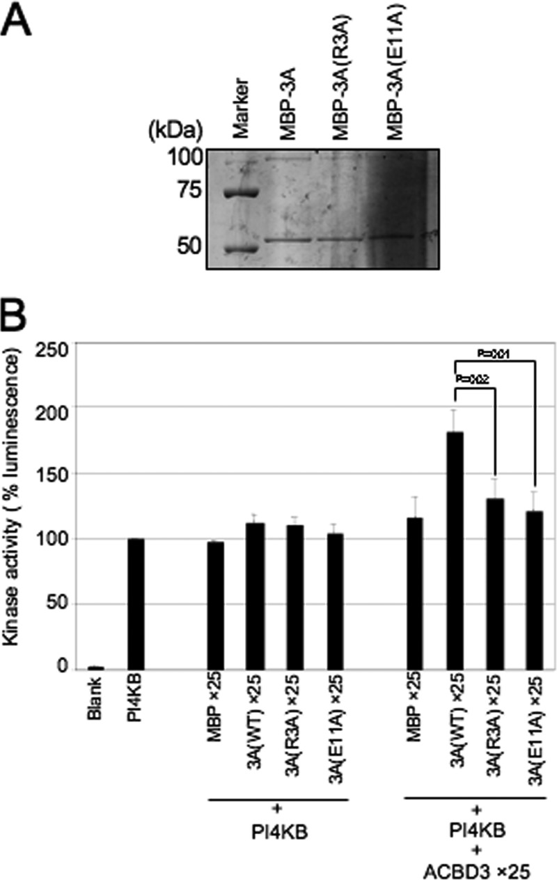
Phosphatidylinositol 4-kinase IIIβ (PI4KB) is a host factor required for the replication of certain picornavirus genomes. We previously showed that nonstructural proteins 2B, 2BC, 2C, 3A, and 3AB of Aichi virus (AiV), a picornavirus, interact with the Golgi protein, acyl-coenzyme A binding domain containing 3 (ACBD3), which interacts with PI4KB. These five viral proteins, ACBD3, PI4KB, and the PI4KB product phosphatidylinositol 4-phosphate (PI4P) colocalize to the AiV RNA replication sites (J. Sasaki et al., EMBO J. 31:754-766, 2012). We here examined the roles of these viral and cellular molecules in the formation of AiV replication complexes. Immunofluorescence microscopy revealed that treatment of AiV polyprotein-expressing cells with a small interfering RNA targeting ACBD3 abolished colocalization of the viral 2B, 2C, and 3A proteins with PI4KB. A PI4KB-specific inhibitor also prevented their colocalization. Virus RNA replication increased the level of cellular PI4P without affecting that of PI4KB, and individual expression of 2B, 2BC, 2C, 3A, or 3AB stimulated PI4P generation. These results suggest that the viral protein/ACBD3/PI4KB complex plays an important role in forming the functional replication complex by enhancing PI4P synthesis. Of the viral proteins, 3A and 3AB were shown to stimulate the in vitro kinase activity of PI4KB through forming a 3A or 3AB/ACBD3/PI4KB complex, whereas the ACBD3-mediated PI4KB activation by 2B and 2C remains to be demonstrated.

Importance: The phosphatidylinositol 4-kinase PI4KB is a host factor required for the replication of certain picornavirus genomes. Aichi virus, a picornavirus belonging to the genus Kobuvirus, forms a complex comprising one of the viral nonstructural proteins 2B, 2BC, 2C, 3A, and 3AB, the Golgi protein ACBD3, and PI4KB to synthesize PI4P at the sites for viral RNA replication. However, the roles of this protein complex in forming the replication complex are unknown. This study showed that virus RNA replication and individual viral proteins enhance the level of cellular PI4P, and suggested that the viral protein/ACBD3/PI4KB complex plays an important role in forming a functional replication complex. Thus, the present study provides a new example of modulation of cellular lipid metabolism by viruses to support the replication of their genomes.


Figures

FIG 1
FIG 1
Processing of the polyproteins expressed by AiV replicon RNA and pCMV-polyprotein. Vero cells were transfected with AiV replicon RNA, pCMV-polyprotein, or pcDNA, and cell lysates prepared 5 and 24 h after transfection were subjected to immunoblotting with anti-2B, anti-2C, or anti-3A antibodies.
FIG 2
FIG 2
Transient expression of the AiV polyprotein induces the formation of the membrane structures containing viral proteins, ACBD3, PI4KB, and PI4P similar to those present in cells with replicating AiV RNA genomes. (A) Vero cells were mock electroporated or electroporated with replicon RNA. Four hours after electroporation, the cells were fixed and double stained with antibodies against ACBD3 (green) and 2B, 2C, or 3A (red). (B, C, and D) Vero cells were transfected with a plasmid (pCMV-polyprotein) that expresses the full-length polyprotein. After 24 h, the cells were immunostained with antibodies against ACBD3 (B), PI4KB (C), PI4P (D) (green), and 2B, 2C, or 3A (red). Scale bars, 20 μm.
FIG 3
FIG 3
ACBD3 knockdown abolishes the colocalization of PI4KB with viral proteins. (A and B) Vero cells were transfected with control siRNA (A) or ACBD3-siRNA (B) and cultured for 72 h before transfection with pCMV-polyprotein. After 24 h, the cells were fixed and triple stained with antibodies against ACBD3 (red), PI4KB (green), and 2B, 2C, or 3A (blue). The panels in the column on the far right show visible light micrographs of cells. Asterisks indicate cells with decreased ACBD3 expression. Scale bars, 20 μm. (C) Western blotting shows that decreased ACBD3 expression did not affect the levels of PI4KB or α-tubulin.
FIG 4
FIG 4
Inhibiting PI4KB activity disrupts PI4KB-, ACBD3-, and viral protein-positive membrane structures. (A, B, and C) Vero cells were transfected with pCMV-polyprotein and then cultured in medium containing 5 μM T00127-HEV1, a PI4KB-specific inhibitor. After 24 h, the cells were fixed and double stained with antibodies against ACBD3 (A), PI4KB (B), or PI4P (C) (green), and 2B, 2C, or 3A (red). Scale bars, 20 μm.
FIG 5
FIG 5
AiV replication and AiV membrane protein expression increase cellular PI4P levels. (A) Vero cells were mock electroporated or electroporated with replicon RNA. PI4P levels were determined 6 h after electroporation. Blots from three independent experiments (left blots 1 to 3) are shown with 0.5- to 20-pmol PI4P standards (middle blots) and 20-pmol PIPn controls (right blots). (B) Lysates prepared from the cells used in panel A were subjected to immunoblotting with antibodies against PI4KB, 2B, or α-tubulin. (C) Vero cells were transfected with pcDNA or the indicated constructs encoding a viral protein. At 24 h (for L, 2B, 2C, or 3A) or 48 h (for 2BC or 3AB) after transfection, each PI4P level was measured as described in panel A and expressed as the value relative to that for pcDNA. (D) Experiments similar to those described in panel C were performed by adding T00127-HEV1 to the cultured medium. The data are means ± the SD for at least six independent experiments. *, P < 0.01; **, P < 0.001; ***, P < 0.0001. (E) Western blotting of cell lysates from panel C.
FIG 6
FIG 6
Viral proteins 3A and 3AB stimulate PI4KB activity in the presence of ACBD3. (A) Purified proteins were electrophoresed through a 10% SDS-polyacrylamide gel and stained with Coomassie brilliant blue. Arrowheads and an arrow indicate each purified protein and BSA contained in the PI4KB elution buffer, respectively. (B) In vitro kinase assays. The kinase activity of PI4KB (2 ng) was assayed in the presence or absence of the indicated molar excess of ACBD3 and MBP or MBP-fused viral proteins compared to PI4KB. The concentration of T00127-HEV1 in the reaction mixtures was 5 μM. Luminescence generated by PI4KB alone was defined as 100%. The experiments were repeated at least three times, and the data are means ± the SD. **, P < 0.001. (C) MBP pulldown assay. MBP or MBP-fused viral proteins immobilized on amylose resins were incubated with purified FLAG-tagged ACBD3, and proteins binding to the resin were analyzed using SDS-PAGE, followed by immunoblotting with anti-FLAG antibody (upper panel). Proteins bound to the membrane were stained with Coomassie brilliant blue (lower panel). Asterisks indicate MBP or MBP-fused viral proteins.
FIG 7
FIG 7
In vitro kinase assay of two 3A mutants. (A) Purified MBP-3A and its MBP-3A mutant fusion proteins were electrophoresed through a 10% SDS-polyacrylamide gel and stained with Coomassie brilliant blue. (B) In vitro kinase assay to examine the ability of the 3A mutants to stimulate PI4KB. The data are presented as described in Fig. 6B.

Similar articles

Cited by

References

    1. Aldabe R, Barco A, Carrasco L. 1996. Membrane permeabilization by poliovirus proteins 2B and 2BC. J. Biol. Chem. 271:23134–23137. 10.1074/jbc.271.38.23134 - DOI - PubMed
    1. Cho MW, Teterina N, Egger D, Bienz K, Ehrenfeld E. 1994. Membrane rearrangement and vesicle induction by recombinant poliovirus 2C and 2BC in human cells. Virology 202:129–145. 10.1006/viro.1994.1329 - DOI - PubMed
    1. Egger D, Teterina N, Ehrenfeld E, Bienz K. 2000. Formation of the poliovirus replication complex requires coupled viral translation, vesicle production, and viral RNA synthesis. J. Virol. 74:6570–6580. 10.1128/JVI.74.14.6570-6580.2000 - DOI - PMC - PubMed
    1. Knox C, Moffat K, Ali S, Ryan M, Wileman T. 2005. Foot-and-mouth disease virus replication sites form next to the nucleus and close to the Golgi apparatus, but exclude marker proteins associated with host membrane compartments. J. Gen. Virol. 86:687–696. 10.1099/vir.0.80208-0 - DOI - PubMed
    1. Krogerus C, Samuilova O, Pöyry T, Jokitalo E, Hyypiä T. 2007. Intracellular localization and effects of individually expressed human parechovirus 1 nonstructural proteins. J. Gen. Virol. 88:831–841. 10.1099/vir.0.82201-0 - DOI - PubMed

Publication types

MeSH terms

Substances

LinkOut - more resources