. 2014 Jun;88(12):6576-85.
doi: 10.1128/JVI.00780-14. Epub 2014 Mar 26.

Identification of a novel rhabdovirus in Spodoptera frugiperda cell lines

Affiliations

Identification of a novel rhabdovirus in Spodoptera frugiperda cell lines

Hailun Ma et al. J Virol. 2014 Jun.

Abstract

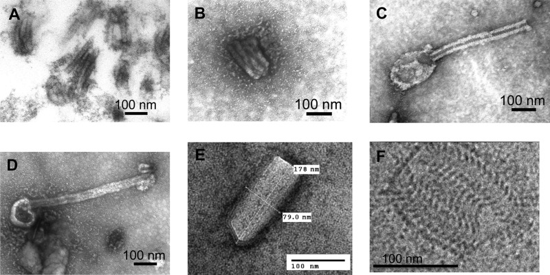
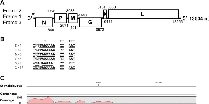
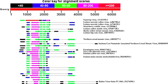
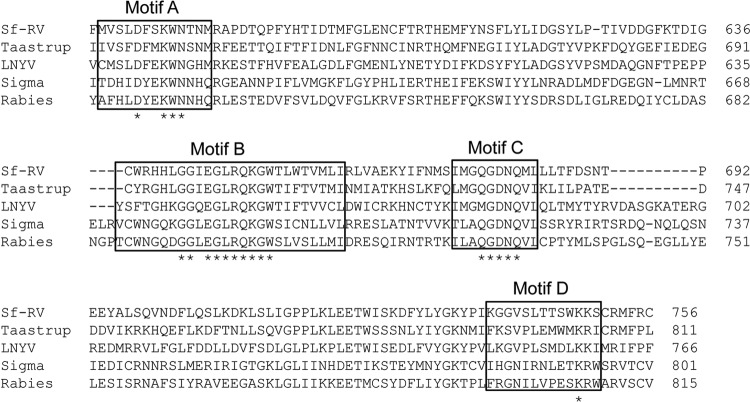
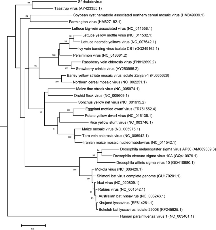
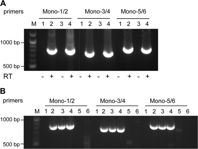
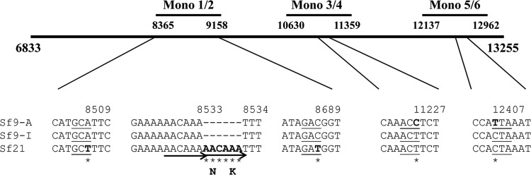
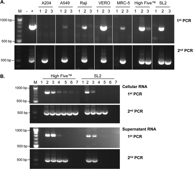
The Sf9 cell line, derived from Spodoptera frugiperda, is used as a cell substrate for biological products, and no viruses have been reported in this cell line after extensive testing. We used degenerate PCR assays and massively parallel sequencing (MPS) to identify a novel RNA virus belonging to the order Mononegavirales in Sf9 cells. Sequence analysis of the assembled virus genome showed the presence of five open reading frames (ORFs) corresponding to the genes for the N, P, M, G, and L proteins in other rhabdoviruses and an unknown ORF of 111 amino acids located between the G- and L-protein genes. BLAST searches indicated that the S. frugiperda rhabdovirus (Sf-rhabdovirus) was related in a limited region of the L-protein gene to Taastrup virus, a newly discovered member of the Mononegavirales from a leafhopper (Hemiptera), and also to plant rhabdoviruses, particularly in the genus Cytorhabdovirus. Phylogenetic analysis of sequences in the L-protein gene indicated that Sf-rhabdovirus is a novel virus that branched with Taastrup virus. Rhabdovirus morphology was confirmed by transmission electron microscopy of filtered supernatant samples from Sf9 cells. Infectivity studies indicated potential transient infection by Sf-rhabdovirus in other insect cell lines, but there was no evidence of entry or virus replication in human cell lines. Sf-rhabdovirus sequences were also found in the Sf21 parental cell line of Sf9 cells but not in other insect cell lines, such as BT1-TN-5B1-4 (Tn5; High Five) cells and Schneider's Drosophila line 2 [D.Mel.(2); SL2] cells, indicating a species-specific infection. The results indicate that conventional methods may be complemented by state-of-the-art technologies with extensive bioinformatics analysis for identification of novel viruses.

Importance: The Spodoptera frugiperda Sf9 cell line is used as a cell substrate for the development and manufacture of biological products. Extensive testing has not previously identified any viruses in this cell line. This paper reports on the identification and characterization of a novel rhabdovirus in Sf9 cells. This was accomplished through the use of next-generation sequencing platforms, de novo assembly tools, and extensive bioinformatics analysis. Rhabdovirus identification was further confirmed by transmission electron microscopy. Infectivity studies showed the lack of replication of Sf-rhabdovirus in human cell lines. The overall study highlights the use of a combinatorial testing approach including conventional methods and new technologies for evaluation of cell lines for unexpected viruses and use of comprehensive bioinformatics strategies for obtaining confident next-generation sequencing results.


Figures

FIG 1
FIG 1
EM analysis of Sf9 cell supernatant. (A to D) Images of pelleted virus from Sf9 cell supernatant (SGS Vitrology). (A) A thin section stained with 2% (wt/vol) ethanolic uranyl acetate and Reynolds' lead citrate; (B to D) negative staining with 2% (wt/vol) ammonium molybdate. (E) Negative staining of pelleted virus after fixation in 2% paraformaldehyde and staining with 2% ammonium molybdate (Rocky Mountain Laboratories, NIAID, NIH). (F) Cryo-EM (NanoImaging, Inc.).
FIG 2
FIG 2
Genome analysis of Sf-rhabdovirus. (A) Genomic organization of Sf-rhabdovirus. The nucleotide positions of the start and end of each ORF are labeled above and below the boxes, respectively, and the reading frames and putative proteins are indicated. (B) Intergenic sequences from the plus strand corresponding to putative gene junction regions are shown. The putative polyadenylation signal, the untranscribed intergenic sequence, and the putative transcription start site are indicated I, II, and III, respectively; consensus sequences are underlined and in bold. (C) Mapping of reads obtained from MPS transcriptome data of Sf9 cells against the Sf-rhabdovirus genome sequence. The consensus sequence and coverage obtained using CLC genomics workbench software are shown.
FIG 3
FIG 3
TBLASTX analysis of the L-protein gene of Sf-rhabdovirus. The whole sequence corresponding to the L protein was queried against the NCBI nucleotide database for viruses. Taastrup virus was the most relevant virus, with 25% coverage and 31% to 47% similarity. The color key for the alignment scores and the results of the top hits are shown. Selected viruses are labeled with their GenBank accession numbers for reference.
FIG 4
FIG 4
Alignment of domain III of the L protein of Sf-rhabdovirus (Sf-RV) with L proteins of some other viruses in the family Rhabdoviridae. The amino acid alignments of Sf-rhabdovirus (GenBank accession number KF947078) with Taastrup virus (Taastrup; GenBank accession number AY423355), sigma virus HAP23 from Drosophila melanogaster (Sigma; GenBank accession number ACU65438), lettuce necrotic yellows virus (LNYV; GenBank accession number YP_425092), and rabies virus strain PV-2061 (Rabies; GenBank accession number JX276550) are shown. Conserved motifs (motifs A to D) are boxed, and identical amino acids are indicated (*). Amino acid positions are indicated from the start of the L protein.
FIG 5
FIG 5
Molecular phylogenetic analysis of Sf-rhabdovirus. The evolutionary history was inferred by using the maximum likelihood method based on the WAG model (14). The tree with the highest log likelihood (−15,561.1192) is shown. The percentages of replicate trees in which the associated taxa clustered together in the bootstrap test (1,000 replicates) are shown next to the branches (39) (values higher than 70% are indicated). The initial tree(s) for the heuristic search was obtained automatically (default settings) by applying neighbor-joining (NJ) (41) and BioNJ (42) algorithms to a matrix of pairwise distances estimated using a JTT model and then selecting the topology with superior log likelihood value. A discrete gamma distribution was used to model evolutionary rate differences among sites (5 categories [+G, parameter = 0.9171]). The tree is drawn to scale, with branch lengths measured as the number of substitutions per site. The analysis involved 33 amino acid sequences. All positions containing gaps and missing data were eliminated. There were a total of 338 positions in the final data set. Evolutionary analyses were conducted in the MEGA (version 5.1) program (15). Human parainfluenza virus was used as an outlier.
FIG 6
FIG 6
RT-PCR analysis of Sf-rhabdovirus-specific sequences in insect cell lines. (A) RNAs isolated from cultured Sf9 cells were analyzed by RT-PCR using the indicated primer sets in the absence (−) and presence (+) of RT. Lanes 1 and 2, cultured Sf9 cells; lanes 3 and 4, 1,000×-concentrated Sf9 cell supernatant RNA; lane M, 100-bp marker, with selected sizes indicated on the left. (B) RT-PCR analysis of RNAs from frozen insect cells. Insect cell lines were analyzed by RT-PCR using the indicated primer sets. Lanes 2 and 3, Sf9 cells from ATCC and Invitrogen, respectively; lanes 4, Sf21 cells; lanes 5, High Five cells, lanes 6, SL2 cells; lane 1, no-template negative control; lane M, 100-bp marker, with selected sizes indicated on the left.
FIG 7
FIG 7
Comparative analysis of Sf-rhabdovirus L-protein gene sequences in Sf9 and Sf21 cells. Nucleotide sequences of Sf-rhabdovirus were obtained from 2 independent RT-PCR amplifications, using primers Mono-1/Mono-2, Mono-3/Mono-4, and Mono-5/Mono-6, from Sf9 cells from ATCC and Sf9 cells from Invitrogen (Sf9-A and Sf9-I, respectively) and cells of the Sf21 cell line. Sequences amplified from cells of each cell line in two independent PCRs were identical. The sequence alignment of the three cell lines is shown. Underlining, codons with mutations; asterisks, mutation positions; arrow, direct repeats; dashes, deleted bases. The nucleotide position on the viral genome is indicated from the 3′ terminus.
FIG 8
FIG 8
RT-PCR analysis of Sf-rhabdovirus sequences in inoculated target cells. RNAs prepared from cells inoculated with filtered supernatant from Sf9 cells were analyzed by RT-PCR using primers Mono-1/Mono-2 and nested primers Mono-1i/Mono-2i. (A) Cells were inoculated with Sf9 cell supernatant, and RNA was prepared on day 2 and day 20 after inoculation (lanes 2 and 3, respectively). Lanes 1, RNA prepared on day 2 from cells inoculated with complete medium as a control; lane −, no-template negative control; lane +, Sf9 cell cDNA-positive control. (B) RT-PCR analysis of cells inoculated with Sf9 cell supernatant. RNAs were prepared from cells, and filtered supernatant was obtained from inoculated High Five cells and SL2 cells on day 2, day 6, day 20, day 34, day 49, and day 62 (lanes 2 to 7, respectively). Lanes 1, RT-PCR analysis of RNAs prepared on day 2 from cells inoculated with complete medium; lane M, 100-bp marker, with selected sizes indicated on the left. Results of the first and second PCR amplifications are shown.

Similar articles

Cited by

References

    1. U.S. Food and Drug Administration. 2013. Vaccines, blood & biologics. U.S. Food and Drug Administration, Bethesda, MD: http://www.fda.gov/BiologicsBloodVaccines/Vaccines/ApprovedProducts/UCM0...
    1. U.S. Food and Drug Administration. 2010. Guidance for industry: characterization and qualification of cell substrates and other biological materials used in the production of viral vaccines for infectious disease indications. U.S. Food and Drug Administration, Bethesda, MD: http://www.fda.gov/downloads/biologicsbloodvaccines/guidancecompliancere...
    1. Victoria JG, Wang C, Jones MS, Jaing C, McLoughlin K, Gardner S, Delwart EL. 2010. Viral nucleic acids in live-attenuated vaccines: detection of minority variants and an adventitious virus. J. Virol. 84:6033–6040. 10.1128/JVI.02690-09 - DOI - PMC - PubMed
    1. U.S. Food and Drug Administration. 2012. FDA briefing document: Vaccines and Related Biological Products Advisory Committee meeting: cell lines derived from human tumors for vaccine manufacture. U.S. Food and Drug Administration, Bethesda, MD: http://www.fda.gov/downloads/AdvisoryCommittees/CommitteesMeetingMateria...
    1. Schneider I. 1972. Cell lines derived from late embryonic stages of Drosophila melanogaster. J. Embryol. Exp. Morphol. 27:353–365 - PubMed

Publication types

Associated data