. 2014 Jun;25(11):1755-68.
doi: 10.1091/mbc.E14-01-0675. Epub 2014 Mar 26.

LMTK1 regulates dendritic formation by regulating movement of Rab11A-positive endosomes

Affiliations

LMTK1 regulates dendritic formation by regulating movement of Rab11A-positive endosomes

Tetsuya Takano et al. Mol Biol Cell. 2014 Jun.

Abstract

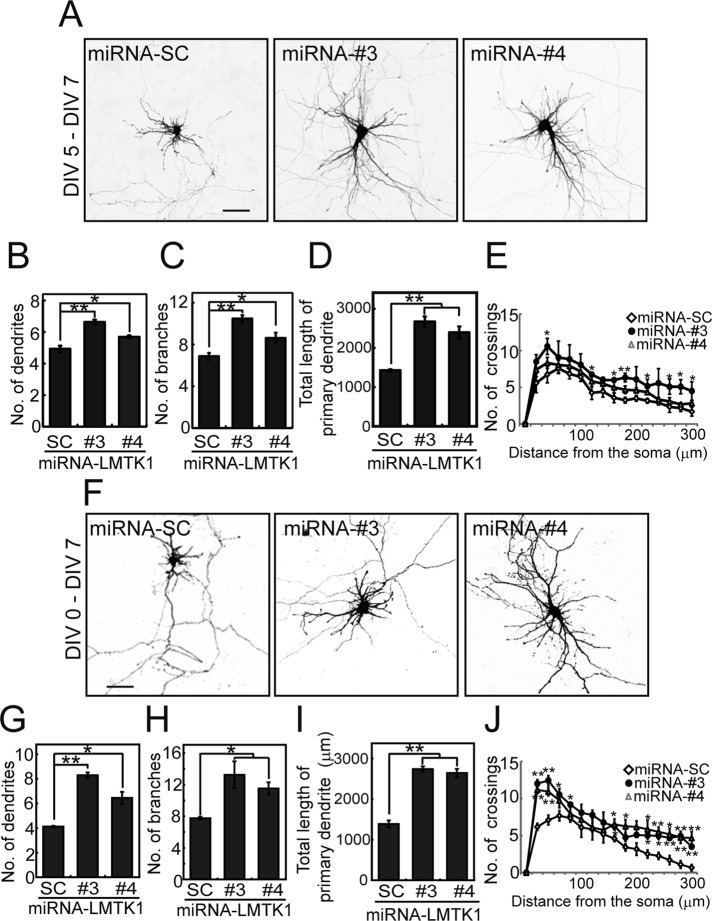
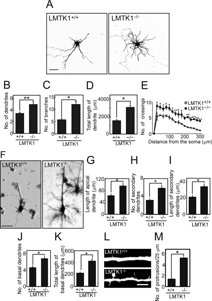
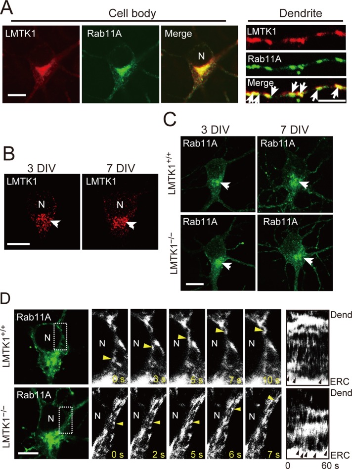
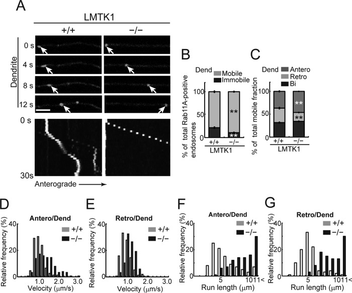
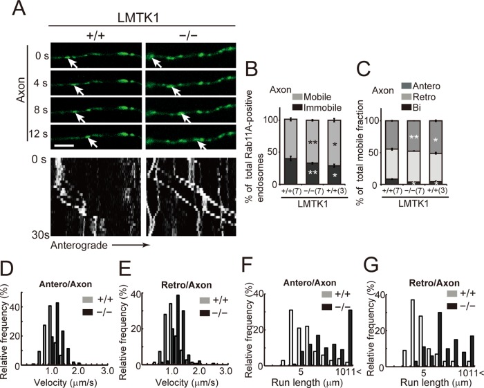
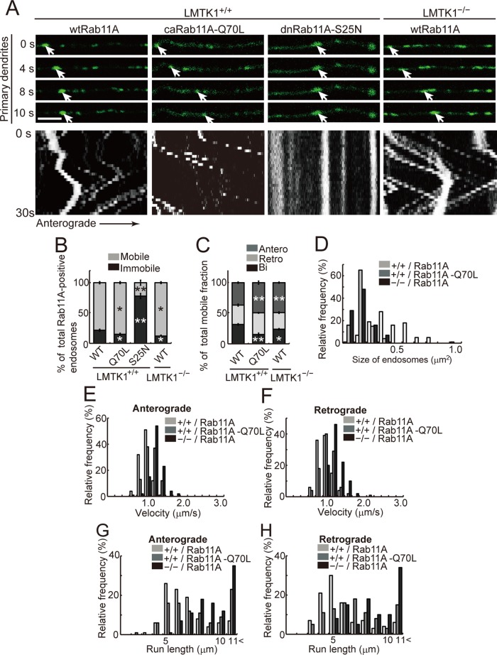
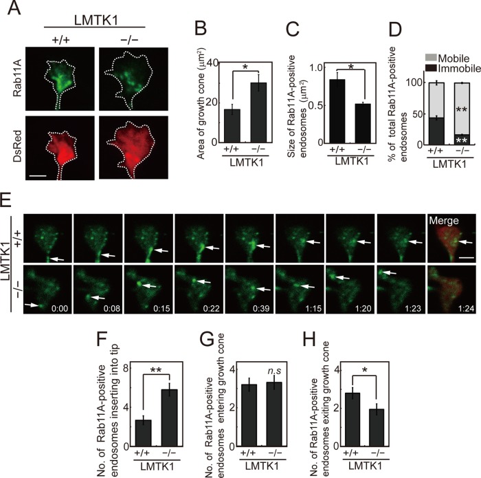
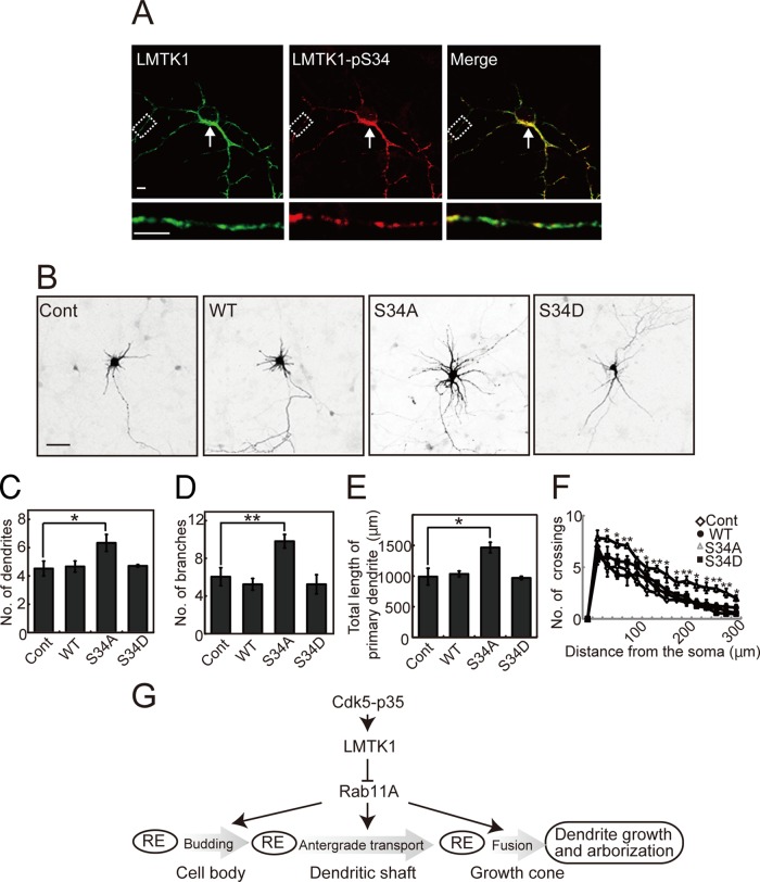
Neurons extend two types of neurites-axons and dendrites-that differ in structure and function. Although it is well understood that the cytoskeleton plays a pivotal role in neurite differentiation and extension, the mechanisms by which membrane components are supplied to growing axons or dendrites is largely unknown. We previously reported that the membrane supply to axons is regulated by lemur kinase 1 (LMTK1) through Rab11A-positive endosomes. Here we investigate the role of LMTK1 in dendrite formation. Down-regulation of LMTK1 increases dendrite growth and branching of cerebral cortical neurons in vitro and in vivo. LMTK1 knockout significantly enhances the prevalence, velocity, and run length of anterograde movement of Rab11A-positive endosomes to levels similar to those expressing constitutively active Rab11A-Q70L. Rab11A-positive endosome dynamics also increases in the cell body and growth cone of LMTK1-deficient neurons. Moreover, a nonphosphorylatable LMTK1 mutant (Ser34Ala, a Cdk5 phosphorylation site) dramatically promotes dendrite growth. Thus LMTK1 negatively controls dendritic formation by regulating Rab11A-positive endosomal trafficking in a Cdk5-dependent manner, indicating the Cdk5-LMTK1-Rab11A pathway as a regulatory mechanism of dendrite development as well as axon outgrowth.


Figures

FIGURE 1:
FIGURE 1:
Down-regulation of LMTK1 increases dendritic arborization in cortical neurons. (A) Effect of LMTK1 knockdown by miRNAs on dendritic arborization. Cortical neurons were transfected with a plasmid encoding EmGFP-miRNA-SC, -miRNA-LMTK1#3, or -miRNA-LMTK1#4 at 5 DIV and were observed at 7 DIV. Bar, 50 μm. (B–D) The number of dendrites (B) or branches (C) and the total length of the primary dendrite (D) in miRNA-expressing neurons (n = 30 each for miRNA-SC and miRNA-LMTK1#3, and n = 28 for miRNA-LMTK1#4). (E) Sholl analysis of neurons transfected with a plasmid encoding miRNA-SC, miRNA-LMTK1#3, or miRNA-LMTK1#4 (n = 12 each). (F) Dendritic arborization of cortical neurons transfected with plasmids encoding miRNAs for LMTK1. Cortical neurons were transfected with a plasmid encoding miRNA-SC, miRNA-LMTK1#3, or miRNA-LMTK1#4 at 0 DIV, and neurites were analyzed at 7 DIV. Bar, 50 μm. (G–I) The number of dendrites (G), branches (H), and total length of the primary dendrite (I) in miRNA-expressing neurons (n = 30 for miRNA-SC, n = 21 for miRNA-LMTK1#3, and n = 30 for miRNA-LMTK1#4). (J) Sholl analysis of neurons transfected with a plasmid encoding miRNA-SC, miRNA-LMTK1#3, or miRNA-LMTK1#4 (n = 12 each). Data are the mean ± SEM of three independent experiments. *p < 0.05, **p < 0.01.
FIGURE 2:
FIGURE 2:
LMTK1/− neurons have longer dendrites and more branches. (A) Cortical neurons from LMTK1+/+ (left) or LMTK1−/− (right) brains were transfected with a plasmid encoding EGFP at 6 DIV and observed at 7 DIV. Bar, 50 μm. (B–D) The number of dendrites (B), branches (C), and total length of the primary dendrite (D) of LMTK1+/+ or LMTK1−/− neurons in cultures at 7 DIV (n = 45 each). (E) Sholl analysis of LMTK1+/+ or LMTK1−/− neurons (n = 12 each). (F) Golgi staining of layer V neurons in the cerebral cortex of LMTK1+/+ (left) and LMTK1−/− (right) mouse brain at postnatal day 7. Bar, 40 μm. (G–I) The length of the apical dendrite (G) and the number (H) and length (I) of secondary dendrites extending from the apical dendrite (n = 20 each for LMTK1+/+ and LMTK1−/− neurons). (J, K) The number (J) and length (K) of basal dendrites (n = 20 each for LMTK1+/+ and LMTK1−/− neurons). (L) Protrusions along an apical dendrite of LMTK1+/+ and LMTK1−/− neurons. Cultured cortical neurons from LMTK1+/+ (top) or LMTK1−/− (bottom) brains were transfected with a plasmid encoding EGFP at 6 DIV and observed at 7 DIV. Bar, 5 μm. (M) The number of dendritic protrusions per 25-μm length of dendrite (n = 30 each for LMTK1+/+ and LMTK1−/− neurons). Data are the mean ± SEM of three independent experiments. *p < 0.05, **p < 0.01, Student's t test.
FIGURE 3:
FIGURE 3:
Localization of LMTK1 and Rab11A in the cell body and dendrites and movement of Rab11A-positive endosomes in the cell body of cortical neurons. (A) Colocalization of LMTK1 and Rab11A in the cell body and dendrites. Plasmids encoding LMTK1 and EGFP-Rab11A were cotransfected into primary cortical neurons at 0 DIV, and cells were visualized by immunostaining with anti-LMTK1 at 7 DIV (left). Middle, Rab11A. Right, merged image; dendrites in which LMTK1 also colocalized with Rab11A (arrows). Bar, 10 μm (cell body), 5 μm (dendrites). (B) Localization of endogenous LMTK1 at 3 (left) or 7 DIV (right). Endogenous LMTK1 was immunostained with anti-LMTK1. Bar, 10 μm. (C) Localization of Rab11A in LMTK1+/+ (top) or LMTK1−/− (bottom) cortical neurons at 3 DIV (left) or 7 DIV (right). Bar, 10 μm. (D) Movement of Rab11A-positive endosomes in the cell body of LMTK1+/+ (top) or LMTK1−/− (bottom) neurons. Neurons were transfected with a plasmid encoding EGFP-Rab11A at 6 DIV. Movement of Rab11A-positive endosomes was observed with real-time imaging. High-magnification images of the area indicated by the dotted line are shown for five frames with 1- to 3-s intervals. The movement of Rab11A-positive endosomes is labeled by arrowheads. Right, kymographs of Rab11A-positive endosomes budding from pericentriolar ERC and moving toward the dendrites. Arrowheads indicate budding of endosomes. N, nucleus. Bar, 20 μm.
FIGURE 4:
FIGURE 4:
Dynamics of Rab11A-positive endosomes in dendrites of LMTK1−/− neurons. (A) Movement of Rab11A-positive endosomes in a dendrite of a LMTK1+/+ or LMTK1−/− neuron. Neurons were transfected with a plasmid encoding EGFP-Rab11A. Movement of Rab11A-positive endosomes in secondary dendrites was observed with real-time imaging. Sequential frames for 12 s with a 4-s interval are shown for LMTK1+/+ and LMTK1−/−. The cell body is on the left side. The movement of a typical Rab11A-positive endosome is indicated with arrows. Bottom, kymographs of Rab11A-positive endosomes moving in dendrites. Bar, 5 μm. (B) Percentage of moving (light gray) or stationary (dark gray) Rab11A-positive endosomes in dendrites. (C) Percentage of Rab11A-positive endosomes moving anterogradely (gray), retrogradely (light gray), or bidirectionally (dark gray) in dendrites (n = 381 endosomes in LMTK1+/+ neurons and n = 432 endosomes in LMTK1−/− neurons). (D, E) Velocity of Rab11A-positive endosomes in dendrites. Relative frequency of Rab11A-positive endosomes moving at the indicated velocities in the anterograde (D) or retrograde (E) direction in dendrites of LMTK1+/+ (light gray) or LMTK1−/− (dark gray) neurons (n = 150 endosomes each for LMTK1+/+ and LMTK1−/−). (F, G) Run length of Rab11A-positive endosomes in dendrites. The distance that Rab11A-positive endosomes moved without pausing was measured in a 20-μm observation window. Relative frequency of Rab11A-positive endosomes moving at the indicated run length in the anterograde (F) or retrograde (G) direction in dendrites of LMTK1+/+ (light gray) or LMTK1−/− (dark gray) neurons (n = 100 endosomes each for LMTK1+/+ and LMTK1−/−).
FIGURE 5:
FIGURE 5:
Dynamics of Rab11A-positive endosomes in axons of LMTK1/ neurons. (A) Movement of Rab11A-positive endosomes in axons of LMTK1+/+ or LMTK1/ neurons. Typical anterograde movement of a Rab11A-positive endosome is indicated with arrows. The cell body is on the left side. Bottom, kymographs of Rab11A-positive endosomes moving in axon. Bar, 5 μm. (B) Percentage of moving (light gray) or stationary (dark gray) Rab11A-positive endosomes at 3 DIV (3) and 7 DIV (7) in axons. (C) Percentage of Rab11A-positive vesicles moving anterogradely (gray), retrogradely (light gray), and bidirectionally (dark gray) in axons of LMTK1+/+ or LMTK1/ neurons (n = 305 endosomes for LMTK1+/+ and n = 301 endosomes for LMTK1/). (D, E) Velocity of Rab11A-positive endosomes in axons. Relative frequency of Rab11A-positive endosomes moving at the indicated velocities in the anterograde (D) or retrograde (E) direction (n = 150 endosomes each for LMTK1+/+ and LMTK1/). (F, G) Run length of Rab11A-positive endosomes in LMTK1+/+ or LMTK1/ neurons. Relative frequency of Rab11A-positive endosomes moving at the indicated run length in the anterograde (F) or retrograde (G) direction (n = 100 endosomes each for LMTK1+/+ and LMTK1/). Data are the mean ± SEM of three independent experiments (*p < 0.05, **p < 0.01, Student's t test).
FIGURE 6:
FIGURE 6:
Movement of caRab11A-Q70L–positive endosomes in LMTK1+/+ neurons and wtRab11A-positive endosomes in LMTK1/ neurons. (A) A plasmid encoding EGFP-wtRab11A, EGFP-caRab11A-Q70L, or EGFP-dnRab11A-S25N was transfected into LMTK1+/+ neurons, and movement of the Rab11A-positive endosomes was analyzed. Sequential frames over 10 s are shown in dendrites of LMTK1+/+ neurons. Right, EGFP-Rab11A transfected into LMTK1/ neurons. The cell body is to the left. The movement of typical Rab11A-positive endosomes is indicated with arrows. Bottom, kymographs of Rab11A-positive endosomes moving in dendrites. (B) Percentage of moving (light gray) or stationary (dark gray) Rab11A-positive endosomes in dendrites. (C) Percentage of Rab11A-positive endosomes moving anterogradely (gray), retrogradely (light gray), or bidirectionally (dark gray) in dendrites (n = 347 endosomes for LMTK1+/+ neurons expressing wtRab11A, n = 349 endosomes for LMTK1+/+ neurons expressing caRab11A-Q70L, n = 174 endosomes for LMTK1+/+ neurons expressing dnRab11A-S25N, and n = 329 endosomes for LMTK1/ neurons expressing wtRab11A). (D) The size of Rab11A-positive endosomes in primary dendrites. Relative frequency of Rab11A-positive endosomes of the indicated sizes. (E, F) The velocity of Rab11A-positive endosomes in primary dendrites. Relative frequency of Rab11A-positive endosomes moving at the indicated velocities in the anterograde (E) or retrograde (F) direction (n = 150 endosomes each for LMTK1+/+ neurons expressing wtRab11A or caRab11A-Q70L and LMTK1/ neurons expressing wtRab11A). (G, H) Run length of wtRab11A, caRab11A-Q70L–positive endosomes in LMTK1+/+ neurons, and Rab11A-positive endosomes in LMTK1/ neurons. Relative frequency of Rab11A-positive endosomes moving at the indicated run length in the anterograde (G) or retrograde (H) direction (n = 100 endosomes each for LMTK1+/+ neurons expressing wtRab11A or caRab11A-Q70L, and LMTK1/ neurons expressing wtRab11A). Data are the mean ± SEM of three independent experiments (*p < 0.05, **p < 0.01, Student's t test).
FIGURE 7:
FIGURE 7:
Movement of Rab11A-positive endosomes in growth cones of LMTK1+/+ or LMTK1/ neurons. (A) Growth cone of cortical neurons, which were prepared from LMTK1+/+ (left) or LMTK1/ (right) mouse brains cotransfected with a plasmid encoding EGFP-Rab11A (top) or DsRed (bottom) at 6 DIV. Rab11A-positive endosomes were observed at 7 DIV. Bar, 2.5 μm. (B) The growth cone area of LMTK1+/+ or LMTK1/ neurons (n = 30 each for LMTK1+/+ or LMTK1/ neurons). (C) The size of Rab11A-positive recycling endosomes in growth cones of LMTK1+/+ or LMTK1/ neurons (n = 30 endosomes each). (D) Percentage of mobile (light gray) or stationary (dark gray) Rab11A-positive endosomes in growth cones (n = 321 endosomes in LMTK1+/+ neurons and n = 250 endosomes in LMTK1/ neurons). (E) Movement of Rab11A-positive endosomes in growth cones of LMTK1/ or LMTK1+/+ neurons. Neurons prepared from LMTK1+/+ (top) or LMTK1/ (bottom) mouse brains were cotransfected with a plasmid encoding EGFP-Rab11A or DsRed. Movements of Rab11A-positive endosomes were analyzed with real-time imaging. Sequential frames are shown over 84 s for LMTK1+/+ (top) and LMTK1/ (bottom). The movement of a typical Rab11A-positive endosome is indicated with arrows. (F–H) The number of Rab11A-positive endosomes that were inserted into the plasma membrane at the tip of the growth cone (F), entered the growth cone from the dendritic shaft (G), or exited the growth cone (H) over 160 s (n = 165 endosomes in LMTK1+/+ neurons and n = 185 endosomes in LMTK1/ neurons). Data are the mean ± SEM of three independent experiments (*p < 0.05; **p < 0.01; n.s., not significant, Student's t test).
FIGURE 8:
FIGURE 8:
Nonphosphorylatable mutant S34A of LMTK1 enhances dendritic arborization. (A) Immunofluorescence staining of primary cortical neurons with anti-pS34. Cortical neurons expressing LMTK1-myc were double labeled with anti-myc (left) and anti-pS34 (middle). Right, merged images. Bottom, higher-magnification views of dendrites. Bar, 5 μm. (B) Effect of LMTK1-Ser34 mutants on dendritic arborization. A plasmid encoding LMTK1-WT, -S34A, or -S34D was cotransfected into cortical neurons along with DsRed at 0 DIV, and images of DsRed are shown at 7 DIV. Bar, 50 μm. (C–E) The number of dendrites (C) or branches (D) and total length of the primary dendrite (E) in the LMTK1-expressing neurons (n = 21 each). (F) Sholl analysis of neurons transfected with a plasmid encoding LMTK1-WT, -S34A, or -S34D (n = 12 each). Data are the mean ± SEM of three independent experiments (*p < 0.05, **p < 0.01, Student's t test). (G) Scheme representing the role of LMTK1 in dendrite formation. Cdk5 regulates LMTK1 by phosphorylation at Ser-34. LMTK1 inhibits Rab11A activity, which promotes budding of endocytic vesicles in the cell body, their transport in dendritic shaft, and fusion in the growth cone. The Cdk5-LMTK1-Rab11A pathway is a regulatory mechanism for dendrite formation, as well as axon outgrowth, via membrane supply.

References

    1. Arimura N, Kaibuchi K. Neuronal polarity: from extracellular signals to intracellular mechanisms. Nat Rev Neurosci. 2007;8:194–205. - PubMed
    1. Ascano M, Richmond A, Borden P, Kuruvilla R. Axonal targeting of Trk receptors via transcytosis regulates sensitivity to neurotrophin responses. J Neurosci. 2009;29:11674–11685. - PMC - PubMed
    1. Baas PW, Deitch JS, Black MM, Banker GA. Polarity orientation of microtubules in hippocampal neurons: uniformity in the axon and nonuniformity in the dendrite. Proc Natl Acad Sci USA. 1988;85:8335–8339. - PMC - PubMed
    1. Baker SJ, Sumerson R, Reddy CD, Berrebi AS, Flynn DC, Reddy EP. Characterization of an alternatively spliced AATYK mRNA: expression pattern of AATYK in the brain and neuronal cells. Oncogene. 2001;20:1015–1021. - PubMed
    1. Bellon A. New genes associated with schizophrenia in neurite formation: a review of cell culture experiments. Mol Psychiatry. 2007;12:620–629. - PubMed

Publication types

MeSH terms

LinkOut - more resources