Review
. 2014 Mar 14:8:33.
doi: 10.3389/fnsys.2014.00033. eCollection 2014.

Disrupting neuronal transmission: mechanism of DBS?

Affiliations
Review

Disrupting neuronal transmission: mechanism of DBS?

Satomi Chiken et al. Front Syst Neurosci. .

Abstract

Applying high-frequency stimulation (HFS) to deep brain structure, known as deep brain stimulation (DBS), has now been recognized an effective therapeutic option for a wide range of neurological and psychiatric disorders. DBS targeting the basal ganglia thalamo-cortical loop, especially the internal segment of the globus pallidus (GPi), subthalamic nucleus (STN) and thalamus, has been widely employed as a successful surgical therapy for movement disorders, such as Parkinson's disease, dystonia and tremor. However, the neurophysiological mechanism underling the action of DBS remains unclear and is still under debate: does DBS inhibit or excite local neuronal elements? In this review, we will examine this question and propose the alternative interpretation: DBS dissociates inputs and outputs, resulting in disruption of abnormal signal transmission.

Keywords: basal ganglia; cortico-basal ganglia loop; deep brain stimulation; electrophysiology; globus pallidus; subthalamic nucleus.


Figures

Figure 1
Figure 1
Deep brain stimulation (DBS) inhibits local neuronal firing. (A) Responses of an internal pallidal (GPi) neuron to local GPi repetitive high-frequency stimulation (HFS; 30 μA, 100 Hz, 10 pulses) in a normal monkey. Raw traces of spike discharges after removing the stimulus artifacts by the template subtraction method (1) and raster and peristimulus time histogram (PSTHs; 100 trials; binwidth, 1 ms) (2) are shown. Arrows indicate the timing of local stimulation. Spontaneous discharges of the GPi neuron were completely inhibited by the stimulation. (B) Effect of local injection of gabazine (GABAA receptor antagonist) in the vicinity of the recorded GPi neuron on inhibition of spontaneous activity induced by GPi-HFS. The inhibition was abolished after gabazine injection. Modified from Chiken and Nambu (2013).
Figure 2
Figure 2
Directly evoked spikes of GPi neurons were inhibited during GPi-HFS. (A) Raw traces showing directly evoked spikes of a GPi neuron by stimulus pulses during GPi-HFS (40 μA, 100 Hz, 10 pulses) in a normal monkey. Traces with long (top) and short (bottom) time scales are shown. Arrows with dotted lines indicate the timing of local stimulation (time 0 in the bottom traces). Filled arrowheads indicate directly evoked spikes. GPi-HFS failed to evoke spikes (open arrowheads; from 6th to 10th stimuli). (B) Effects of local gabazine injection on the inhibition of direct evoked GPi responses. Gabazine injection decreased failure rate, and each stimulus successfully evoked spikes (5th, 9th, and 10th stimuli). Modified from Chiken and Nambu (2013).
Figure 3
Figure 3
GPi-DBS disrupts information flow through the GPi. (A) Schematic diagram showing the cortico-basal ganglia pathway and stimulating (Stim.) and recording (Rec.) sites in the electrophysiological experiments (left), along with a typical response pattern (right) in the (GPi) to cortical stimulation (Cx Stim.) with early excitation, inhibition, and late excitation, which are mediated by the (1) cortico-subthalamo (STN)-GPi hyperdirect, (2) striato-GPi direct, and (3) striato-external pallido (GPe)-STN-GPi indirect pathways, respectively. (B) Effects of local GPi-HFS on cortically evoked responses of a GPi neuron in a normal monkey. PSTH (100 trials) in response to the single pulse stimulation (arrowhead with dotted line) of the primary motor cortex (Cx) (1) and PSTH in response to Cx stimulation (arrowhead with dotted line) during GPi-HFS (arrows) (2) are shown. Cortical stimulation was applied 50 ms after the initiation of GPi-HFS. The cortically evoked responses were entirely inhibited during GPi-HFS. Modified from Chiken and Nambu (2013).
Figure 4
Figure 4
Both STN-DBS and STN blocking disrupt information flow through the STN. (A) Effects of local STN-DBS on cortically evoked responses of a substantia nigra pars reticulata (SNr) neuron in a normal rat. PSTH (50 trials) in response to the single pulse stimulation of the Cx (arrow) (1) and PSTH in response to Cx stimulation during STN-DBS (2) are shown. The cortically evoked early and late excitation was abolished during STN-DBS, while cortically evoked inhibition was preserved. Modified from Maurice et al. (2003). (B) Effects of STN blocking on cortically evoked responses of a GPi neuron in a normal monkey. PSTH (100 trials) in response to the single pulse stimulation of the Cx (arrow with dotted line) (1) and PSTH in response to Cx stimulation after blocking STN activity by muscimol (GABAA receptor agonist) injection into the STN (2) are shown. The cortically evoked early and late excitation was abolished after injection of muscimol into the STN, while cortically evoked inhibition was preserved. Modified from Nambu et al. (2000). Note that the pattern of cortically evoked responses of a SNr neuron during STN-DBS is similar to that of a GPi neuron after STN blocking.
Figure 5
Figure 5
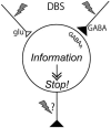
Mechanism underling effectiveness of deep brain stimulation (DBS). DBS activates axon terminals in the stimulated nucleus and induces release of large amount of neurotransmitters, such as GABA and glutamate, and dissociates inputs and outputs in the stimulated nucleus, resulting in disruption of abnormal information flow through the cortico-basal ganglia loop.

References

    1. Abosch A., Kapur S., Lang A. E., Hussey D., Sime E., Miyasaki J., et al. (2003). Stimulation of the subthalamic nucleus in Parkinson’s disease does not produce striatal dopamine release. Neurosurgery 53, 1095–1105 10.1227/01.neu.0000088662.69419.1b - DOI - PubMed
    1. Anderson T. R., Hu B., Iremonger K., Kiss Z. H. (2006). Selective attenuation of afferent synaptic transmission as a mechanism of thalamic deep brain stimulation-induced tremor arrest. J. Neurosci. 26, 841–850 10.1523/jneurosci.3523-05.2006 - DOI - PMC - PubMed
    1. Anderson M. E., Postupna N., Ruffo M. (2003). Effects of high-frequency stimulation in the internal globus pallidus on the activity of thalamic neurons in the awake monkey. J. Neurophysiol. 89, 1150–1160 10.1152/jn.00475.2002 - DOI - PubMed
    1. Aziz T. Z., Peggs D., Sambrook M. A., Crossman A. R. (1991). Lesion of the subthalamic nucleus for the alleviation of 1-methyl-4-phenyl-1,2,3,6-tetrahydropyridine (MPTP)-induced parkinsonism in the primate. Mov. Disord. 6, 288–292 10.1002/mds.870060404 - DOI - PubMed
    1. Bar-Gad I., Elias S., Vaadia E., Bergman H. (2004). Complex locking rather than complete cessation of neuronal activity in the globus pallidus of a 1-methyl-4-phenyl-1,2,3,6-tetrahydropyridine-treated primate in response to pallidal microstimulation. J. Neurosci. 24, 7410–7419 10.1523/jneurosci.1691-04.2004 - DOI - PMC - PubMed

LinkOut - more resources