Review
. 2014 Mar 18:8:25.
doi: 10.3389/fnint.2014.00025. eCollection 2014.

Operant conditioning of spinal reflexes: from basic science to clinical therapy

Affiliations
Review

Operant conditioning of spinal reflexes: from basic science to clinical therapy

Aiko K Thompson et al. Front Integr Neurosci. .

Abstract

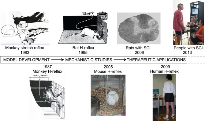
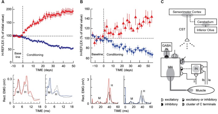
New appreciation of the adaptive capabilities of the nervous system, recent recognition that most spinal cord injuries are incomplete, and progress in enabling regeneration are generating growing interest in novel rehabilitation therapies. Here we review the 35-year evolution of one promising new approach, operant conditioning of spinal reflexes. This work began in the late 1970's as basic science; its purpose was to develop and exploit a uniquely accessible model for studying the acquisition and maintenance of a simple behavior in the mammalian central nervous system (CNS). The model was developed first in monkeys and then in rats, mice, and humans. Studies with it showed that the ostensibly simple behavior (i.e., a larger or smaller reflex) rests on a complex hierarchy of brain and spinal cord plasticity; and current investigations are delineating this plasticity and its interactions with the plasticity that supports other behaviors. In the last decade, the possible therapeutic uses of reflex conditioning have come under study, first in rats and then in humans. The initial results are very exciting, and they are spurring further studies. At the same time, the original basic science purpose and the new clinical purpose are enabling and illuminating each other in unexpected ways. The long course and current state of this work illustrate the practical importance of basic research and the valuable synergy that can develop between basic science questions and clinical needs.

Keywords: H-reflex; learning and memory; locomotion; spinal cord injury; spinal cord plasticity.


Figures

Figure 1
Figure 1
Operant conditioning of spinal reflexes from 1978 to 2013. The work began with model development and progressed to mechanistic studies and then to clinical applications. These three phases have overlapped to a considerable degree and continue to do so. (SCI: spinal cord injury) (Wolpaw et al., ; Wolpaw, ; Chen and Wolpaw, ; Carp et al., ; Chen et al., ; Thompson et al., 2009, 2013b).
Figure 2
Figure 2
H-reflex operant conditioning results in rats (A) and humans (B), and the hierarchy of brain and spinal cord plasticity that underlies H-reflex conditioning (C). (A) As illustrated in Figure 1 (“Rat H-reflex”), in a rat with chronically implanted EMG electrodes and a tibial nerve cuff, the implant wires travel subcutaneously to a head-mounted connector and then through a flexible cable and a commutator to amplifiers and stimulator. The rat moves freely about the cage as soleus muscle activity is monitored 24 h per day. Whenever the absolute (i.e., rectified) value of soleus EMG stays in a specified range for a randomly varying 2.3- to 2.7-s period, a nerve cuff stimulus elicits an M-wave just above threshold and an H-reflex. Top: For the first 10 days (from day –10 to day 0), the rat is exposed to the control mode, in which no reward occurs and the H-reflex is simply measured to determine its initial size. For the next 50 days, it is exposed to the up-conditioning (HRup) or down-conditioning (HRdown) mode, in which a food-pellet reward occurs whenever the H-reflex is above (HRup) or below (HRdown) a criterion value. A rat averages 2000–6000 trials per day, and the criterion is set to provide 500–1000 rewards per day to satisfy the daily requirement (based on body weight). The background EMG and the M-wave stay constant throughout. Successful conditioning (defined as a change of at least 20% in the correct direction) occurs in 75–80% of the rats (the others remain within 20% of their control value). The graphs show average (± SEM) daily H-reflex sizes for 59 successful HRup rats (red upward triangles) and 81 successful HRdown rats (blue downward triangles). In both groups, mode-appropriate change in H-reflex size develops steadily over the 50 days. Bottom: Average absolute post-stimulus EMG for representative days from an HRup rat (left) and an HRdown rat (right) under the control mode (solid) and near the end of HRup or HRdown conditioning (dashed). After conditioning, the H-reflex is larger in the HRup rat and smaller in the HRdown rat, while the background EMG activity and the M-wave have not changed (Updated from Wolpaw, 1997). (B) As illustrated in Figure 1 (“Human H-reflex”), EMG activity is monitored in a person with EMG electrodes over the soleus muscle and tibial nerve-stimulating electrodes in the popliteal fossa. The person maintains a natural standing posture facing a screen that displays the current absolute level of soleus EMG in relation to a specified range. Whenever the absolute value of soleus EMG stays in this range for several sec, tibial nerve stimulation elicits an M-wave just above threshold and an H-reflex. Top: For the first six sessions (i.e., baseline sessions, from day −14 to day 0), the person is exposed to the control mode, in which the H-reflex is simply measured to determine its initial size. For the next 24 sessions (i.e., conditioning sessions, days 0–56, three sessions per week), the person is exposed to the HRup or HRdown conditioning mode, in which, after each conditioning trial, the screen provides immediate feedback indicating whether the H-reflex was above (HRup) or below (HRdown) a criterion value. The person completes 225 conditioning trials per session. The background EMG and the M-wave stay constant throughout the sessions. Successful conditioning occurs in about 80% of the people. The graphs show average (± SEM) daily H-reflex sizes for six successful HRup people (red upward triangles) and eight successful HRdown people (blue downward triangles). In both groups, mode-appropriate change in H-reflex size develops steadily over the 24 conditioning sessions. Bottom: Average peri-stimulus EMG from an HRup subject (left) and an HRdown subject (right) for a baseline session (i.e., control mode) (solid) and for the last HRup or HRdown conditioning session (dashed) (A stimulus artifact occurs at 0 ms) (From Thompson et al., 2009). (C) A hierarchy of brain and spinal cord plasticity underlies H-reflex conditioning. The shaded ovals indicate the spinal and supraspinal sites of definite or probable plasticity associated with operant conditioning of the H-reflex. “MN” is the motoneuron, “CST” is the main corticospinal tract, “IN” is a spinal interneuron, and “GABA IN” is a GABAergic spinal interneuron. Dashed pathways imply the possibility of intervening spinal interneurons. The monosynaptic and probably oligosynaptic H-reflex pathway from groups Ia, II, and Ib afferents to the motoneuron is shown. Definite (dark gray) or probable (light gray) sites of plasticity include: the motoneuron membrane (i.e., firing threshold and axonal conduction velocity); motor unit properties; GABAergic interneurons; GABAergic terminals and C terminals on the motoneuron; the Ia afferent synaptic connection; terminals conveying oligosynaptic groups I and II inhibition or excitation to the motoneuron; sensorimotor cortex; and cerebellum. As described in the text, the data suggest that the reward contingency acts through the inferior olive to guide and maintain plasticity in the cerebellum that guides and maintains plasticity in sensorimotor cortex that (via the CST) guides and maintains plasticity in the spinal cord that is directly responsible for H-reflex change (Modified from Wolpaw, 2010).

References

    1. Abel B. M., Emore E., Thompson A. K. (2011). Operant up-conditioning of the ankle dorsiflexor motor evoked potential in people with multiple sclerosis. Society for Neuroscience 41st Annual Meeting, Program No. 917.919. Washington, D.C.
    1. Barbeau H., Rossignol S. (1987). Recovery of locomotion after chronic spinalization in the adult cat. Brain Res. 412, 84–95 10.1016/0006-8993(87)91442-9 - DOI - PubMed
    1. Barbeau H. (2003). Locomotor training in neurorehabilitation: emerging rehabilitation concepts. Neurorehabil. Neural Repair 17, 3–11 10.1177/0888439002250442 - DOI - PubMed
    1. Bawa P., Stein R. B., Tatton W. G. (1979). Dynamics of a long-latency reflex pathway in the monkey. Biol. Cybern. 34, 107–110 10.1007/bf00365474 - DOI - PubMed
    1. Carp J. S., Chen X. Y., Sheikh H., Wolpaw J. R. (2001a). Operant conditioning of rat H-reflex affects motoneuron axonal conduction velocity. Exp. Brain Res. 136, 269–273 10.1007/s002210000608 - DOI - PubMed

LinkOut - more resources