. 2014 Mar 18:6:44.
doi: 10.3389/fnagi.2014.00044. eCollection 2014.

Transcriptomics of post-stroke angiogenesis in the aged brain

Affiliations

Transcriptomics of post-stroke angiogenesis in the aged brain

Ana Maria Buga et al. Front Aging Neurosci. .

Abstract

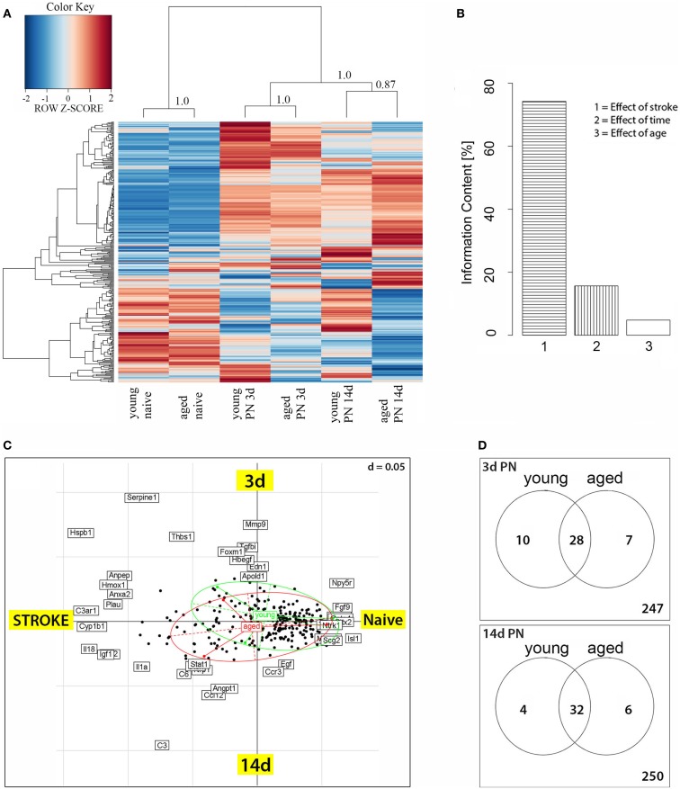
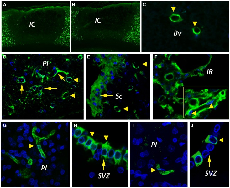
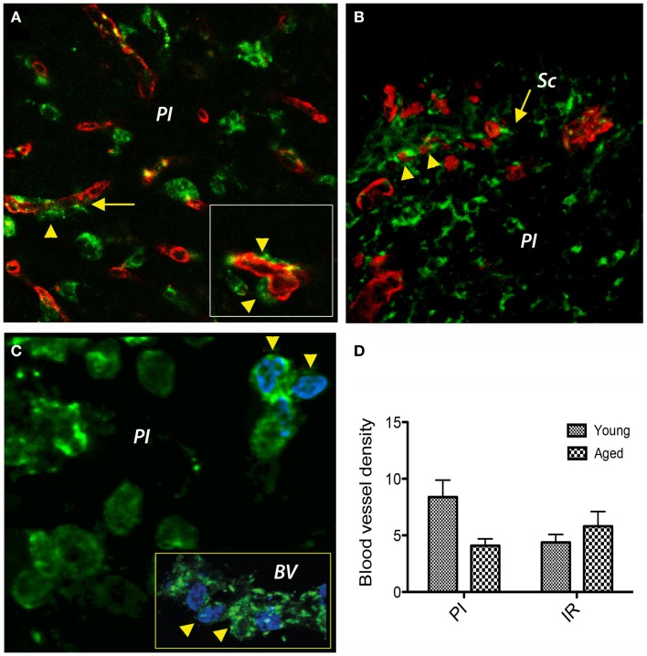
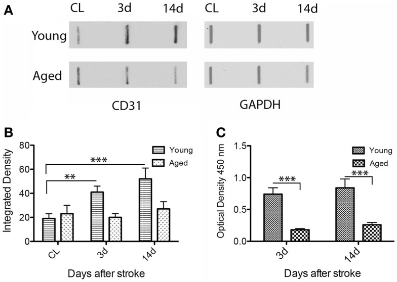
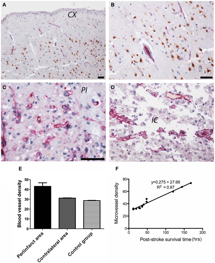
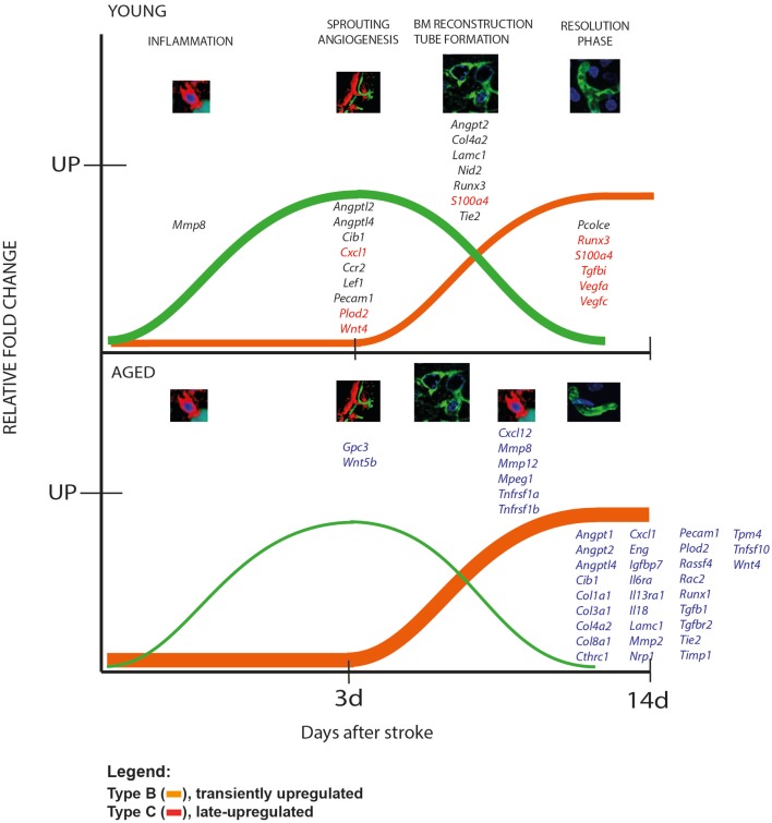
Despite the obvious clinical significance of post-stroke angiogenesis in aged subjects, a detailed transcriptomic analysis of post-stroke angiogenesis has not yet been undertaken in an aged experimental model. In this study, by combining stroke transcriptomics with immunohistochemistry in aged rats and post-stroke patients, we sought to identify an age-specific gene expression pattern that may characterize the angiogenic process after stroke. We found that both young and old infarcted rats initiated vigorous angiogenesis. However, the young rats had a higher vascular density by day 14 post-stroke. "New-for-stroke" genes that were linked to the increased vasculature density in young animals included Angpt2, Angptl2, Angptl4, Cib1, Ccr2, Col4a2, Cxcl1, Lef1, Hhex, Lamc1, Nid2, Pcam1, Plod2, Runx3, Scpep1, S100a4, Tgfbi, and Wnt4, which are required for sprouting angiogenesis, reconstruction of the basal lamina (BL), and the resolution phase. The vast majority of genes involved in sprouting angiogenesis (Angpt2, Angptl4, Cib1, Col8a1, Nrp1, Pcam1, Pttg1ip, Rac2, Runx1, Tnp4, Wnt4); reconstruction of a new BL (Col4a2, Lamc1, Plod2); or tube formation and maturation (Angpt1, Gpc3, Igfbp7, Sparc, Tie2, Tnfsf10), had however, a delayed upregulation in the aged rats. The angiogenic response in aged rats was further diminished by the persistent upregulation of "inflammatory" genes (Cxcl12, Mmp8, Mmp12, Mmp14, Mpeg1, Tnfrsf1a, Tnfrsf1b) and vigorous expression of genes required for the buildup of the fibrotic scar (Cthrc1, Il6ra, Il13ar1, Il18, Mmp2, Rassf4, Tgfb1, Tgfbr2, Timp1). Beyond this barrier, angiogenesis in the aged brains was similar to that in young brains. We also found that the aged human brain is capable of mounting a vigorous angiogenic response after stroke, which most likely reflects the remaining brain plasticity of the aged brain.

Keywords: aging; angiogenesis; stroke; transcriptomics.


Figures

Figure 1
Figure 1
(A) Heatmap of genes differentially expressed between post-stroke and naïve animals. Scaled expression values of all 292 differentially expressed genes are shown for each group with deep blue being the lowest and dark red the highest expression level. The depicted dendrograms cluster samples (top) and genes (left) employing average agglomeration and Euclidian distance measure. Cluster branch stability was tested with the R-package “pvclust”. Approximately unbiased p-values calculated by multiscale bootstrap resampling are shown at cluster branches. (B) Correspondence analysis of differentially expressed genes and samples grouped by animal age. The panel depicts the Eigenvalues of the correspondence analysis and shows that the major factors contributing to the variance of stroketomics analysis were stroke (75%), post-stroke time (15%), and age (10%). (C) Sources of variability were stroke and post-stroke time, which formed the coordinates. The graph shows the distribution of transcripts (black dotes) as a function of treatment (stroke) and post-stroke time. Samples from young (green ellipse) and aged (red ellipse) animals show an extensive overlap as well as some difference in their post-stroke response. (D) Venn diagrams for 3 and 14 days post-stroke penumbra (PN) showing genes that were up- or downregulated exclusively in old or young rats, or in both age groups.
Figure 2
Figure 2
Infarct size and angiogenesis assessment by immunofluorescence. (A,B) By NeuN immunohistochemistry, the cortical infarcts were similar in size in both age groups, i.e., 35 ± 3.1% of total cortical volume in young rats and 40 ± 7.9% in aged rats. (C) In the unlesioned hemisphere, CD31-positive blood vessels (green) were rarely detected. (D) By day 14, new blood vessels were emerging in the peri-infarcted area of young animals. The revascularization process in aged animals was delayed and was characterized by the accumulation of CD31-immunopositivity in the fibrotic scar (E). Beyond the fibrotic scar, however, in a region that we dubbed “islet of regeneration,” vigorous angiogenesis was detected in aged animals as well (F). By day 14, mature BV had developed in the peri-infarcted area of young animals (G). Similarly, new, mature blood vessels were detected in the same areas of the aged rats (I). Please note the unexpected expression of CD31 in the wall of the subventricular zone both in young (H) and aged animals (J). (C–J) are Z-projection images. BV, blood vessel; IC, infarct core; IR, islet of regeneration; PI, peri-infarct; Sc, scar; SVZ, subventricular zone.
Figure 3
Figure 3
Double immunofluorescence prolyl 4-hydroxylase/CD31 and prolyl 4-hydroxylase/BrdU in the peri-infarcted area. (A) Double labeling of cells with a P4Hbeta antibody (green) and CD31 (red) revealed that under hypoxic conditions P4Hb-positive fibroblasts/fibrocytes seems to emanate from the vascular wall (inset, arrowheads) a process that was more evident in young brains. (B) In the aged animals, we noted a preferential agglomeration/accumulation of fibroblasts in the scar region. (C) Double labeling of cells with a P4Hb (green) and the proliferation marker BrdU (blue) in aged rat brains revealed that some proliferating fibrocytes/fibroblasts had BrdU-positive nuclei in the peri-lesional area (arrowheads). Eventually, some co-labeled cells emanated from the capillary wall [(C), inset, arrowheads]. (D) At day 14 post-stroke, the vascular density was higher in the peri-infarcted area of young animals as compared to the similar region of aged rats (twofold). However, beyond the inhibitory fibrotic scar, the vascular density was similar in the two age groups. (A–C) represent Z-projection images.
Figure 4
Figure 4
Quantitative analysis of CD31 expression in the rat model. By dot blot (A) and ELISA analysis (C), CD31 immunoreactivity in the peri-infarcted area was slightly increased from day 3 to day 14 in both age groups. However, CD31 levels in the young animals were clearly higher than those in aged animals both at day 3 (fourfold; p = 0.001) and day 14 (threefold; p = 0.001) both by dot blot (B) and ELISA(C).
Figure 5
Figure 5
Angiogenesis in post-stroke human tissue. (A) In control subjects, CD105 immunoreactivity (red) was present in the endothelial lining of the cortical capillaries in an orderly pattern. Neuronal nuclei are shown in brown. An enlarged view is shown in (B). (C) In the peri-infarcted area, CD105 immunostaining was present in tortuous, branched blood vessels including randomly scattered ECs. (D) In the infarct core, CD105 immunopositivity was present in the endothelium of spared blood vessels. (E) Quantitatively, the highest vascular density (given as number of microvessels/0.7386 mm2) was in the peri-infarcted area as compared to the contralateral side and the control group. (F) We also noted a strong correlation between post-stroke angiogenesis and post-stroke survival time [r(13) = 0.984, p < 0.001]. CX, cortex; PI, peri-infarct; IC, infarct core. Bars, 50 μm.
Figure 6
Figure 6
Patterns of gene expression after stroke in animal model. We distinguished two patterns of gene regulation, transiently upregulated (type B, expression recovered after the acute phase, green line) and “late-upregulated” (type C, expression went up after the acute phase; orange line). Please note that aged animals showed larger numbers of genes that were belatedly upregulated than the young animals. The young rats, in contrast, had a larger number of transiently upregulated including Cxcl1, S100a4, Runx3, Tgfbi, Vegfa, Vegfc.

References

    1. Badan I., Buchhold B., Hamm A., Gratz M., Walker L. C., Platt D., et al. (2003). Accelerated glial reactivity to stroke in aged rats correlates with reduced functional recovery. J. Cereb. Blood Flow Metab. 23, 845–854.10.1097/01.WCB.0000071883.63724.A7 - DOI - PubMed
    1. Bailey J. R., Bland P. W., Tarlton J. F., Peters I., Moorghen M., Sylvester P. A., et al. (2012). IL-13 promotes collagen accumulation in Crohn’s disease fibrosis by down-regulation of fibroblast MMP synthesis: a role for innate lymphoid cells. PLoS ONE 7:e52332.10.1371/journal.pone.0052332 - DOI - PMC - PubMed
    1. Barnett H. J. (2002). Stroke prevention in the elderly. Clin. Exp. Hypertens. 24, 563–57110.1081/CEH-120015333 - DOI - PubMed
    1. Basile D. P., Friedrich J. L., Spahic J., Knipe N., Mang H., Leonard E. C., et al. (2011). Impaired endothelial proliferation and mesenchymal transition contribute to vascular rarefaction following acute kidney injury. Am. J. Physiol. Renal Physiol. 300, 721–733.10.1152/ajprenal.00546.2010 - DOI - PMC - PubMed
    1. Bolstad B. M., Irizarry R. A., Astrand M., Speed T. P. (2003). A comparison of normalization methods for high density oligonucleotide array data based on variance and bias. Bioinformatics 19, 185–193.10.1093/bioinformatics/19.2.185 - DOI - PubMed