. 2014 Feb 20;2(3):311-22.
doi: 10.1016/j.stemcr.2014.01.008. eCollection 2014 Mar 11.

Lgr5-positive supporting cells generate new hair cells in the postnatal cochlea

Affiliations

Lgr5-positive supporting cells generate new hair cells in the postnatal cochlea

Naomi F Bramhall et al. Stem Cell Reports. .

Abstract

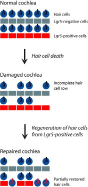
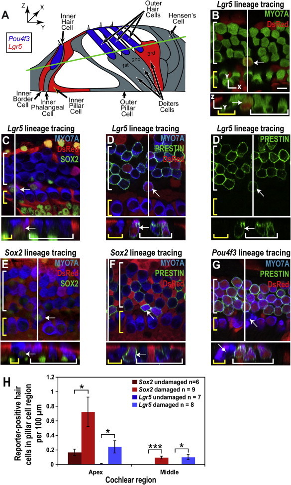
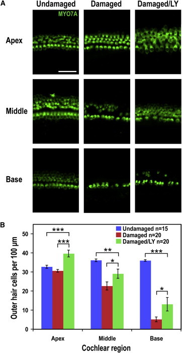
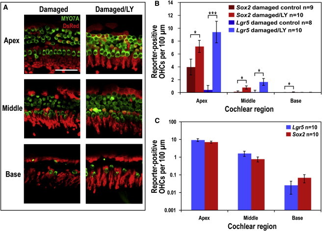
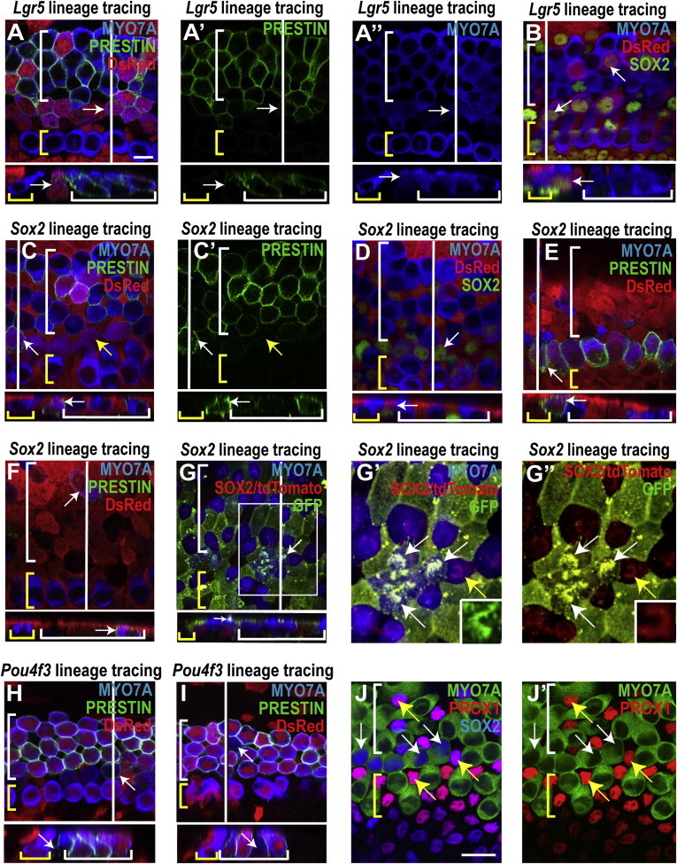
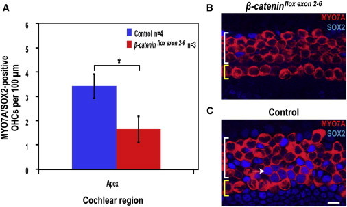
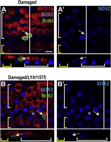
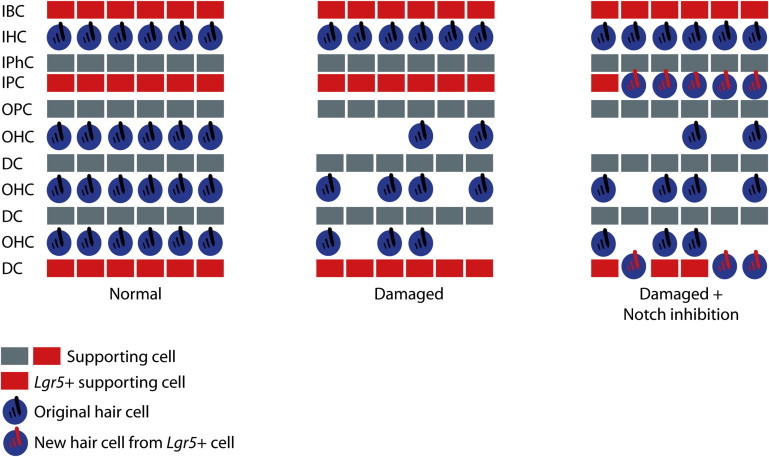
The prevalence of hearing loss after damage to the mammalian cochlea has been thought to be due to a lack of spontaneous regeneration of hair cells, the primary receptor cells for sound. Here, we show that supporting cells, which surround hair cells in the normal cochlear epithelium, differentiate into new hair cells in the neonatal mouse following ototoxic damage. Using lineage tracing, we show that new hair cells, predominantly outer hair cells, arise from Lgr5-expressing inner pillar and third Deiters cells and that new hair cell generation is increased by pharmacological inhibition of Notch. These data suggest that the neonatal mammalian cochlea has some capacity for hair cell regeneration following damage alone and that Lgr5-positive cells act as hair cell progenitors in the cochlea.


Figures

None
Graphical abstract
Figure 1
Figure 1
New Hair Cells in the Pillar Cell Region after Gentamicin Damage (A) Illustration of organ of Corti structure showing the Pou4f3-positive hair cells (blue), the Lgr5-positive supporting cells (red), and the remaining supporting cells in gray. Both the red and gray supporting cells are Sox2 positive. The green line indicates the xy plane from which the confocal slices in (B)–(G) are taken. (B–G) Confocal slices and cross sections from the midapex of neonatal organ of Corti explant cultures, treated with gentamicin and lineage-traced using the CAG-tdTomato reporter, were stained for DsRed (red). A white line on the whole-mount image shows the location of the cross section, and yellow and white brackets indicate IHCs and OHCs, respectively. Arrows point to new reporter-positive (or reporter-negative for Pou4f3) hair cells in the pillar cell region. Scale bar, 10 μm. (B) A reporter-positive hair cell from the Lgr5 lineage (such as those counted in H) was visible in the pillar cell region. (C and D) Reporter staining identified the hair cells marked by the white arrows as derived from Lgr5-positive cells; costaining for SOX2 (C) and location in the pillar cell region indicated that they were newly differentiated, and an OHC phenotype was suggested by the expression of PRESTIN (D). (D′) PRESTIN channel from (D) shows staining in the membrane and cuticular plate of the new hair cell. (E and F) Staining for the Sox2 lineage reporter identified the hair cells marked by the white arrows as derived from supporting cells; their location (pillar cell region) and costaining for SOX2 (E) identified them as newly differentiated cells, and costaining for PRESTIN (F) indicated an OHC identity. (G) The lack of Pou4f3 lineage reporter staining and the location in the pillar region identified the hair cell marked by the white arrow as a new hair cell, and costaining for PRESTIN indicated an OHC identity. (H) Increased numbers of Lgr5 (blue bars) and Sox2 (red bars) reporter-positive hair cells were observed in the pillar cell region of the organ of Corti after gentamicin treatment (mean ± SEM per 100 μm; p < 0.05, ∗∗∗p < 0.001).
Figure 2
Figure 2
Damage followed by Notch Inhibition Leads to an Increase in Hair Cell Numbers (A) Images of hair cells in the apex, middle, and base of an undamaged control, a damaged control, and a damaged/LY411575-treated explant culture show hair cell loss following damage and increased hair cell numbers with LY411575 treatment. Scale bar, 50 μm. (B) Hair cell counts in the apex, middle, and base indicated significantly more OHCs in the damaged/LY411575-treated cultures than in the damaged controls (mean ± SEM per 100 μm; p < 0.05, ∗∗p < 0.01, ∗∗∗p < 0.001).
Figure 3
Figure 3
LY411575 Treatment after Damage Results in Transdifferentiation of Lgr5- and Sox2-Positive Supporting Cells into Hair Cells (A) Images of the three regions of the organ of Corti from a damaged control and a damaged/LY411575-treated explant culture that were Lgr5 lineage traced using the CAG-tdTomato reporter and stained for DsRed (red). Hair cells were stained for MYO7A (green). Many more reporter-positive hair cells (costained for DsRed and MYO7A) were visible in the apical and middle regions of the culture treated with LY411575 than the damaged control. Lgr5 lineage tracing indicated that these reporter-labeled hair cells were derived from Lgr5-positive supporting cells. Scale bar, 50 μm. (B) Reporter-positive OHC counts for Sox2 and Lgr5 lineage tracing of gentamicin/LY411575-treated and control explants showed significantly more reporter-labeled OHCs following LY411575 treatment (mean ± SEM per 100 μm; p < 0.05, ∗∗∗p < 0.001). (C) Reporter-positive OHC counts following gentamicin/LY411575 treatment revealed no significant difference between Sox2 and Lgr5 lineage tracing in any of the cochlear regions, suggesting that lineage tracing with both markers labeled the same population of cells (mean ± SEM per 100 μm, plotted on a logarithmic scale).
Figure 4
Figure 4
New Hair Cells Have Characteristics of Both Supporting Cells and Immature Hair Cells (A and B) In the midapex from lineage-traced organ of Corti explant cultures treated with gentamicin/LY411575, reporter expression (DsRed staining) identified the hair cells indicated by white arrows as derived from Lgr5-positive cells. A white line shows the location of the cross section and yellow and white brackets indicate IHCs and OHCs, respectively. Many of the lineage-tagged hair cells costained for PRESTIN (A), indicating an OHC identity (PRESTIN and MYO7A channels shown in A′ and A″, respectively) and SOX2 (B), confirming that the lineage-tagged hair cells in the pillar and OHC regions were newly differentiated. Scale bar, 10 μm. (C–G) Sox2 lineage tracing using either the CAG-tdTomato reporter stained for DsRed (C–F) or the CAG-tdTomato-EGFP reporter stained for GFP (G). Reporter-positive hair cells marked with white arrows are descended from Sox2-positive cells. (C and D) Lineage-tagged hair cells visible in the pillar and OHC regions of the midapex (white arrows) costained for PRESTIN (C; PRESTIN channel shown in C′), suggesting an OHC phenotype. Other reporter-expressing hair cells were negative for PRESTIN (C, yellow arrows). Lineage tagged hair cells were also costained for SOX2 (D), indicating that they were newly differentiated. (E and F) In the middle region where hair cell damage was extensive, reporter-positive hair cells were observed in what appeared to be the pillar cell region (E) and the OHC area (F). (G) Immature stereocilia bundles were observed on some reporter-positive hair cells in the pillar cell region (white arrow, midapex), further suggesting they were newly differentiated. Note that because the CAG-tdTomato-EGFP reporter has a membrane localization sequence, the GFP signal is highly visible in the membrane-rich stereocilia bundles located atop hair cells that were derived from SOX2-positive supporting cells. SOX2 staining in the nucleus of the cell indicated by the white arrow is consistent with a new hair cell. (G′ and G″) Higher magnification of the region outlined with a white box in (G) shows several GFP-positive stereocilia bundles (white arrows), indicating reporter recombination, whereas the bundles of the original hair cells remain positive for tdTomato (yellow arrow). The insets show stereociliary bundles of individual hair cells, either new, and therefore recombined (G′, GFP), or original (G″, tdTomato). (H and I) The absence of reporter expression in the hair cells marked with white arrows from Pou4f3 lineage-traced cultures using the CAG-tdTomato reporter (red) indicated that they were new hair cells located in the pillar (H) and OHC (I) regions. Costaining for PRESTIN suggested an OHC identity in some of these hair cells (H). (J and J′) SOX2/MYO7A double-positive hair cells in the pillar cell region are PROX1 negative (white arrows), whereas the surrounding supporting cells are SOX2/PROX1 positive (yellow arrows). Scale bar, 20 μm.
Figure 5
Figure 5
Wnt Signaling Is Necessary for Supporting Cell Transdifferentiation (A) Double-positive MYO7A/SOX2 OHC counts for organ of Corti cultures from β-cateninflox(exon2–6);Sox2-Cre-ER pups and their β-cateninflox(exon2–6)-negative littermates (control) after gentamicin and LY411575 treatment show significantly less double-positive OHCs in the apex (mean ± SEM per 100 μm; p < 0.05). (B and C) Images from the apex of the organ of Corti after gentamicin and LY411575 treatment of β-cateninflox(exon2–6);Sox2-Cre-ER cultures (B) and β-cateninflox(exon2–6)-negative controls (C). Yellow and white brackets indicate IHCs and OHCs, respectively. Scale bar, 10 μm. MYO7A/SOX2-positive cells are absent in (B), whereas the white arrow indicates one of several double-positive cells in (C).
Figure 6
Figure 6
Supporting Cells Proliferate to Generate New Hair Cells (A) A gentamicin- and BrdU-treated organ of Corti explant from the midapex showed a pair of triple-labeled hair cells in the pillar cell region. A white line shows the location of the cross section, and yellow and white brackets indicate IHCs and OHCs, respectively. The arrow points to a triple-labeled (BrdU/SOX2/MYO7A) cell in the pillar cell region. Its unusual location and costaining indicated a newly differentiated hair cell generated through proliferation. (A′) The SOX2 channel from (A) is shown. (B) The arrow points to a single triple-labeled cell in the pillar region of a gentamicin/LY411575-treated explant culture, indicating a newly differentiated hair cell generated through proliferation, and the asterisk indicates a BrdU-negative, SOX2-positive hair cell, suggesting generation through transdifferentiation rather than proliferation. (B′) The SOX2 channel from (B) is shown. Scale bar, 10 μm.
Figure 7
Figure 7
Schematic Showing the Normal Organ of Corti, the Organ of Corti following Gentamicin Damage, and the Organ of Corti following Gentamicin Damage and Notch Inhibition Cell types are labeled as follows: inner border cell (IBC), inner hair cell (IHC), inner phalangeal cell (IPhC), inner pillar cell (IPC), outer pillar cell (OPC), outer hair cell (OHC), and Deiters cell (DC). Supporting cells are indicated in gray or red. Red supporting cells are Lgr5 positive. Original hair cells are indicated as blue cells with black stereocilia bundles. Following damage, outer hair cells are missing and are replaced spontaneously or after Notch inhibition by new hair cells derived from Lgr5-positive inner pillar and third Deiters cells (blue cells with red stereocilia bundles).

References

    1. Arnold K., Sarkar A., Yram M.A., Polo J.M., Bronson R., Sengupta S., Seandel M., Geijsen N., Hochedlinger K. Sox2(+) adult stem and progenitor cells are important for tissue regeneration and survival of mice. Cell Stem Cell. 2011;9:317–329. - PMC - PubMed
    1. Balak K.J., Corwin J.T., Jones J.E. Regenerated hair cells can originate from supporting cell progeny: evidence from phototoxicity and laser ablation experiments in the lateral line system. J. Neurosci. 1990;10:2502–2512. - PMC - PubMed
    1. Barker N., van Es J.H., Kuipers J., Kujala P., van den Born M., Cozijnsen M., Haegebarth A., Korving J., Begthel H., Peters P.J., Clevers H. Identification of stem cells in small intestine and colon by marker gene Lgr5. Nature. 2007;449:1003–1007. - PubMed
    1. Bermingham-McDonogh O., Stone J.S., Reh T.A., Rubel E.W. FGFR3 expression during development and regeneration of the chick inner ear sensory epithelia. Dev. Biol. 2001;238:247–259. - PubMed
    1. Bermingham-McDonogh O., Oesterle E.C., Stone J.S., Hume C.R., Huynh H.M., Hayashi T. Expression of Prox1 during mouse cochlear development. J. Comp. Neurol. 2006;496:172–186. - PMC - PubMed

Publication types

MeSH terms

Substances