Comparative Study
. 2014 Jul;177(1):47-63.
doi: 10.1111/cei.12339.

Comparison of peptide-major histocompatibility complex tetramers and dextramers for the identification of antigen-specific T cells

Affiliations
Comparative Study

Comparison of peptide-major histocompatibility complex tetramers and dextramers for the identification of antigen-specific T cells

G Dolton et al. Clin Exp Immunol. 2014 Jul.

Abstract

Fluorochrome-conjugated peptide-major histocompatibility complex (pMHC) multimers are widely used for flow cytometric visualization of antigen-specific T cells. The most common multimers, streptavidin-biotin-based 'tetramers', can be manufactured readily in the laboratory. Unfortunately, there are large differences between the threshold of T cell receptor (TCR) affinity required to capture pMHC tetramers from solution and that which is required for T cell activation. This disparity means that tetramers sometimes fail to stain antigen-specific T cells within a sample, an issue that is particularly problematic when staining tumour-specific, autoimmune or MHC class II-restricted T cells, which often display TCRs of low affinity for pMHC. Here, we compared optimized staining with tetramers and dextramers (dextran-based multimers), with the latter carrying greater numbers of both pMHC and fluorochrome per molecule. Most notably, we find that: (i) dextramers stain more brightly than tetramers; (ii) dextramers outperform tetramers when TCR-pMHC affinity is low; (iii) dextramers outperform tetramers with pMHC class II reagents where there is an absence of co-receptor stabilization; and (iv) dextramer sensitivity is enhanced further by specific protein kinase inhibition. Dextramers are compatible with current state-of-the-art flow cytometry platforms and will probably find particular utility in the fields of autoimmunity and cancer immunology.

Keywords: T cell receptors; T cells; autoimmunity; diabetes; tumour immunology.


Figures

Fig. 1
Fig. 1
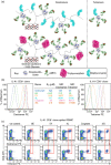
Peptide–major histocompatibility complex (pMHC) dextramers bind CD8+ T cells substantially better than tetramers when T cell receptor (TCR)–pMHC affinity is low. (a) A schematic showing how pMHC dextramers differ from pMHC tetramers. Dextramers have a dextran backbone and carry more fluorochrome and pMHC per molecule. The fluorochrome/pMHC ratio differs between allophycocyanin (APC) and R-phycoerythrin (PE) dextramers. Figure 1(a) is available as a separate high-resolution file (Supporting Information Fig. S3) in the online supplement that accompanies this article. (b) The ILA1 CD8+ T cell clone was stained with PE-conjugated tetramer (right) or dextramer (left) reagents constructed with human leucocyte antigen (HLA)-A2 monomers bearing the wild-type human telomerase reverse transcriptase (hTERT)540–548 peptide (ILAKFLHWL), variants thereof with defined TCR–pMHC-I affinities as indicated, or the HIV-1 Pol476–484 peptide (ILKEPVHGV) as an irrelevant control. (c) Human peripheral blood mononuclear cells (PBMCs) (106) were spiked with clonal ILA1 cells (104) and stained with the indicated tetramers (bottom) or dextramers (top), the viability dye Aqua and monoclonal antibodies against the surface markers CD3, CD4, CD8, CD14 and CD19. Gates were set serially on lymphocytes, single cells and live CD3+CD14CD19 cells prior to display as CD8 versus tetramer/dextramer. In each column, the red box is positioned on the antigen-specific CD8+ T cell population that stains most brightly to aid visual comparison.
Fig. 2
Fig. 2
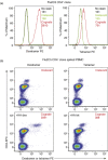
Dextramers made with peptide–major histocompatibility complex (pMHC) class II bind CD4+ T cells substantially better than tetramers. (a) The influenza (Flu)2C5 CD4+ T cell clone was stained with phycoerythrin (PE)-conjugated (right) or dextramer (left) reagents constructed with human leucocyte antigen (HLA)-DR1 monomers bearing the cognate influenza A virus HA307–319 peptide (PKYVKQNTLKLAT; red line) or the HIV-1 p24 Gag299–312 peptide (DRFYKTLRAEQASQ; green line) as an irrelevant control. Mean fluorescence intensity (MFI) values are shown inset for each tetramer/dextramer reagent and unstained cells (black line). (b) Human peripheral blood mononuclear cells (PBMCs) (106) were spiked with clonal Flu2C5 cells (104) and stained with the indicated tetramers (right) or dextramers (left), the viability dye Aqua and monoclonal antibodies against the surface markers CD3, CD4, CD8, CD14 and CD19. Gates were set serially on lymphocytes, single cells and live CD3+CD14CD19 cells prior to display as CD4 versus tetramer/dextramer. MFI values are shown inset.
Fig. 3
Fig. 3
Dextramers bind a type I diabetes-derived CD8+ T cell clone substantially better than tetramers and also distinguish such cells from other T cells in a spiked peripheral blood mononuclear cell (PBMC) setting. (a) The 3F2 CD8+ T cell clone was stained with phycoerythrin (PE)-conjugated tetramer (right) or dextramer (left) reagents constructed with human leucocyte antigen (HLA)-A2 monomers bearing the cognate preproinsulin (PPI)15–24 peptide (ALWGPDPAAA) or the melanoma antigen (MAGE) A3168–176 peptide (YLEYRQVPV) as an irrelevant control. Different amounts of tetramer or dextramer were used as indicated with respect to the monomeric peptide-major histocompatibility complex (pMHC) component. Mean fluorescence intensity (MFI) values are shown for each condition. (b) Human PBMCs (106) were spiked with clonal 3F2 cells (104) and stained with the indicated tetramers (right) or dextramers (left), the viability dye Aqua and monoclonal antibodies against the surface markers CD3, CD4, CD8, CD14 and CD19. Gates were set serially on lymphocytes, single cells and live CD3+CD14CD19 cells prior to display as CD8 versus tetramer/dextramer. Clonal 3F2 cells were easily distinguishable on the basis of brighter CD8 expression. Reagent quantities with respect to the pMHC-I component (black) and MFI values (red) are shown inset.
Fig. 4
Fig. 4
Superior ex vivo detection of antigen-specific CD8+ T cells with dextramers. Human leucocyte antigen (HLA)-A2+ peripheral blood mononuclear cells (PBMCs) from non-type I diabetes (T1D) donors (left, middle) or T1D patients (right) were stained with tetramer or dextramer reagents constructed with HLA-A2 monomers bearing the Epstein–Barr virus (EBV) BMLF-1259–267 peptide (GLCTLVAML; left), the MART-126–35 heteroclitic peptide (ELAGIGILTV; middle) or the preproinsulin (PPI)15–24 peptide (ALWGPDPAAA; right). Each symbol and line combination denotes a different donor. Gates were set serially on lymphocytes, single cells and live CD3+CD14CD19 cells prior to analysis as CD8 versus tetramer/dextramer. Detected antigen-specific CD8+ T cell frequencies (top) and mean fluorescence intensity (MFI) values (bottom) are shown. Representative flow cytometry plots are shown in Supporting Information Fig. S2. The difference in MFI and percentage of CD8+ T cells detected between dextramers and tetramers is significantly different (P ≤ 0·01: paired, two-tailed t-test).
Fig. 5
Fig. 5
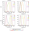
Peptide–major histocompatibility complex (pMHC) dextramer staining is enhanced by protein kinase inhibitor (PKI) treatment. The ILA1 CD8+ T cell clone was stained with phycoerythrin (PE)-conjugated tetramer (right) or dextramer (left) reagents constructed with human leucocyte antigen (HLA)-A2+ monomers bearing the cognate human telomerase reverse transcriptase (hTERT)540–548 peptide (ILAKFLHWL), variants thereof with defined T cell receptor (TCR)-pMHC-I affinities as indicated in the key, or the HIV-1 Pol476–484 peptide (ILKEPVHGV) as an irrelevant control after pretreatment with (bottom) or without (top) 50 nM dasatinib. Mean fluorescence intensity (MFI) values are shown inset.
Fig. 6
Fig. 6
Superior recovery of antigen-specific CD8+ T cells with peptide–major histocompatibility complex (pMHC) dextramer + protein kinase inhibitor (PKI). Human peripheral blood mononuclear cells (PBMCs) (106) were spiked with clonal ILA1 cells (104), pretreated with or without 50 nM dasatinib (+/− PKI) and then stained with phycoerythrin (PE)-conjugated tetramer (lower) or dextramer (upper) reagents as indicated (details in Fig. 5), followed by the viability dye Aqua and monoclonal antibodies against the surface markers CD3, CD4, CD8, CD14 and CD19. Gates were set serially on lymphocytes, single cells and live CD3+CD14CD19 cells prior to display as CD8 versus tetramer/dextramer. In each column, the red box is positioned on the antigen-specific CD8+ T cell population that stains most brightly to aid visual comparison.
Fig. 7
Fig. 7
Improved staining of autoimmune T cell clones with peptide–major histocompatibility complex (pMHC) dextramer and protein kinase inhibitor (PKI). The 4C6 and 1E6 CD8+ T cell clones were stained with phycoerythrin (PE)-conjugated tetramer or dextramer reagents constructed with human leucocyte antigen (HLA)-A24 monomers bearing the LWMRLLPLL peptide or HLA-A2 monomers bearing the ALWGPDPAAA peptide, respectively, after pretreatment with or without 50 nM dasatinib (+/− PKI). Irrelevant allotype-matched control reagents were constructed with HLA-A24–AYAQKIFKIL and HLA-A2–NLVPMVATV monomers. FMO = fluorescence minus one.
Fig. 8
Fig. 8
Superior ex vivo detection of antigen-specific CD8+ T cells with a combinatorial staining approach using a protein kinase inhibitor (PKI) and peptide–major histocompatibility complex (pMHC) dextramer. Human leucocyte antigen (HLA)-A2+ peripheral blood mononuclear cells (PBMCs) from healthy controls were stained with phycoerythrin (PE)-conjugated tetramer (right) or dextramer (left) reagents constructed with HLA-A2 monomers bearing the MART-126–35 heteroclitic peptide (ELAGIGILTV) after pretreatment with or without 50 nM dasatinib (+/− PKI). Gates were set serially on lymphocytes, single cells and live CD3+CD14CD19 cells prior to display as CD8 versus tetramer/dextramer. Detected antigen-specific CD8+ T cell frequencies (black) and mean fluorescence intensity (MFI) values (red) are shown inset.
Fig. 9
Fig. 9
Peptide–major histocompatibility complex (pMHC) dextramers are compatible with high-resolution flow cytometry. Human leucocyte antigen (HLA)-A2+ peripheral blood mononuclear cells (PBMCs) from healthy controls were stained with phycoerythrin (PE)-conjugated or dextramer reagents constructed with HLA-A2 monomers bearing the CMV pp65495–503 peptide (NLVPMVATV), followed by the viability dye ViViD and monoclonal antibodies against the surface markers CD3, CD8, CD14, CD19, CD27, CD45RO, CD57, CD69 and CCR7. Gates were set serially on lymphocytes, single cells and live CD3+CD14CD19 cells (a) prior to display as CD8 versus tetramer/dextramer (b,c). Boolean gating was carried out to exclude artefacts and fluorochrome aggregates as indicated (a). Yellow or green dots superimposed on density plots showing the phenotypical profile of the overall CD8+ T cell population depict individual antigen-specific CD8+ T cells stained with dextramer or tetramer, respectively (d–i). The following bivariate analyses are shown: CD27 versus CD45RO (d,e); CD57 versus CCR7 (f,g); and CD69 versus CCR7 (h,i).
Fig. 10
Fig. 10
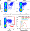
Dextramers can be used to detect activated T cells during an intracellular cytokine assay. A 10-day-old enriched T cell line specific for the influenza epitope, GILGFVFTL (matrix protein residues 58–66), was cultured with or without cognate peptide for 5 h in the presence of monensin and brefeldin A. Cells were subsequently stained with cognate [human leucocyte antigen (HLA)-A2 GILGFVFTL] or irrelevant (HLA-A2–ALWGPDPAAA) dextramer or tetramer, cell surface markers (CD8, CD3, CD19, CD14), a viability stain and intracellularly for interferon (IFN)-γ. Unstimulated cells were stained with cognate dextramer (a) or tetramer (b), with the gate shown based on staining with an irrelevant dextramer or tetramer. The percentage of staining of IFN-γ+ cells (c: red gate, based on a fluorescence minus 1) with cognate dextramer (red) or tetramer (green) is based upon the staining of the same IFN-γ+ cells with an irrelevant dextramer (blue) or tetramer (orange) (d). The percentage of gated cells shown inset (a–c).
Fig. 11
Fig. 11
Similar visual staining patterns with peptide–major histocompatibility complex (pMHC) dextramers and tetramers. The NLV2F3 CD8+ T cell clone was stained with 4′,6-diamidino-2-phenylindole (DAPI) (blue), followed by Alexa488-conjugated tetramer or fluorescein isothiocyanate (FITC)-conjugated dextramer reagents constructed with human leucocyte antigen (HLA)-A2 monomers bearing the cytomeglovirus (CMV) pp65495–503 peptide (NLVPMVATV). Staining conditions are indicated; dasatinib was used at a concentration of 50 nM. Cells were visualized using a Leica DM LB2 fluorescent microscope.

References

    1. Wooldridge L, Lissina A, Cole DK, van den Berg HA, Price DA, Sewell AK. Tricks with tetramers: how to get the most from multimeric peptide-MHC. Immunology. 2009;126:147–164. - PMC - PubMed
    1. Klenerman P, Cerundolo V, Dunbar PR. Tracking T cells with tetramers: new tales from new tools. Nat Rev Immunol. 2002;2:263–272. - PubMed
    1. Newell EW, Sigal N, Bendall SC, Nolan GP, Davis MM. Cytometry by time-of-flight shows combinatorial cytokine expression and virus-specific cell niches within a continuum of CD8+ T cell phenotypes. Immunity. 2012;36:142–152. - PMC - PubMed
    1. Altman JD, Moss PA, Goulder PJ, et al. Phenotypic analysis of antigen-specific T lymphocytes. Science. 1996;274:94–96. - PubMed
    1. Batard P, Peterson DA, Devevre E, et al. Dextramers: new generation of fluorescent MHC class I/peptide multimers for visualization of antigen-specific CD8+ T cells. J Immunol Methods. 2006;310:136–148. - PubMed

Publication types

MeSH terms