. 2015 Jan;179(1):39-49.
doi: 10.1111/cei.12342.

Loading of nuclear autoantigens prototypically recognized by systemic lupus erythematosus sera into late apoptotic vesicles requires intact microtubules and myosin light chain kinase activity

Affiliations

Loading of nuclear autoantigens prototypically recognized by systemic lupus erythematosus sera into late apoptotic vesicles requires intact microtubules and myosin light chain kinase activity

M Zirngibl et al. Clin Exp Immunol. 2015 Jan.

Abstract

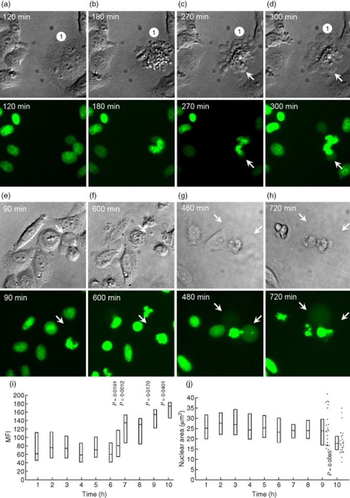
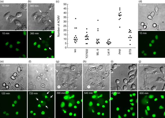
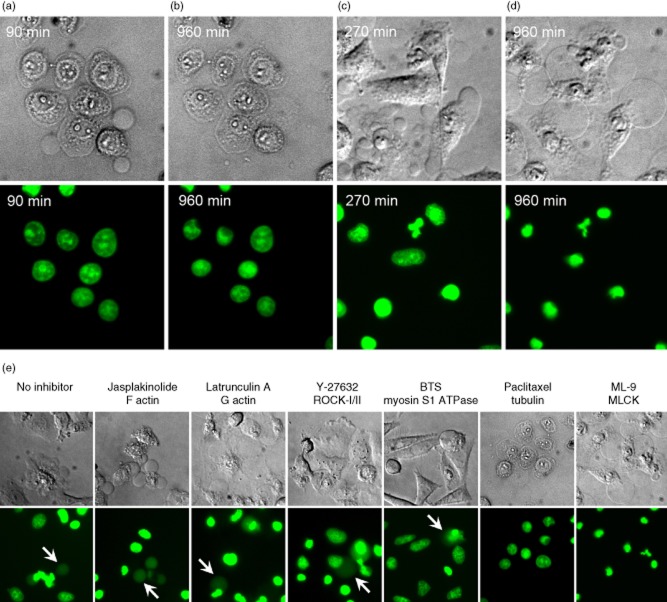
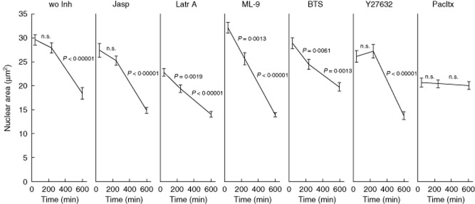
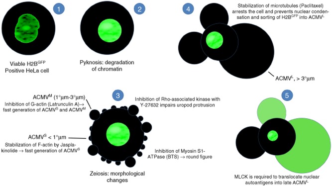
Most cases of systemic lupus erythematosus (SLE) are characterized by an impaired clearance of apoptotic cells in various tissues. Non-cleared apoptotic waste is considered an immunogen driving the autoimmune response in patients with SLE. During the execution of apoptosis, membrane blebs are formed and filled with cellular components. Here, we evaluate the cytoskeletal pathway(s) responsible for the loading of SLE prototypic nuclear autoantigens into the apoptotic cell-derived membranous vesicles (ACMV) generated during late phases of apoptosis. HeLa cells expressing a fusion protein of histone H2B with green fluorescent protein (GFP) were irradiated with ultraviolet (UV)-B to induce apoptosis. The appearance and trafficking of chromatin-derived material was monitored by fluorescence microscopy. Specific inhibitors of cytoskeletal pathways were employed to identify the motile elements involved in translocation and trafficking of the nuclear components. We observed that immediately after their appearance the ACMV did not contain histone H2B(GFP) ; in this phase the fluorescence was contained in the nuclear remnants and the cytoplasm. Within consecutive minutes the ACMV were loaded with chromatin-derived material, whereas the loading of simultaneously created ACMV with histone H2B(GFP) was not uniform. Some ACMV were preferentially filled and, consequently, showed a remarkably higher histone H2B(GFP) accumulation. Inhibitors of the cytoskeleton revealed that functional microtubules and myosin light chain kinase are required for nuclear shrinkage and loading of nuclear material into the ACMV, respectively.

Keywords: apoptosis; blebbing; nuclear autoantigens; systemic lupus erythematosus.


Figures

Fig. 1
Fig. 1
Redistribution of histone 2B-green fluorescent protein (H2BGFP) during ultraviolet (UV)-B-induced apoptosis in HeLa cells. Adherent HeLa cells expressing fluorescent H2BGFP histone were cultured to a density of 3 × 10⁴ cells/cm2. After UV-B irradiation with 900 mJ/cm2 the distribution of H2BGFP was monitored by video-microscopy. (a–d) The characteristic nuclear changes that had been described for adherent cells undergoing apoptosis. After 270 min the cell marked with ‘1’ had undergone waves of formation of apoptotic cell-derived membranous vesicles (ACMV). None of these contained detectable amounts of H2BGFP; 30 min later two of the vesicles were filled with substantial amounts of H2BGFP (arrows). Note that (1) not all ACMV contain degraded chromatin (H2BGFP); (2) ACMV are formed initially without nuclear material, and are loaded in a second step (arrows). (e,f) Some large ACMVL remain unloaded even 10 h after their formation. (g,h) In very late phases of apoptosis some ACMVL which had been loaded with H2BGFP disintegrate and release their load into the culture supernatant, most probably during secondary necrosis. (i) mean fluorescence intensity (MFI) of the nuclei during the 10 h after irradiation. Note a significant increase of the MFI starting 6 h after irradiation (Student's t-test of cellular MFI at t = 0 versus t = × hours after irradiation). (j) nuclear area during the 10 h after irradiation. Note a significant shrinkage of the nuclei 9 h after irradiation (Student's t-test of cellular nuclear area at t = 0 versus t = × hours after irradiation).
Fig. 2
Fig. 2
F- and G-actin, p160ROCK and myosin are not required for loading into apoptotic cell-derived membranous vesicles (ACMV)L of histone 2B-green fluorescent protein (H2BGFP). Culturing and induction of apoptosis in HeLa cells was performed as described in Fig. 1. (a,b) HeLa cells were incubated with the F-actin inhibitor jasplakinolide. (a) Ten min after addition of the inhibitor, the cells start to form large numbers of ACMVS/M. (b) This leads to a loss of cell volume followed by shrinkage of the cell corpses. Whereas the inhibition of F-actin causes quick and dramatic changes in plasma membrane appearance, the creation and the loading of ACMVL with H2BGFP remains unaffected as to be observed 360 min after irradiation (arrows). (c) ACMV count 90 min after induction of apoptosis. HeLa cells incubated with jasplakinolide (Jasp) show a significant higher number of released ACMVS/M. HeLa cells treated with latrunculin A (Lat A) show no significant change in the ACMV count (most ACMV were observed transiently during the first 10–60 min (see d). (d,e,f) HeLa cells were incubated with the G-actin inhibitor latrunculin A. (d) Ten min after addition of the inhibitor, the cells start to form small ACMVS/M. (e) After 120 min the nuclei appear plastically with a flat cytoplasmic rim. (f) Note that loading of ACMVL with histone H2BGFP fusion protein remains unaffected as to be observed 720 min after irradiation (arrows). (g,h) HeLa cells were incubated with the p160ROCK inhibitor Y-27632; (g) 390 min after addition of the inhibitor, some cells display abnormalities in uropod protrusion (arrow). (g,h) Note that loading of ACMVL with H2BGFP remains unaffected (1). (i,j) HeLa cells were incubated with the myosin inhibitor N-benzyl-p-toluene sulphonamide (BTS); (i) 210 min after addition of BTS, most cells adopted a round shape but preserved their capabilities to move. (j) Note that ACMVL were still loaded with H2BGFP. Small, medium-sized and large apoptotic cell-derived membranous vesicles are marked ACMVS/M/l, respectively.
Fig. 3
Fig. 3
Microtubules and myosin light chain kinase (MLCK) are required for nuclear shrinkage and loading of apoptotic cell-derived membranous vesicles (ACMV)L with histone 2B-green fluorescent protein (H2BGFP), respectively. (a,b) HeLa cells incubated with an agent stabilizing microtubules (paclitaxel) are arrested at an early stage of apoptosis. (b) Nuclear shrinkage is impaired as well as the sorting of nuclear autoantigens into late ACMVL. (c,d) The formation of ACMVL is not affected during apoptosis in the presence of 1-(5-chloronaphthalene-1-sulphonyl)-1H-hexahydro-1,4-diazepine (ML-9), an inhibitor of myosin light chain kinase (MLCK). (d) Nuclear shrinkage and chromatin condensation is performed regularly. However, sorting of nuclear autoantigens into late ACMVL cannot be observed. (e) In the absence of inhibitors, and in the presence of jasplakinolide, latrunculin A, Y-27632 and N-benzyl-p-toluene sulphonamide (BTS) histone H2BGFP-positive ACMVL are readily observed (arrows). Paclitaxel conserves the cell at an early state of cell death. Nuclear shrinkage and chromatin condensation is arrested. Cells incubated with ML-9 regularly generate ACMVL; translocation of nuclear autoantigens into these was not detected.
Fig. 4
Fig. 4
Morphometric measurement of nuclear size 30, 240 and 600 min after irradiation. Y-27632 and jasplakinolide do not have any influence on nuclear shrinkage. 1-(5-chloronaphthalene-1-sulphonyl)-1H-hexahydro-1,4-diazepine (ML-9) and N-benzyl-p-toluene sulphonamide (BTS) significantly accelerate and decelerate nuclear shrinkage, respectively. Cells treated with latrunculin A or paclitaxel (Pacltx) already display a significantly smaller nuclear area 30 min after treatment. Whereas cells treated with latrunculin A continue shrinkage, the nuclei of paclitaxel-treated cells maintain their size for at least 10 h.
Fig. 5
Fig. 5
Synopsis: schematic display of the translocation in apoptotic cells of nuclear autoantigens. HeLa cells were transfected with pBOS-histone 2B-green fluorescent protein (H2BGFP) to label histone H2B as a representative of nuclear proteins serving as autoantigens in patients with systemic lupus erythematosus (SLE). After induction of apoptosis via irradiation with ultraviolet (UV)-B, processing and translocation of H2B was monitored employing fluorescence microscopy. The transfected cells were incubated with inhibitors of cell dynamics to identify the cytoskeletal proteins involved in the trafficking of H2B. (1) Viable H2BGFP expressing cell with a regularly configured nucleus. (2) Apoptosis starts with chromatin condensation and degradation (pyknosis). (3) In early apoptosis, the cells undergo dramatic morphological changes. They shrink and generate apoptotic cell-derived membranous vesicles (ACMV), zeiosis. Initially small (< 1 μm) and intermediate (1–3 μm) ACMVS/M are generated. These ACMV do not contain substantial amounts of H2BGFP. Several inhibitors of cell dynamics have impact on the morphology of the dying cell. p160ROCK (Y-27632) is required for uropod protrusion. Inhibition of F-actin (jasplakinolide) or G-actin (latrunculin A) leads to the early generation of ACMVS and ACMVM. Inhibition of myosin S1 ATPase [N-benzyl-p-toluene sulphonamide (BTS)] causes rounding of the cell. (4) However, all the above listed inhibitors do not have any impact on formation of late ACMVL and the translocation of H2B into these. Inhibition of microtubules (paclitaxel) arrests the apoptotic process at an early time-point. It impairs nuclear shrinkage as well as formation of ACMVL and translocation of H2BGFP. (5) Inhibition of myosin light chain kinase (MLCK) by 1-(5-chloronaphthalene-1-sulphonyl)-1H-hexahydro-1,4-diazepine (ML-9) does not preclude the formation of ACMVL, but impairs their loading with histone H2BGFP.

Similar articles

Cited by

References

    1. Hasegawa M, Sato S, Kikuchi K, Takehara K. Antigen specificity of antihistone antibodies in systemic sclerosis. Ann Rheum Dis. 1998;57:470–475. - PMC - PubMed
    1. Wayaku T, Hasegawa M, Kaji K, et al. Antigen specificity of antihistone antibodies in connective tissue disease patients with anti-U1RNP antibodies. Rheumatol Int. 2007;28:113–119. - PubMed
    1. Ostensen M, Fredriksen K, Kass E, Rekvig OP. Identification of antihistone antibodies in subsets of juvenile chronic arthritis. Ann Rheum Dis. 1989;48:114–117. - PMC - PubMed
    1. Leak AM, Woo P. Juvenile chronic arthritis, chronic iridocyclitis, and reactivity to histones. Ann Rheum Dis. 1991;50:653–657. - PMC - PubMed
    1. Gompertz NR, Isenberg DA, Turner BM. Correlation between clinical features of systemic lupus erythematosus and levels of antihistone antibodies of the IgG, IgA, and IgM isotypes. Ann Rheum Dis. 1990;49:524–527. - PMC - PubMed

Publication types

MeSH terms