. 2014 May;28(5):745-57.
doi: 10.1210/me.2013-1335. Epub 2014 Mar 27.

The ability of thyroid hormone receptors to sense t4 as an agonist depends on receptor isoform and on cellular cofactors

Affiliations

The ability of thyroid hormone receptors to sense t4 as an agonist depends on receptor isoform and on cellular cofactors

Amy Schroeder et al. Mol Endocrinol. 2014 May.

Abstract

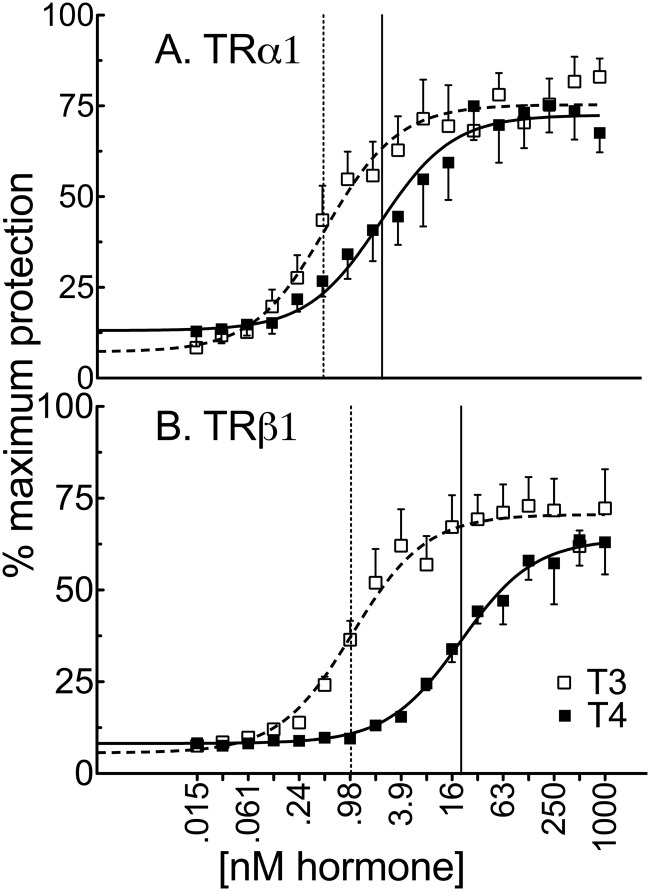
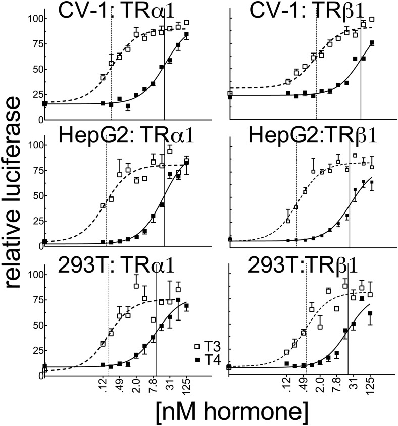
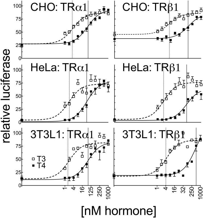
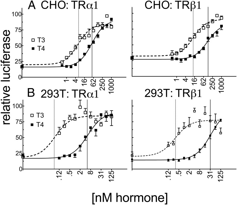
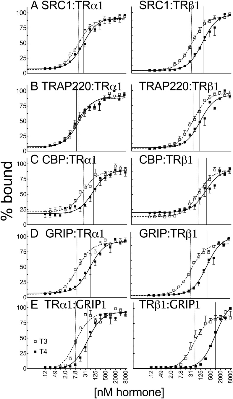
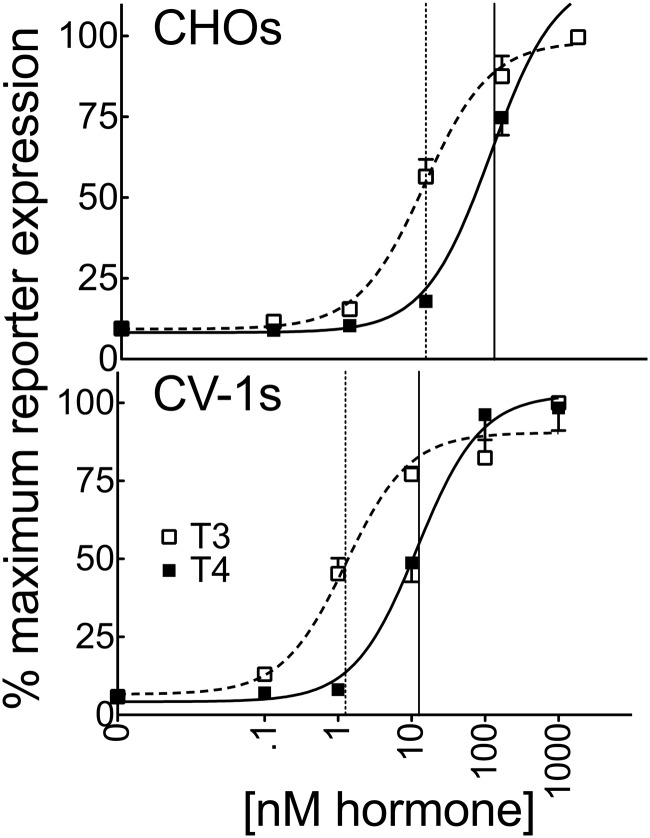
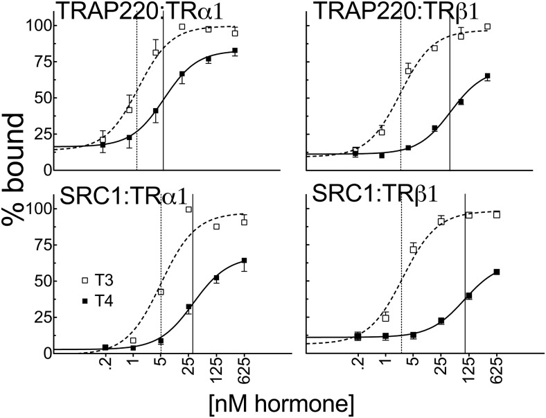
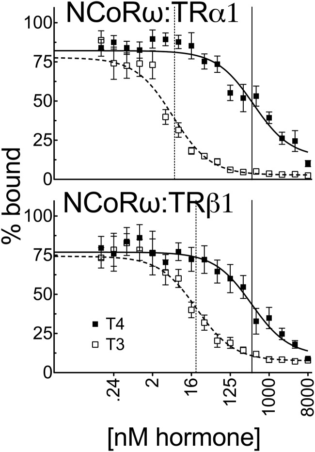
T4 (3,5,3',5'-tetraiodo-l-thyronine) is classically viewed as a prohormone that must be converted to the T3 (3,5,3'-triiodo-l-thyronine) form for biological activity. We first determined that the ability of reporter genes to respond to T4 and to T3 differed for the different thyroid hormone receptor (TR) isoforms, with TRα1 generally more responsive to T4 than was TRβ1. The response to T4 vs T3 also differed dramatically in different cell types in a manner that could not be attributed to differences in deiodinase activity or in hormone affinity, leading us to examine the role of TR coregulators in this phenomenon. Unexpectedly, several coactivators, such as steroid receptor coactivator-1 (SRC1) and thyroid hormone receptor-associated protein 220 (TRAP220), were recruited to TRα1 nearly equally by T4 as by T3 in vitro, indicating that TRα1 possesses an innate potential to respond efficiently to T4 as an agonist. In contrast, release of corepressors, such as the nuclear receptor coreceptor NCoRω, from TRα1 by T4 was relatively inefficient, requiring considerably higher concentrations of this ligand than did coactivator recruitment. Our results suggest that cells, by altering the repertoire and abundance of corepressors and coactivators expressed, may regulate their ability to respond to T4, raising the possibility that T4 may function directly as a hormone in specific cellular or physiological contexts.


Figures

Figure 1.
Figure 1.
TRα1 (A) exhibits a greater affinity for T4 vs T3 than does TRβ1 (B). Radiolabeled in vitro transcribed and translated TRs were incubated with 0.025 U (TRα1) or 0.05 U (TRβ1) of elastase and the indicated concentrations of vehicle, T3, or T4 for 10 minutes. The samples were analyzed by SDS-PAGE, and the percent recovery of the receptor protease-resistant core (maximum = 100%) is indicated for each T3 or T4 concentration. Error bars indicate SEMs of at least 3 biological replicate experiments. Vertical dashed and solid lines represent EC50s T3 and T4, respectively.
Figure 2.
Figure 2.
Very low endogenous expression of TRs in CV-1 and CHO cells induced very low basal activity on a DR4-CON-tk reporter. CHO (A) or CV-1 cells (B) were transiently transfected via lipofection with an empty expression vector (EV) or expression vectors containing TRα1 or TRβ1, a consensus DR4-CON-tk-luciferase reporter, and β-galactosidase control in hormone-depleted medium. After 24 hours in hormone-depleted medium, the cells were exposed to the indicated T3 or T4 concentrations. Cells were incubated an additional 24 hours before harvest, and lysates were analyzed for luciferase activity and β-galactosidase levels. Luciferase values were normalized against the β-galactosidase control for each sample. Error bars indicate SEMs for at least 3 biological replicate experiments.
Figure 3.
Figure 3.
The ability of TRα1 to respond to T4 vs T3 differs in CV-1, HepG2, and HEK293T cells and substantially exceeds observed affinity differences determined in vitro. CV-1, HepG2, and HEK293T cells were transiently transfected as described for Figure 2 with the consensus DR4-CON-tk-luciferase reporter. Luciferase values were normalized against the β-galactosidase control for each sample and plotted as percent T3 maximum. Error bars indicate SEMs of at least 3 biological replicate experiments. Unfortunately, we could not measure the TR protein levels expressed in these transfected cells due to limitations arising from the relatively low levels of expression and technical limitations as to the commercially available anti-TR antibodies. Vertical dashed and solid lines represent EC50s T3 and T4, respectively.
Figure 4.
Figure 4.
The ability of TRα1 to respond to T4 vs T3 differs much less dramatically in CHO, HeLa, and 3T3L1 cells. CHO, HeLa, and 3T3–L1 cells were transiently transfected in the same manner as in Figure 3 with the DR4-CON-tk-luciferase reporter. Error bars indicate SEMs of at least 3 biological replicate experiments. Vertical dashed and solid lines represent EC50s T3 and T4, respectively.
Figure 5.
Figure 5.
A known nonconsensus TRE exhibits an ability similar to that of the consensus DR4 TRE to respond to T4 vs T3 in CHO and HEK293T cells. A, CHO cells were transiently transfected as described for Figure 3 except with a DR4-M-tk-luciferase reporter derived from the murine leukemia virus. Luciferase values were normalized against the β-galactosidase control for each sample and plotted as percent T3 maximum. Error bars indicate SEMs of at least 3 replicate experiments. B, Same experiment as for panel A was repeated using HEK293T cells. Vertical dashed and solid lines represent EC50s T3 and T4, respectively.
Figure 6.
Figure 6.
T4 recruits certain coactivators to TRα1 with potency that was nearly equal to that of T3 in vitro. Full-length in vitro–transcribed and translated, 35S-labeled human TRα1 or TRβ1 were incubated with glutathione-agarose bead immobilized GST fusions of coactivator proteins (LXXLL receptor interaction motifs) indicated within each panel: SRC1 (amino acids 568–891) (A), TRAP220 (486–723) (B), CBP (1–451) (C), and GRIP (544–767) (D). Incubations were conducted in the presence of the indicated T3 or T4 concentration. The immobilized GST constructs were washed, and the nuclear receptors remaining bound to each construct were eluted. The resulting coactivator-TR complexes were characterized by SDS-PAGE and phosphorimager analysis and plotted as the percent maximum binding for each coactivator. Error bars indicate SEs of at least 3 replicate experiments. E, Reverse experiment was conducted with full-length in vitro–transcribed and translated 35S-labeled GRIP1 with immobilized full-length GST-TRα1 or GST-TRβ1, with vehicle alone or the indicated T3 or T4 hormone concentration. Vertical dashed and solid lines represent EC50s T3 and T4, respectively.
Figure 7.
Figure 7.
SRC1 is recruited efficiently by both T3 and T4 to TRα1 in CHO and CV-1 cells by a mammalian 2-hybrid reaction. An expression vector encoding a Gal4 DNA binding domain (GBD)–SRC1 (LXXLL domains) fusion and an expression vector encoding a Gal4 activation domain–TRα1 fusion was transfected into CHO or CV-1 cells together with a GAL-17-mer luciferase reporter as described previously. The graphs represent means of 4 replicates. Error bars indicate SEMs. Vertical dashed and solid lines represent EC50s T3 and T4, respectively.
Figure 8.
Figure 8.
T4 and T3 recruit certain coactivators to TR-DNA complexes with almost the same potency as do uncomplexed TRs. A DR4 consensus TRE shows that TRα1 homodimers bound to DNA require less T4 than does TRβ1 to supershift TRAP220 or SRC1. EMSAs were conducted by incubating human TRα1 or TRβ1 derived from a recombinant baculovirus/SF9 cell system with 1 pmol of 32P-labeled TA-DR4 oligonucleotide probe and 400 ng of glutathione-purified GST-coactivator proteins (TRAP220 [486–723] or SRC1 [568–891]) in the presence of the indicated hormone concentrations or vehicle alone. The resulting TR-DNA complexes were resolved by native acrylamide gel electrophoresis and were visualized using phosphorimager analysis. The TR-DNA complexes were not supershifted by GST alone and were quantified relative to the amount of unshifted TR homodimer. The graphs represent a mean of n > 3 replicates. Error bars indicate SEMs. Vertical dashed and solid lines represent EC50s T3 and T4, respectively.
Figure 9.
Figure 9.
A much greater concentration of T4 than T3 is required to release NCoRω from TRs in vitro. Full-length 35S-labeled human TRα1 or TRβ1 was incubated with immobilized GST fusions of NCoRω (amino acids 1817–2453) in the presence of increasing T3 or T4 as indicated for coactivators in Figure 6. The resulting corepressor-TR complexes were washed, and the corepressor remaining bound at each hormone concentration was released by incubation with soluble glutathione and was characterized by SDS-PAGE and phosphorimager analysis. The graphs represent means of n > 3 replicates. Error bars indicate SEMs. Vertical dashed and solid lines represent EC50s T3 and T4, respectively.

Similar articles

Cited by

References

    1. Larsen PR, Davies TF, Schlumberger MJ, Hay ID. Thyroid physiology and diagnostic evaluation of patients with thyroid disorders. In: Larsen PR, Kronenberg HM, Melmed S, Polonsky KS, eds. Williams Textbook of Endocrinology. 10th ed Philadelphia: Saunders; 2003:331–373.
    1. Yen PM. Physiological and molecular basis of thyroid hormone action. Physiol Rev. 2001;81:1097–1142. - PubMed
    1. Cheng SY, Leonard JL, Davis PJ. Molecular aspects of thyroid hormone actions. Endocr Rev. 2010;31:139–170. - PMC - PubMed
    1. Flamant F, Baxter JD, Forrest D, et al. International Union of Pharmacology. LIX. The pharmacology and classification of the nuclear receptor superfamily: thyroid hormone receptors. Pharmacol Rev. 2006;58:705–711. - PubMed
    1. Wu Y, Koenig RJ. Gene regulation by thyroid hormone. Trends Endocrinol Metab. 2000;11:207–211. - PubMed

Publication types

MeSH terms