. 2015 Mar;67(3):409-25.
doi: 10.1111/jphp.12248. Epub 2014 Mar 27.

Laser-engineered dissolving microneedle arrays for protein delivery: potential for enhanced intradermal vaccination

Affiliations

Laser-engineered dissolving microneedle arrays for protein delivery: potential for enhanced intradermal vaccination

Maelíosa T C McCrudden et al. J Pharm Pharmacol. 2015 Mar.

Abstract

Objectives: We aimed to highlight the utility of novel dissolving microneedle (MN)-based delivery systems for enhanced transdermal protein delivery. Vaccination remains the most accepted and effective approach in offering protection from infectious diseases. In recent years, much interest has focused on the possibility of using minimally invasive MN technologies to replace conventional hypodermic vaccine injections.

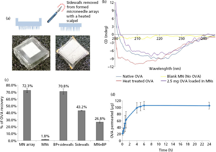
Methods: The focus of this study was exploitation of dissolving MN array devices fabricated from 20% w/w poly(methyl vinyl ether/maleic acid) using a micromoulding technique, for the facilitated delivery of a model antigen, ovalbumin (OVA).

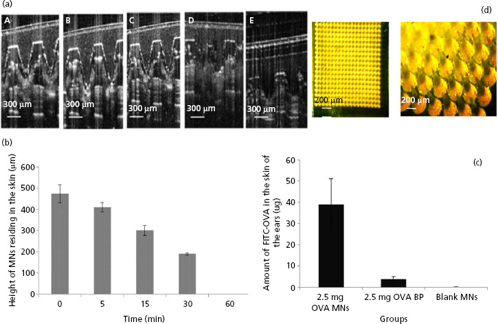
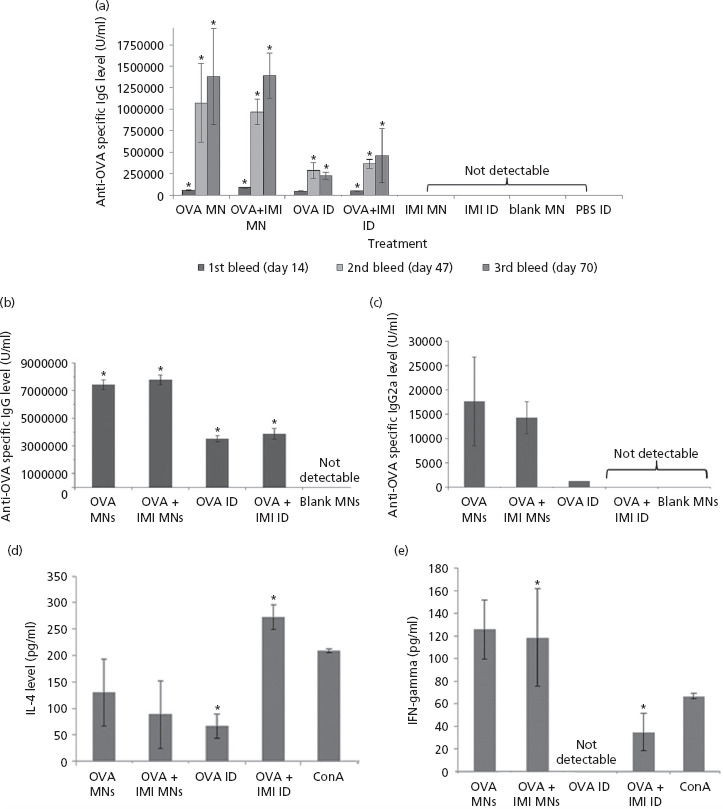
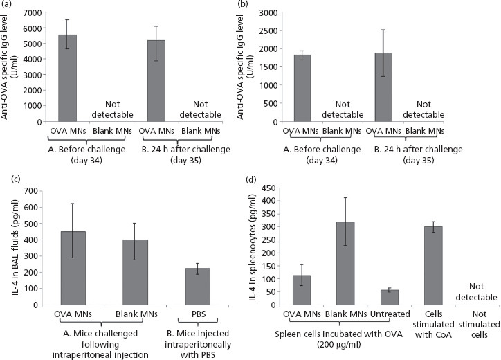
Key findings: A series of in-vitro and in-vivo experiments were designed to demonstrate that MN arrays loaded with OVA penetrated the stratum corneum and delivered their payload systemically. The latter was evidenced by the activation of both humoral and cellular inflammatory responses in mice, indicated by the production of immunoglobulins (IgG, IgG1, IgG2a) and inflammatory cytokines, specifically interferon-gamma and interleukin-4. Importantly, the structural integrity of the OVA following incorporation into the MN arrays was maintained.

Conclusion: While enhanced manufacturing strategies are required to improve delivery efficiency and reduce waste, dissolving MN are a promising candidate for 'reduced-risk' vaccination and protein delivery strategies.

Keywords: laser-engineering; microneedles; protein delivery; vaccination.


Figures

Figure 1
Figure 1
(a) Digital photograph image of a microneedle (MN) array with sidewalls (i) and (ii) after removal of the sidewalls using a hot scalpel blade. (b) Circular dichroism (CD) spectra of ovalbumin (OVA). OVA which had been encapsulated into and released from MN prepared from 20% w/v poly(methyl vinyl ether/maleic acid) (PMVE/MA) at loading of 2.5 mg/array (violet) and CD spectra of OVA standard solution (dark blue), heat-treated OVA solution (red) and non-OVA loaded MN (control, blank MN) (yellow) are presented here. (c) Determination of the loading capacity of the MN device and distribution of OVA within the MN array. MN arrays were loaded with OVA at a concentration of 2.5 mg OVA per device. The content of OVA was measured in either the whole MN array (MN array) or within isolated parts of the MN device, that is, the needles of the array only (MNs), the baseplate of the array including the sidewalls (BP + sidewalls), the sidewalls only (sidewalls) and the MN array including the baseplate but lacking the sidewalls (MN + BP). The results are expressed as the % mean ± SD, (n = 5 MN arrays) of the nominal OVA content recovered from the MN array. (d) Cumulative amount (μg) of OVA which permeated across dermatomed neonatal porcine skin after release from PMVE/MA MN arrays loaded with OVA at a concentration of 2.5 mg per array (means ± SD, n = 7).
Figure 2
Figure 2
(a) Optical coherence tomography (OCT) real-time in-vivo visualisation of microneedle (MN), fabricated from 20% w/v poly(methyl vinyl ether/maleic acid) (PMVE/MA) and loaded with 2.5 mg ovalbumin (OVA), inserted into the ear of mice for increasing application times: i.e. 1 min (A), 5 min (B), 15 min (C), 30 min (D) and (E) 60 min. (b) Dissolution profile of MN arrays (calculated as reduction of MN height) inserted into mouse skin at increasing application times (mean ± SD, n = 15). (c) The amount (μg) of fluorescently labelled OVA (FITC-OVA) quantified in the excised ear skin of mice that were treated with MN arrays loaded with 2.5 mg FITC-OVA (2.5 mg OVA MNs); baseplates (arrays lacking needles) loaded with 2.5 mg FITC-OVA (2.5 mg OVA BP) or blank MN arrays containing no FITC-OVA (blank MNs). Data are presented as mean ± standard deviation (SD), n = 4. (d) A representative light micrograph of a portion of a MN array prepared from aqueous blends of 20% w/v PMVE/MA and loaded with 2.5 mg of FITC-OVA. The scale bar represents 200 μm.
Figure 3
Figure 3
(a) Total anti-ovalbumin (OVA) serum IgG levels after immunisation using either microneedle (MN) or conventional intradermal administration (ID). Mice were treated with OVA-loaded MN, with or without IMI (OVA MN and OVA + IMI), IMI plus MN (IMI MN) or blank MN (blank MN). Mice treated by conventional ID injections received OVA (OVA ID), OVA plus IMI (OVA + IMI ID), IMI (IMI ID) and PBS (PBS ID). Animals were bled on days 14, 47 and 70. Data are presented as mean ± standard deviation (SD), n = 4. (For reference to the statistical significances, please refer to the main text.) Humoral and cellular immune responses elicited in mice following immunisation via MN or ID was determined in sera (b, c) or in cultured splenocytes (d, e). The titres of total anti-OVA IgG subclasses (b, c), as well as IL-4 (d) and IFN- γ (e) cytokines are presented. Mice were treated with OVA-loaded MN, with or without IMI (OVA MN and OVA + IMI), IMI plus MN (IMI MN) or blank MN (blank MN). Mice treated by ID were injected with solutions of OVA (OVA ID) or OVA plus IMI (OVA IMI). Data are presented as means ± SD. In the case of IgG1, n = 4; IgG2a, n = 2; IFN- γ, n = 2; IL-4, n = 4.
Figure 4
Figure 4
Levels of anti- ovalbumin (OVA)-specific total IgG (a) and IgG1 (b) of C57BL/6 mice before and after intraperitoneal challenge with OVA. Mice were immunised twice (at day 0 and day 14) with microneedle (MN) arrays loaded with 2.5 mg OVA/array or MN arrays devoid of OVA (blank MNs) as a control group. At day 34 before challenge, mice were bled to determine the levels of antibodies in their systems. Mice were then challenged intraperitoneal with 2 mg OVA in a volume of 100 μl phosphate-buffered saline (PBS) and blood samples were collected 24 h after challenge to monitor the levels of antibodies produced in response to the challenge. Brace A indicates the levels pre-challenge while Brace B indicates the total IgG levels and the IgG1 levels post-challenge. Data are presented as mean ± SD, n = 3. Detection of the Th2 cytokine, IL-4, in BAL fluids (c) and in cultured splenocytes (d). C57BL/6 mice were immunised twice with OVA loaded MN arrays at 2.5 mg/array, then challenged intraperitoneal with OVA (2 mg in 100 μl PBS). The control group consisted of mice treated with control MN arrays (blank MNs) before challenge. A second control group were injected with 100 μl PBS (PBS) only. Data are represented as mean ± standard deviation (SD), n = 3.

References

    1. Donnelly RF, et al. Microneedle-Mediated Transdermal and Intradermal Drug Delivery. New Jersey: John Wiley & Sons, 2012.
    1. Ito Y, et al. Transdermal insulin application system with dissolving microneedles. Diabetes Technol Ther 2012; 14: 891–899. - PubMed
    1. Liu S, et al. The development and characteristics of novel microneedle arrays fabricated from hyaluronic acid, and their application in the transdermal delivery of insulin. J Control Release 2012; 161: 933–941. - PubMed
    1. Donnelly RF, et al. Processing difficulties and instability of carbohydrate microneedle arrays. Drug Dev Ind Pharm 2009; 35: 1242–1254. - PMC - PubMed
    1. Lee JW, et al. Dissolving microneedles for transdermal drug delivery. Biomaterials 2008; 29: 2113–2124. - PMC - PubMed

Publication types