. 2014 Jul;142(3):396-405.
doi: 10.1111/imm.12264.

Melanoma cell lysate induces CCR7 expression and in vivo migration to draining lymph nodes of therapeutic human dendritic cells

Affiliations

Melanoma cell lysate induces CCR7 expression and in vivo migration to draining lymph nodes of therapeutic human dendritic cells

Fermín E González et al. Immunology. 2014 Jul.

Abstract

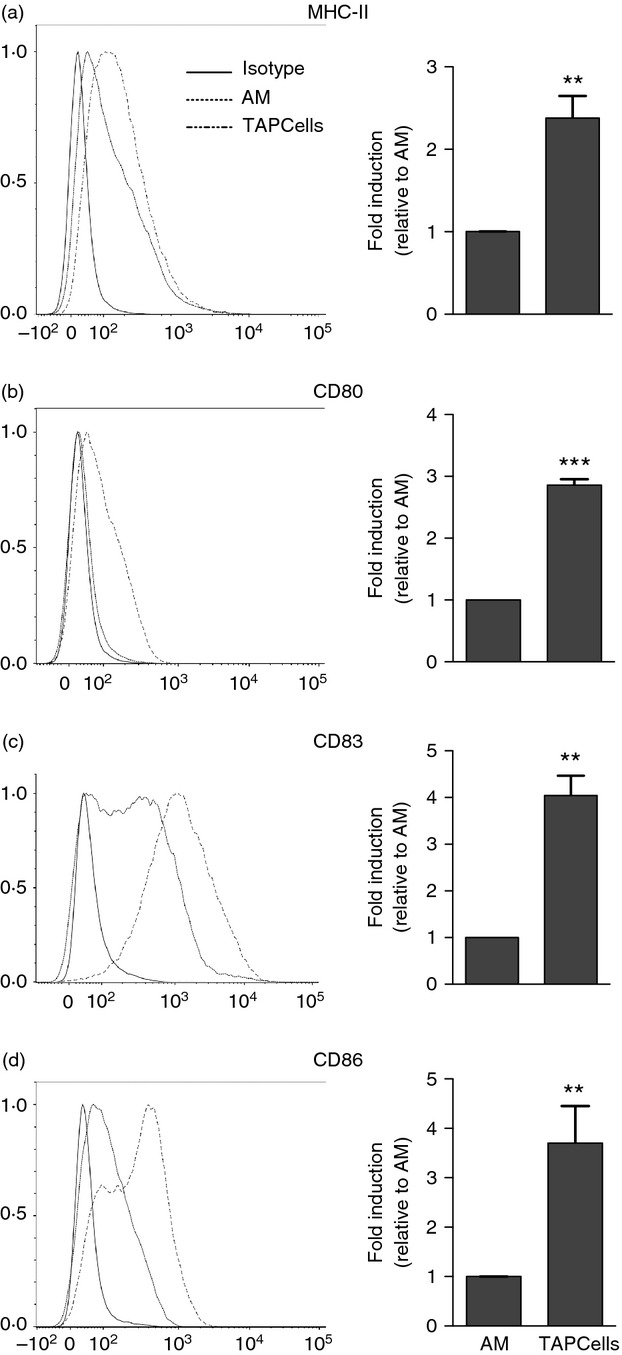
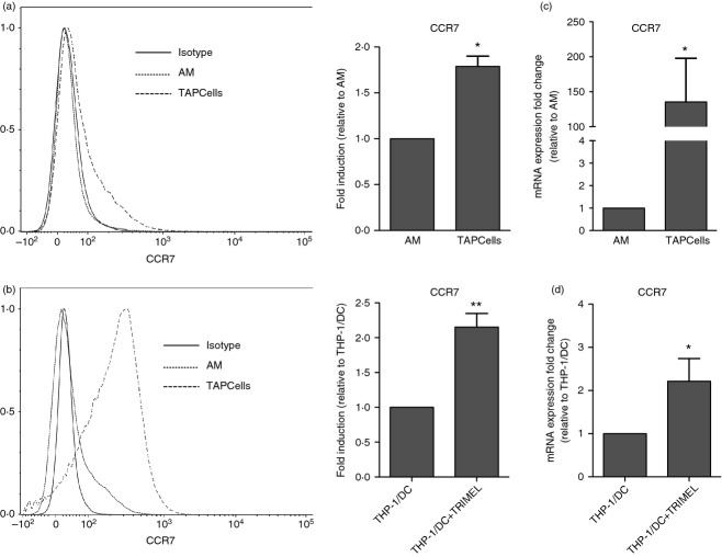
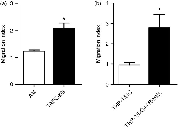
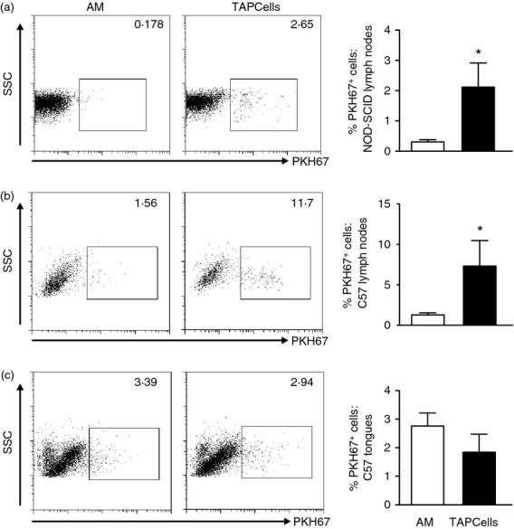
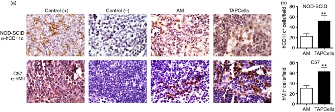
We have previously reported a novel method for the production of tumour-antigen-presenting cells (referred to as TAPCells) that are currently being used in cancer therapy, using an allogeneic melanoma-derived cell lysate (referred to as TRIMEL) as an antigen provider and activation factor. It was recently demonstrated that TAPCell-based immunotherapy induces T-cell-mediated immune responses resulting in improved long-term survival of stage IV melanoma patients. Clinically, dendritic cell (DC) migration from injected sites to lymph nodes is an important requirement for an effective anti-tumour immunization. This mobilization of DCs is mainly driven by the C-C chemokine receptor type 7 (CCR7), which is up-regulated on mature DCs. Using flow cytometry and immunohistochemistry, we investigated if TRIMEL was capable of inducing the expression of the CCR7 on TAPCells and enhancing their migration in vitro, as well as their in vivo relocation to lymph nodes in an ectopic xenograft animal model. Our results confirmed that TRIMEL induces a phenotypic maturation and increases the expression of surface CCR7 on melanoma patient-derived DCs, and also on the monocytic/macrophage cell line THP-1. Moreover, in vitro assays showed that TRIMEL-stimulated DCs and THP-1 cells were capable of migrating specifically in the presence of the CCR7 ligand CCL19. Finally, we demonstrated that TAPCells could migrate in vivo from the injection site into the draining lymph nodes. This work contributes to an increased understanding of the biology of DCs produced ex vivo allowing the design of new strategies for effective DC-based vaccines for treating aggressive melanomas.

Keywords: CCR7; dendritic cells; immunotherapy; melanoma; migration; tumour lysates.


Figures

Figure 1
Figure 1
TRIMEL induces a mature dendritic cell (DC) phenotype in tumour-antigen-presenting cells (TAPCells). Representative histograms and statistical quantifications comparing the expression of (a) MHC-II, (b) CD80, (c) CD83 and (d) CD86 between activated monocytes (AMs) and TAPCells. The quantification of the expression of the maturation marker considered the mean fluorescence intensity of TAPCells in relation to AMs. The expression of surface markers on AMs and TAPCells was assessed by flow cytometry (CD11c+ cells were gated). Data in the bars represent at least four independent experiments with PBMC derived from different melanoma patients. Bars indicate SEM; **P < 0·01; and ***P < 0·001.
Figure 2
Figure 2
TRIMEL induces the expression of the chemokine receptor CCR7 on tumour-antigen-presenting cells (TAPCells) and THP-1 cells. Representative histograms of CCR7 surface expression on (a) activated monocytes (AMs) and TAPCells after 24 hr of stimulation with TRIMEL (100 μg/ml) or medium, and (b) on THP-1 cells previously stimulated with granulocyte–macrophage colony-stimulating factor and interleukin-4 for 5 days and additionally stimulated or non-stimulated with TRIMEL (100 μg/ml) for 24 hr [THP-1/dendritic cell (DC) + TRIMEL and THP-1/DC, respectively]. Quantification of the CCR7 mRNA expression (increasing in mean fluorescence intensity value) after TRIMEL stimulation in (c) TAPCells in relation to AMs and in (d) THP-1/DC like cells (THP-1/DC + TRIMEL cells) or controls (THP-1/DC cells). Cell surface protein expression on AMs and TAPCells was evaluated with flow cytometry of CD11c-positive cells. Data for flow cytometry and mRNA quantification were obtained from at least three independent experiments using peripheral blood mononuclear cells from different melanoma patients. Bars indicate SEM; *P < 0·05; and **P < 0·01.
Figure 3
Figure 3
TRIMEL induces the in vitro transmigration of tumour-antigen-presenting cells (TAPCells) and THP-1 cells. Evaluation of the CCL19-dependent, transmigratory activity of TRIMEL-activated cells. (a) Transmigration of monocyte-derived antigen-presenting cells obtained from different melanoma patients in the presence (TAPCells) or absence (activated monocytes; AMs) of TRIMEL. (b) The transmigration of THP-1/dendritic cell-like cells with or without TRIMEL stimulation. Transmigration capability of different cells was tested with a chemotaxis assay in a 48-well transwell chamber. Lower wells were loaded with CCL19 (10 ng/μl) or alternatively with only the medium, and the upper wells were loaded with 50 000 cells/well per condition and incubated for 4 hr at 37°. Data were obtained from at least three independent experiments. The migration index values correspond to the difference in transmigration between cells in the presence or absence of CCL19. Bars indicate SEM; *P < 0·05.
Figure 4
Figure 4
Tumour-antigen-presenting cells (TAPCells) can migrate in vivo to draining lymph nodes in an ectopic xenograft mouse model. NOD-SCID and natural killer cell-depleted C57BL/6 mice, as described in the Material and methods, were injected submucosally with PKH67-stained activated monocytes (AMs) or TAPCells (2 × 10⁶ cells/mouse) in the middle of the tongue. The animals were killed after 24 hr and cervical lymph nodes (NOD-SCID and C57BL/6 mice) and tongues (C57BL/6 mice) were recovered and processed. Representative dot plots show PKH67+ human cell populations (left panels) and graphics show quantification of the gated positive cells for each strain and organ (right panel). (a) PKH67+ cells in NOD-SCID lymph nodes (five per mouse). (b) PKH67+ cells in C57BL/6 mice lymph nodes (five per mouse). (c) PKH67+ cells in C57BL/6 tongues (one per mouse). Data obtained from three independent experiments. Bars indicate SEM; *P < 0·05.
Figure 5
Figure 5
Immunohistochemical detection of human cells in lymph nodes from NOD-SCID and natural killer (NK) cell-depleted C57BL/6 mice injected with tumour-antigen-presenting cells (TAPCells). (a) Cervical lymph nodes derived from activated monocytes (AMs), TAPCells and PBS-injected NOD-SCID mice (top row) and NK cell-depleted C57BL/6 mice (bottom row) were stained with an anti-human CD11c monoclonal antibody (NOD-SCID mice) and an anti-mitochondria polyclonal antibody (C57BL/6 mice) and then analysed by immunohistochemistry. (a) Stained tissues. (b) Positive cell quantification in NOD-SCID injected mice (upper figure) and C57BL/6 injected mice (lower figure). Human tonsils were used as positive controls [Control (+)] and spleens (α-hCD11c) and lymph nodes (α-hMit) from PBS-injected animals were used as negative controls [Control (−)]. Eight representative photographic fields for each strain and condition were counted using ImageJ software (NIH, Bethesda, MD). Bars indicate SEM; **P < 0·01.

Similar articles

Cited by

References

    1. Palucka K, Banchereau J. Cancer immunotherapy via dendritic cells. Nat Rev Cancer. 2012;12:265–77. - PMC - PubMed
    1. Schmitt N, Morita R, Bourdery L, Bentebibel SE, Zurawski SM, Banchereau J, Ueno H. Human dendritic cells induce the differentiation of interleukin-21-producing T follicular helper-like cells through interleukin-12. Immunity. 2009;31:158–69. - PMC - PubMed
    1. Steinman RM, Idoyaga J. Features of the dendritic cell lineage. Immunol Rev. 2010;234:5–17. - PubMed
    1. Cheong C, Matos I, Choi JH, et al. Microbial stimulation fully differentiates monocytes to DC-SIGN/CD209+ dendritic cells for immune T cell areas. Cell. 2010;143:416–29. - PMC - PubMed
    1. Hart DN. Dendritic cells: unique leukocyte populations which control the primary immune response. Blood. 1997;90:3245–87. - PubMed

Publication types

MeSH terms

LinkOut - more resources