. 2014 Mar 28:14:63.
doi: 10.1186/1471-2148-14-63.

The ubiquilin gene family: evolutionary patterns and functional insights

Affiliations

The ubiquilin gene family: evolutionary patterns and functional insights

Ignacio Marín. BMC Evol Biol. .

Abstract

Background: Ubiquilins are proteins that function as ubiquitin receptors in eukaryotes. Mutations in two ubiquilin-encoding genes have been linked to the genesis of neurodegenerative diseases. However, ubiquilin functions are still poorly understood.

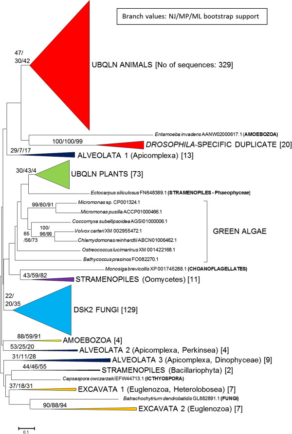
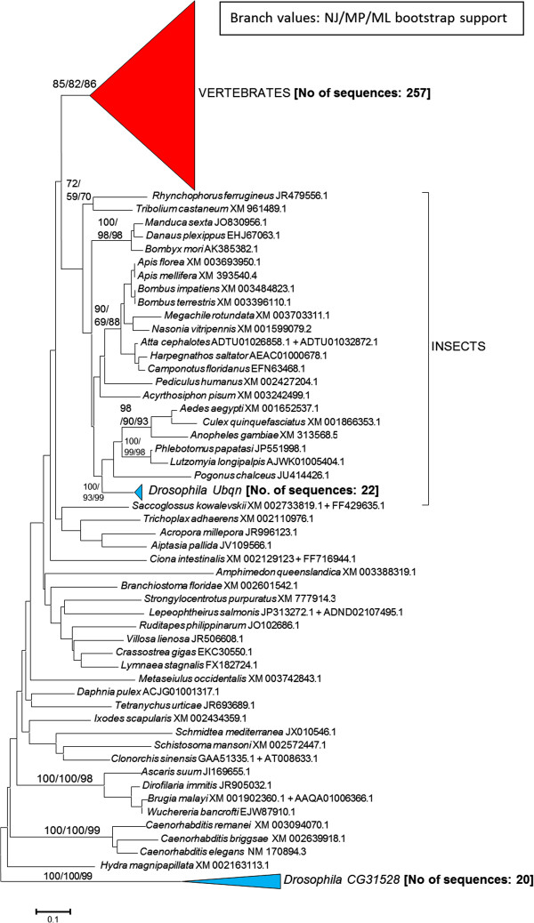
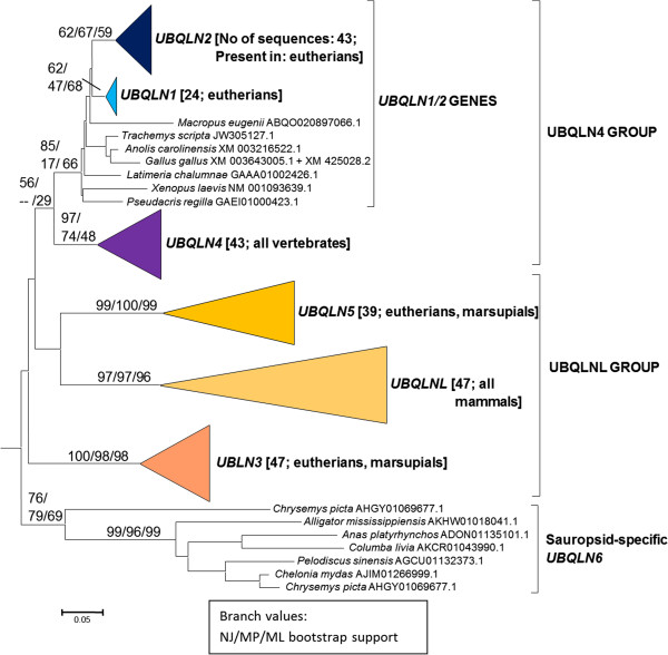
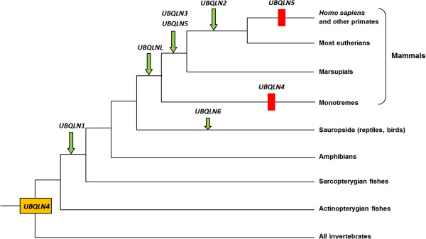
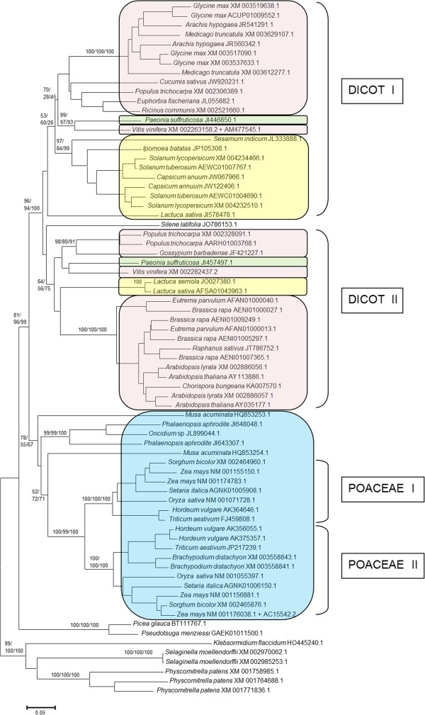
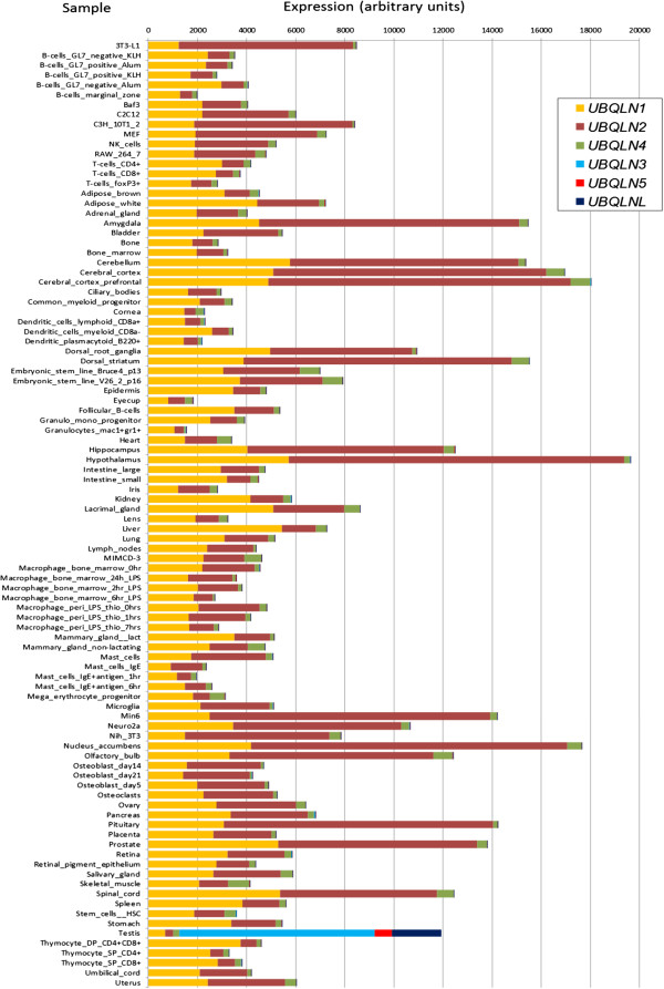
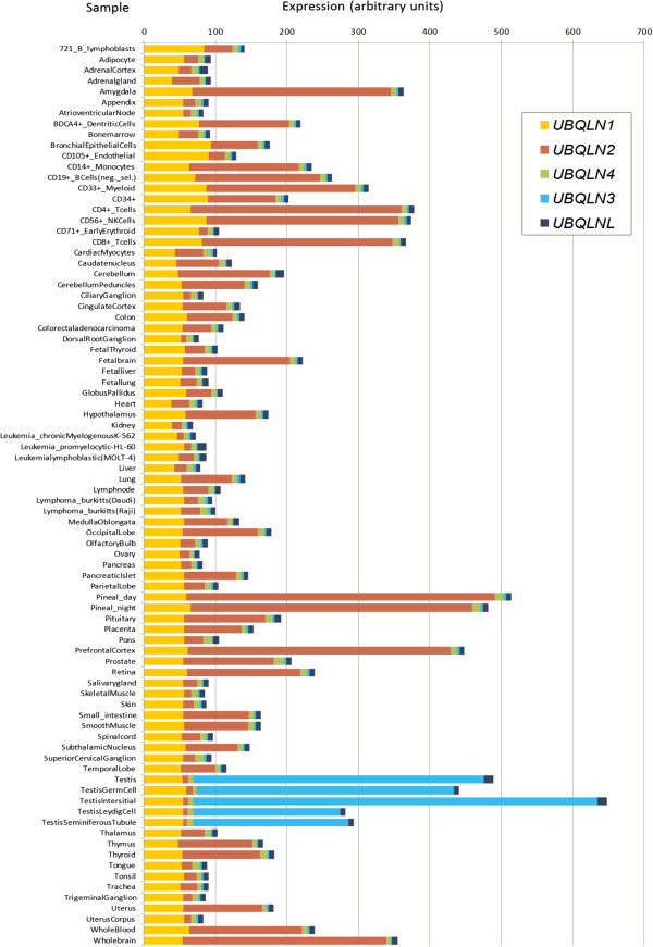
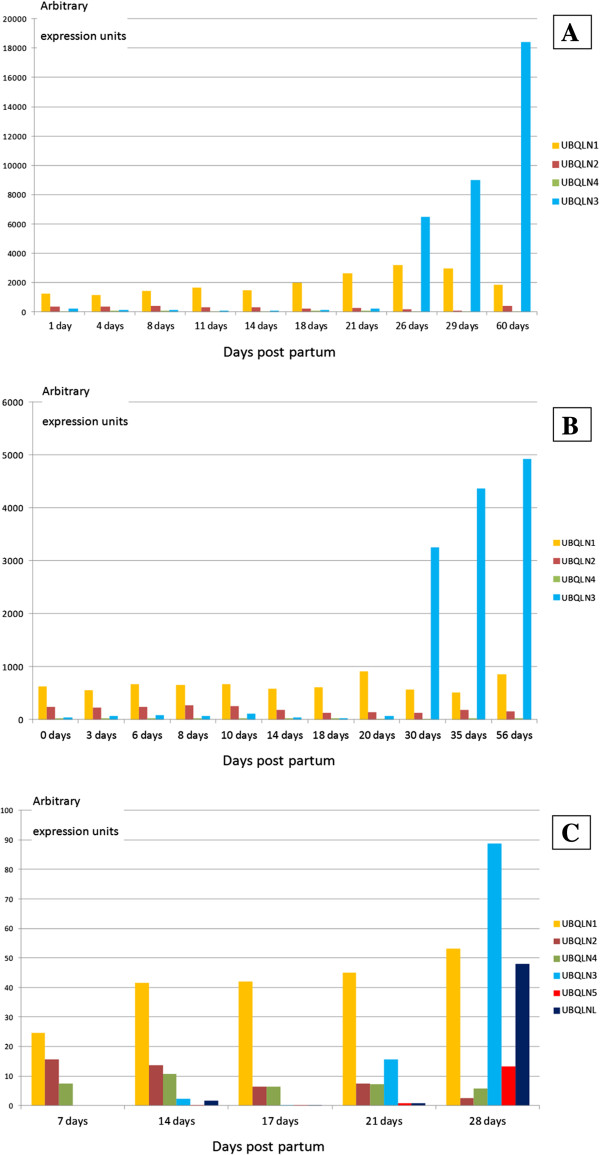
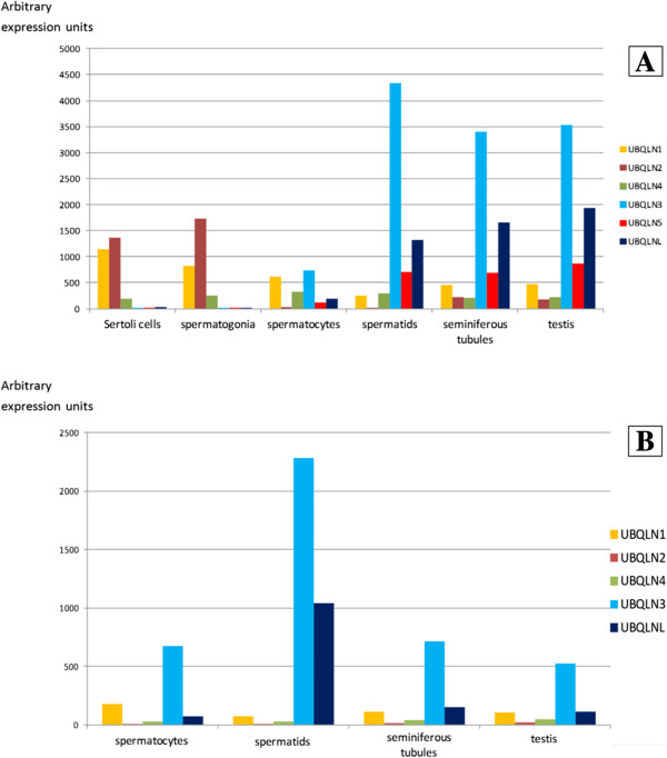
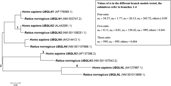
Results: In this study, evolutionary and functional data are combined to determine the origin and diversification of the ubiquilin gene family and to characterize novel potential roles of ubiquilins in mammalian species, including humans. The analysis of more than six hundred sequences allowed characterizing ubiquilin diversity in all the main eukaryotic groups. Many organisms (e. g. fungi, many animals) have single ubiquilin genes, but duplications in animal, plant, alveolate and excavate species are described. Seven different ubiquilins have been detected in vertebrates. Two of them, here called UBQLN5 and UBQLN6, had not been hitherto described. Significantly, marsupial and eutherian mammals have the most complex ubiquilin gene families, composed of up to 6 genes. This exceptional mammalian-specific expansion is the result of the recent emergence of four new genes, three of them (UBQLN3, UBQLN5 and UBQLNL) with precise testis-specific expression patterns that indicate roles in the postmeiotic stages of spermatogenesis. A gene with related features has independently arisen in species of the Drosophila genus. Positive selection acting on some mammalian ubiquilins has been detected.

Conclusions: The ubiquilin gene family is highly conserved in eukaryotes. The infrequent lineage-specific amplifications observed may be linked to the emergence of novel functions in particular tissues.


Figures

Figure 1
Figure 1
Summary tree indicating the distribution of ubiquilins in eukaryotes. This is the neighbor-joining (NJ) tree, but the maximum-parsimony (MP) and maximum-likelihood (ML) dendrograms were similar enough as to allow all the results to be drawn together. The tree is drawn to scale, with branch lengths in the same units as those of the evolutionary distances used to infer the phylogenetic tree. A scale bar is shown below the tree. The numbers in the branches indicate bootstrap supports (in percentages) for the three methods of phylogenetic reconstruction (as follows: NJ/MP/ML). For simplicity, only the most relevant boostrap values are indicated. In brackets, the number of sequences within each group. These groups were made by putting together all the sequences that belonged to related species, in order to deduce the minimum number of groups for each eukaryotic class. Thus, all plant can be put in a single group, animal sequences can be classified into two groups, etc. For alveolates, stramenopiles and excavates, the main phyla that can be found within each group are indicated (in parentheses). A more detailed view of this tree can be found in Additional file 2.
Figure 2
Figure 2
Animal ubiquilins. Bootstrap support (NJ/MP/ML) and number of sequences per group (in brackets) as in Figure 1. Bootstrap values are shown only for internal branches with consistent support. Scale bar as in Figure 1.
Figure 3
Figure 3
Classification of vertebrate ubiquilins. This figure corresponds to the subtree indicated as a triangle labelled “Vertebrates” in Figure 2, which has been expanded here, to show all the main types of ubiquilins. In brackets, the number of sequences in each group and the different types of vertebrates that have each ubiquilin gene. Bootstrap support (NJ/MP/ML) as in previous figures. Scale bar as in Figure 1.
Figure 4
Figure 4
Most parsimonious hypothesis to explain the diversification of the ubiquilin genes in animals. Arrow: origin of a new gene. Rectangle: gene loss. A single gene, which would be orthologous to vertebrate UBQLN4 was present when animals emerged.
Figure 5
Figure 5
Plant ubiquilins. Two ancient duplications are indicated as Dicot I/II and Poaceae I/II (see text). Colors are applied to some main angiosperm classes (pink: rosid dicots; green: saxifragal dicots; yellow: asterid dicots; blue: monocots).
Figure 6
Figure 6
Ubiquilin gene expression in different organs, tissues or cell types of wild-type mice. Data, in arbitrary expression units, obtained from the BioGPS database [57]. The key names of the samples used in BioGPS are indicated. Although most names are autoexplicative, additional details of these samples can be found at http://www.biogps.org. Notice the qualitative difference found for testis respect to the other samples. In testis, most of the expression observed derives from UBQLNL group genes (UBQLN3, UBQLN5, UBQLNL) which are not expressed, or at very low levels, in all other tissues.
Figure 7
Figure 7
Expression of ubiquilins in normal human tissues, organs or cell types. Data also from BioGPS. Again, all testis-derived samples show a very high level of expression of UBQLNL group genes (UBQLN3 and UBQLNL; UBQLN5 is absent in humans).
Figure 8
Figure 8
Expression of ubiquilin genes in mouse testis, at different days post partum. Data, in arbitrary expression units, derived from Schultz et al.[58](Panel A), Shima et al.[59](Panel B) and Laiho et al.[60] (Panel C).
Figure 9
Figure 9
Expression of ubiquilin genes in particular testis samples in mouse (Panel A) and human (B). Data from Chalmel et al.[62], again in arbitrary expression units.
Figure 10
Figure 10
Dendrogram corresponding to the five human ubiquilin genes and their orthologs in rat. In parenthesis, accession numbers of the sequences. The tree shown here was obtained with the NJ method, but ML and MP analyses generated exactly the same topology. Branches 1–4 are those in which potential positive selection (ω > 1) was detected in PAML analyses. The observed ω values in the three different branch models [63] tested are also indicated. See text for details.

References

    1. Vallen EA, Ho W, Winey M, Rose MD. Genetic interactions between CDC31 and KAR1, two genes required for duplication of the microtubule organizing center in Saccharomyces cerevisiae. Genetics. 1994;137:407–422. - PMC - PubMed
    1. Biggins S, Ivanovska I, Rose MD. Yeast ubiquitin-like genes are involved in duplication of the microtubule organizing center. J Cell Biol. 1996;133:1331–1346. doi: 10.1083/jcb.133.6.1331. - DOI - PMC - PubMed
    1. Funakoshi M, Geley S, Hunt T, Nishimoto T, Kobayashi H. Identification of XDRP1; a Xenopus protein related to yeast Dsk2p binds to the N-terminus of cyclin A and inhibits its degradation. EMBO J. 1999;18:5009–5018. doi: 10.1093/emboj/18.18.5009. - DOI - PMC - PubMed
    1. Wu AL, Wang J, Zheleznyak A, Brown EJ. Ubiquitin-related proteins regulate interaction of vimentin intermediate filaments with the plasma membrane. Mol Cell. 1999;4:619–625. doi: 10.1016/S1097-2765(00)80212-9. - DOI - PubMed
    1. Kaye FJ, Modi S, Ivanovska I, Koonin EV, Thress K, Kubo A, Kornbluth S, Rose MD. A family of ubiquitin-like proteins binds the ATPase domain of Hsp70-like Stch. FEBS Lett. 2000;467:348–355. doi: 10.1016/S0014-5793(00)01135-2. - DOI - PubMed

Publication types

Substances