Case Reports
. 2014 Apr;64(2):148-56.

Endometrial decidualization and deciduosis in aged rhesus macaques (Macaca mulatta)

Affiliations
Case Reports

Endometrial decidualization and deciduosis in aged rhesus macaques (Macaca mulatta)

Amanda P Beck et al. Comp Med. 2014 Apr.

Abstract

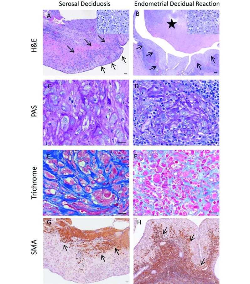
Superficial decidualization of the endometrial stroma is an essential feature of the implantation stage of pregnancy in rhesus macaques and other primates. Decidualization involves proliferation of the endometrial stromal cells, with differentiation into morphologically distinct decidual cells. Previous reports involving nonpregnant rhesus monkeys have described local- ized and widespread endometrial decidualization in response to administration of progesterone and synthetic progestogens. Ectopic decidua or 'deciduosis' describes the condition in which groups of decidual cells are located outside of the endometrium, most often in the ovaries, uterus and cervix but also in various other organs. In humans, most cases of deciduosis are associated with normal pregnancy, and ectopic decidua can be found in the ovary in nearly all term pregnancies. Here we describe pronounced endometrial decidualization in 2 rhesus macaques. Both macaques had been treated long-term with medroxyprogesterone acetate for presumed endometriosis, which was confirmed in one of the macaques at postmortem examination. In one animal, florid extrauterine and peritoneal serosal decidualization was admixed multifocally with carcinomatosis from a primary colonic adenocarcinoma. Cells constituting endometrial and serosal decidualization reactions were immunopositive for the stromal markers CD10, collagen IV, smooth muscle actin, and vimentin and immunonegative for cytokeratin. In contrast, carcinomatous foci were cytokeratin-positive. To our knowledge, this report describes the first cases of serosal peritoneal decidualization in rhesus macaques. The concurrent presentation of serosal peritoneal decidualization with carcinomatosis is unique.


Figures

Figure 1.
Figure 1.
(A) Case 1. Within the caudal peritoneal cavity, the abdominal serosa contains extensive multifocal to coalescing, cream-colored, nodular to linear, flat lesions that enclosed the uterus, ovaries, bladder (white asterisk) and distal rectum (black asterisk). (B) Case 1. The uterus contains a focal intraluminal mural mass (arrow) accompanied by endometrial hemorrhage and thickening. (C) Case 2. The uterine endometrium is diffusely and markedly thickened with an irregular yellow to brown surface covered by small amount of blood and yellow–brown fluid.
Figure 2.
Figure 2.
Histopathology and immunohistochemical features of (A, C, E, G; same site) the colonic adenocarcinoma and (B, D, F, H; same site) a metastatic focus of colonic adenocarcinoma from case 1. (A) The primary tumor is an invasive, transmural colonic adenocarcinoma, which extends through the basement membrane (arrow). Hematoxylin and eosin stain; scale bar, 200 μm. (B) A metastatic focus of colonic adenocarcinoma is present on the serosal surface of the large intestine (arrows), distinct from the primary tumor. Hematoxylin and eosin stain; scale bar, 200 μm. (C) Neoplastic glands of the primary colonic carcinoma are strongly positive for cytokeratin AE1/AE3 immunostain. The arrow indicates the comparable site in panel A. Scale bar, 200 μm. (D) Similarly, neoplastic glands of the focus of serosal carcinomatosis are strongly positive for cytokeratin AE1/AE3 immunostain. Scale bar, 50 μm. (E) Within the primary colonic carcinoma and (F) metastatic focus of carcinomatosis, both neoplastic epithelium and stroma are negative for CD10 immunostain. Scale bar, 50 μm. Collagen IV staining is immunopositive in stroma but not epithelial cells within the (G) primary carcinoma and (H) focus of serosal carcinomatosis. Scale bar, 50 μm.
Figure 3.
Figure 3.
Histopathology and immunohistochemical features of the (A, C, E, G; same site) serosal and (B, D, F, H; same site) endometrial decidual reaction from case 1. (A) The abdominal serosa contains extensive nodular lesions (arrows) with morphology distinct from those of the carcinomatous foci noted above. Inset, Nodules consist of nests of spindloid to large polygonal cells with abundant, often granular cytoplasm, and variably sized nuclei. Cells are embedded in varying amounts of collagenous or eosinophilic stroma and resemble those within the uterine endometrium. Hematoxylin and eosin stain; scale bar: 50 μm (main image), 20 μm (inset). (B) The uterine displays a band of extensive endometrial decidualization (arrows), continuous with a similarly decidualized partly necrotic endometrial mass (asterisk). Inset, Decidual cells are round to polygonal, with variably distinct cell borders, moderate amounts of pale eosinophilic highly vacuolated or granular cytoplasm and centrally placed round to oval nuclei with finely stippled chromatin and a single basophilic nucleolus. Hematoxylin and eosin stain; scale bar: 200 μm (main image), 20 μm (inset). The stroma of both the (C) endometrial decidual reaction and (D) uterine serosal decidual reaction are strongly periodic acid–Schiff-positive. Scale bar, 20 μm. (E, F). The stroma of both the (E) endometrial and (F) uterine serosal decidual reactions are strongly positive with Masson trichrome stain. Scale bar, 20 μm. Decidual cells were immunopositive for smooth muscle actin (SMA) in both the (G) endometrial and (H) serosal decidual foci. Intensity of immunostaining for SMA decreased as decidual cells evolved from spindloid to polygonal morphology (G, H: arrows). Scale bar, 50 μm.
Figure 4.
Figure 4.
Additional immunohistochemical features of the (A, C, E) serosal and (B, D, F) endometrial decidual reactions from case 1. (A, B) The serosal and endometrial decidual reactions are immunonegative for AE1/AE3. Scale bar, 50 μm. Decidualized stromal cells within the serosal and endometrial decidual reactions displaymembranous immunoreactivity for (C, D) CD10 and (E, F) collagen IV. Scale bar, 20 μm.

References

    1. Abee CR, Mansfield K, Tardif S, Morris T, editors. 2012. . Nonhuman primates in biomedical research. Volume 2: diseases. Waltham (MA): Academic Press.
    1. Arnold DL, Nera EA, Stapley R, Tolnai G, Claman P, Hayward S, Tryphonas H, Bryce F. 1996. Prevalence of endometriosis in rhesus (Macaca mulatta) monkeys ingesting PCB (Aroclor 1254): review and evaluation. Fundam Appl Toxicol 31:42–55 - PubMed
    1. Assaf BT, Miller AD. 2012. Pleural endometriosis in an aged rhesus macaque (Macaca mulatta): a histopathologic and immunohistochemical study. Vet Pathol 49:636–641 - PMC - PubMed
    1. Benagiano G, Pera A, Primiero FM. 2000. The endometrium and hormonal contraceptives. Hum Reprod 15 Suppl 1:101–118 - PubMed
    1. Bertens APMG, Helmond FA, Hein PR. 1982. Endometriosis in rhesus monkeys. Lab Anim 16:281–284 - PubMed

Publication types

LinkOut - more resources