. 2014 Apr:16:44-53.
doi: 10.1016/j.dnarep.2014.01.015. Epub 2014 Feb 27.

Overexpression of DNA ligase III in mitochondria protects cells against oxidative stress and improves mitochondrial DNA base excision repair

Affiliations

Overexpression of DNA ligase III in mitochondria protects cells against oxidative stress and improves mitochondrial DNA base excision repair

Mansour Akbari et al. DNA Repair (Amst). 2014 Apr.

Abstract

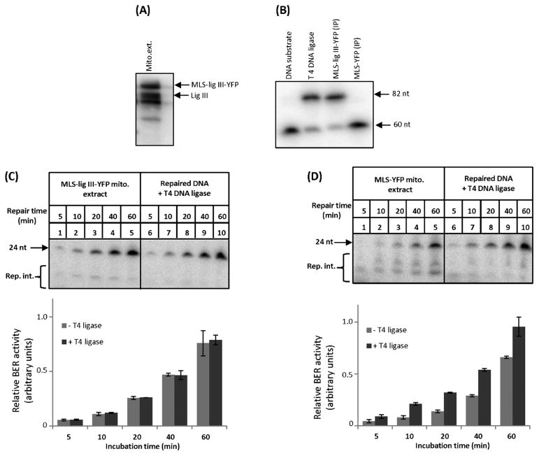
Base excision repair (BER) is the most prominent DNA repair pathway in human mitochondria. BER also results in a temporary generation of AP-sites, single-strand breaks and nucleotide gaps. Thus, incomplete BER can result in the generation of DNA repair intermediates that can disrupt mitochondrial DNA replication and transcription and generate mutations. We carried out BER analysis in highly purified mitochondrial extracts from human cell lines U2OS and HeLa, and mouse brain using a circular DNA substrate containing a lesion at a specific position. We found that DNA ligation is significantly slower than the preceding mitochondrial BER steps. Overexpression of DNA ligase III in mitochondria improved the rate of overall BER, increased cell survival after menadione induced oxidative stress and reduced autophagy following the inhibition of the mitochondrial electron transport chain complex I by rotenone. Our results suggest that the amount of DNA ligase III in mitochondria may be critical for cell survival following prolonged oxidative stress, and demonstrate a functional link between mitochondrial DNA damage and repair, cell survival upon oxidative stress, and removal of dysfunctional mitochondria by autophagy.

Keywords: Autophagy; Cell survival; Mitochondrial DNA repair intermediates; Oxidative stress.


Conflict of interest statement

statement The authors declare no conflict of interest.

Figures

Fig. 1
Fig. 1
Representative image of Western blot analysis of mitochondrial extracts prepared from proteinase K treated, purified mitochondria. 20 μg total cell extracts and 50 μg mitochondrial extracts were used in each experiment. Antibodies against TOPO IIα, Ku 86, lamin and PCNA were used to assess contamination of the mitochondrial extracts with nuclear proteins. VDAC-1 and COX IV were used as the marker of the outer- and the inner-mitochondrial membrane proteins, respectively, and Polγ and TFAM as the core mitochondrial nucleoid proteins.
Fig. 2
Fig. 2
BER analysis of mitochondrial extracts. BER analysis was carried out in duplicate using two independently prepared mitochondrial extracts from HeLa (A) and U2OS (B) cell lines. The increased level of the upper band after T4 DNA ligase treatment relative to the untreated samples indicates the presence of unligated DNA repair intermediates (rep. int.) in the sample. (C) BER analysis of mitochondrial extracts from mouse brain as per panels A and B. The bar charts show the relative intensity of the upper band (fully repaired 24 nucleotides) to the lower bands (repair intermediates) in each lane. The error bars show standard deviation of the mean. Absence of detectable repair products in G:C control DNA substrate indicates site specific repair of uracil in U:G DNA substrate.
Fig. 3
Fig. 3
Overexpression of DNA ligase III in mitochondria. U2OS cells stably expressing DNA ligase III fused with YFP and the mitochondrial localization signal of SOD2 (MLS-lig III-YFP, panels I and II) or MLS-YFP alone as control (III and IV), were established. Representative images of live MLS-lig III-YFP and MLS-YFP cells pre-incubated with MitoTracker Red showing mitochondrial localization of the fusion proteins (see merged images). Panels II and IV are high magnification, z-stacked image of the mitochondrial network in a MLS-lig III-YFP and a MLS-YFP cell, respectively.
Fig. 4
Fig. 4
Mitochondrial extracts from MLS-lig III-YFP cells show increased rate of BER compared with control cells. (A) Western blot analysis of mitochondria isolated from MLS-lig III-YFP cells using antibody against DNA ligase III. (B) To test whether MLS-lig III-YFP fusion protein is catalytically active, MLS-ligase III-YFP was immunoprecipitated (IP) from mitochondrial extracts using an antibody against GFP (also recognizes YFP). The IP was assayed for DNA ligase activity for 1 h, and DNA was separated in denaturing polyacrylamide gel. T4 DNA ligase and the IP from mitochondrial extracts of MLS-YFP cells were used as positive and negative controls, respectively. Upper band corresponds to the fully ligated 82 nucleotides substrate (82 nt), and the lower band are unligated 60 nucleotide end-labeled substrate. (C) BER analysis of mitochondrial extracts from MLS-lig III-YFP, and (D) MLS-YFP cells.
Fig. 5
Fig. 5
Overexpression of DNA ligase III in mitochondria enhances cell survival following menadione-induced oxidative stress. (A) Sensitivity of the cells to menadione was measured using a WST-1 assay after treatment for 3 h. Cells were allowed to recover for 19 h before WST-1 analysis. (B) Colony forming ability assay. Cells were treated with 10 μM menadione for 3 h. After six days, cells were fixed and stained with crystal violet and the colonies were counted. Colony forming ability of the MLS-lig III-YFP cells following menadione treatment was significantly higher than the control U2OS and MLS-YFP cells. The experiment was performed three times, and each experiment was carried out in triplicate. Data are presented as mean ±SEM. *p < 0.05 one-way analysis of variance (ANOVA) followed by Bonferroni’s post hoc test.
Fig. 6
Fig. 6
Rotenone-induced autophagy is lower in MLS-lig III-YFP cells. Western blots of MLS-YFP and MLS-lig III-YFP cells for the autophagosome marker LC3B. Cells were treated with 5 μM rotenone (rot. autophagy inducer) or mock treated with DMSO (<0.1%). The experiment was carried out in triplicate. The ratio of the lower band (LC3B II) to the upper band (LC3B I) was used to calculate the rotenone-induced autophagy activity. Data are presented as mean ± SEM. *p < 0.05 one-way ANOVA with Bonferroni’s post hoc test. Autophagy activity in MLS-lig III-YFP cells treated with rotenone was significantly lower than the control cells exposed to rotenone.

References

    1. Lauritzen KH, Moldestad O, Eide L, Carlsen H, Nesse G, Storm JF, Mansuy IM, Bergersen LH, Klungland A. Mitochondrial DNA toxicity in forebrain neurons causes apoptosis, neurodegeneration, and impaired behavior. Mol Cell Biol. 2010;30:1357–1367. - PMC - PubMed
    1. McFarland R, Taylor RW, Turnbull DM. Mitochondrial disease – its impact, etiology, and pathology. Curr Top Dev Biol. 2007;77:113–155. - PubMed
    1. Simsek D, Furda A, Gao Y, Artus J, Brunet E, Hadjantonakis AK, Van Houten B, Shuman S, McKinnon PJ, Jasin M. Crucial role for DNA ligase III in mitochondria but not in Xrcc1-dependent repair. Nature. 2011;471:245–248. - PMC - PubMed
    1. Holt IJ, Harding AE, Morgan-Hughes JA. Deletions of muscle mitochondrial DNA in patients with mitochondrial myopathies. Nature. 1988;331:717–719. - PubMed
    1. Zeviani M, Moraes CT, DiMauro S, Nakase H, Bonilla E, Schon EA, Rowland LP. Deletions of mitochondrial DNA in Kearns–Sayre syndrome. Neurology. 1988;38:1339–1346. - PubMed

Publication types

MeSH terms