. 2014 Mar 15;5(5):1290-303.
doi: 10.18632/oncotarget.1789.

Arsenic-induced sub-lethal stress reprograms human bronchial epithelial cells to CD61¯ cancer stem cells

Affiliations

Arsenic-induced sub-lethal stress reprograms human bronchial epithelial cells to CD61¯ cancer stem cells

Qingshan Chang et al. Oncotarget. .

Abstract

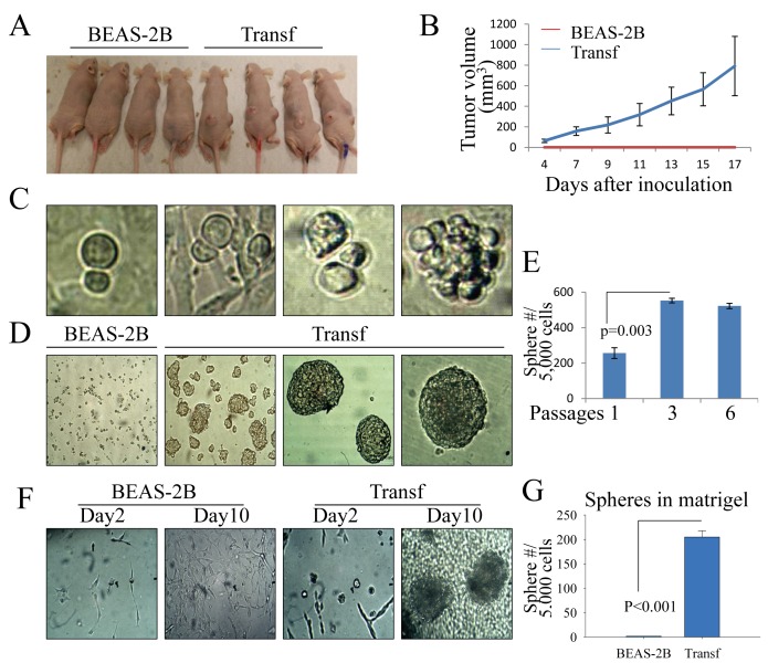
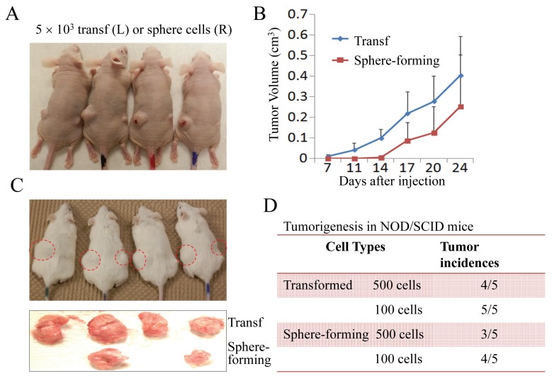
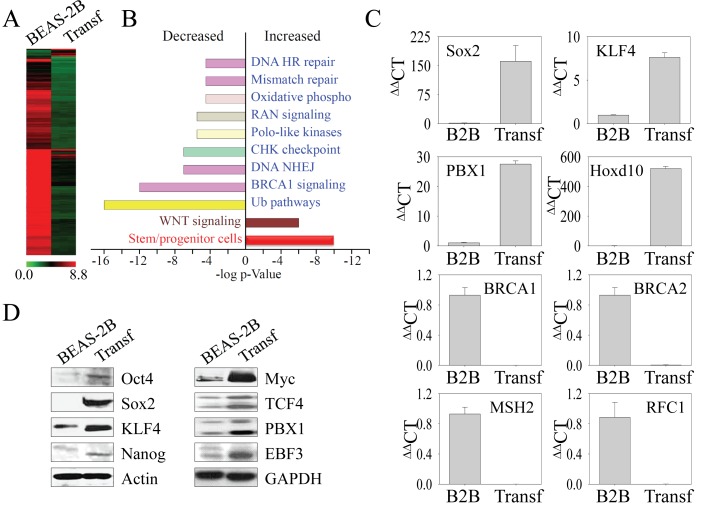
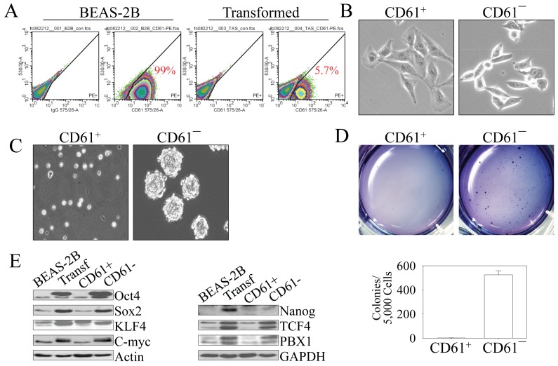
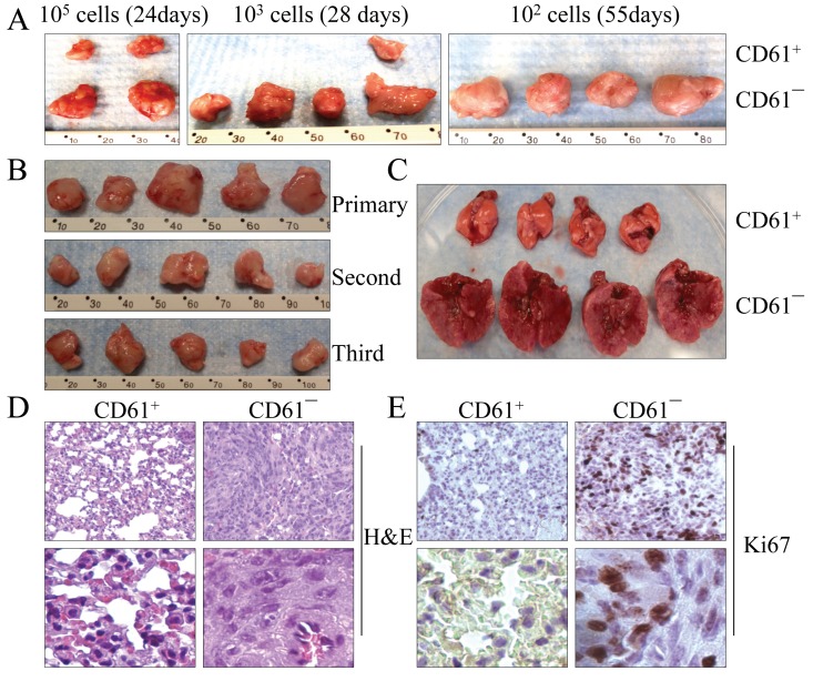
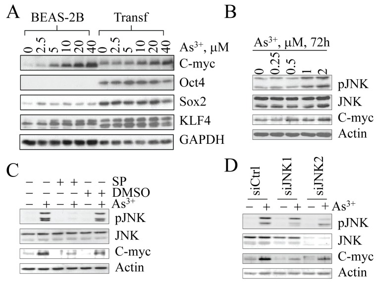
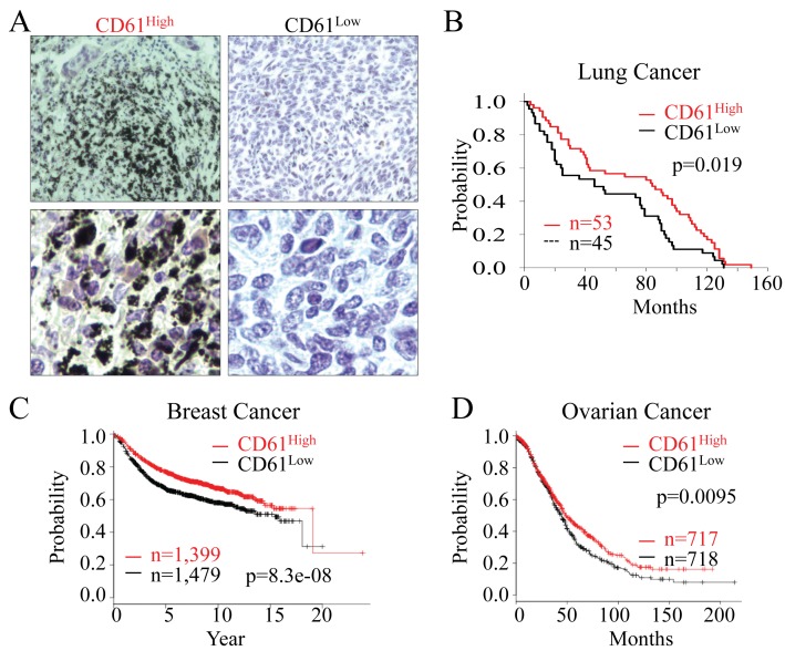
In the present report, we demonstrate that sub-lethal stress induced by consecutive exposure to 0.25 µM arsenic (As3+) for six months can trigger reprogramming of the human bronchial epithelial cell (BEAS-2B) to form cancer stem cells (CSCs) without forced introduction of the stemness transcription factors. These CSCs formed from As3+-induced sub-lethal stress featured with an increased expression of the endogenous stemness genes, including Oct4, Sox2, Klf4, Myc, and others that are associated with the pluripotency and self-renewal of the CSCs. Flow cytometry analysis indicated that 90% of the CSC cells are CD61¯, whereas 100% of the parental cells are CD61+. These CD61¯ CSCs are highly tumorigenic and metastatic to the lung in xenotransplantation tests in NOD/SCID Il2rγ-/- mice. Additional tests also revealed that the CD61¯ CSCs showed a significant decrease in the expression of the genes important for DNA repair and oxidative phosphorylation. To determine the clinical relevance of the above findings, we stratified human lung cancers based on the level of CD61 protein and found that CD61low cancer correlates with poorer survival of the patients. Such a correlation was also observed in human breast cancer and ovarian cancer. Taken together, our findings suggest that in addition to the traditional approaches of enforced introduction of the exogenous stemness circuit transcription factors, sub-lethal stress induced by consecutive low dose As3+ is also able to convert non-stem cells to the CSCs.


Figures

Figure 1
Figure 1. The transformed cells induced by As3+ have features of CSCs
(A & B) Tumorigenicity of the parental (BEAS-2B) and transformed (Transf) cells was determined by injecting 1 × 10⁶ cells subcutaneously into the flank of 6-week-old male BALB/c nude mice. The image shows tumor sizes 17 days after injection. (C) Asymmetric division of the transformed cells. (D) Tumor sphere formation assay for the parental (BEAS-2B) and transformed (Transf) cells. (E) The sphere-forming cells were enriched after the passage of tumor spheres. (F) Three-dimensional (3D) culture of the parental (BEAS-2B) and transformed (Transf) cells in a Matrigel matrix. Tumor spheres were visible for the transformed cells 10 days after initial seeding of the cells in Matrigel matrix. (G) Quantification of the tumor spheres of the parental cells and the transformed cells in 3D culture.
Figure 2
Figure 2. The transformed cells and sphere-forming cells are tumorigenic in vivo
(A) The nude mice were subcutaneously injected with 5,000 transformed cells and sphere-forming cells at the left (L) and right (R) sides, respectively. (B) Tumor volumes were measured on the indicated days. (C) Tumorigenicity assay of the transformed cells and sphere-forming cells in NOD/SCID Il2rγ−/− mice. (D) Tumor incidence rates of the transformed cells and sphere-forming cells in NOD/SCID mice after 45 days of injection with 100 or 500 cells.
Figure 3
Figure 3. The transformed cells induced by As3+ express genes for the stemness of CSCs
(A) Gene expression profiles were compared between parental (BEAS-2B) and the transformed (Transf) cells using Affymetric Human Gene 1.0 ST Arrays. (B) Ingenuity Pathway assay for the differentially expressed genes in the transformed cells compared to the parental cells based on BioRank Scores. (C) Verification of the differentially expressed genes by real-time PCR. (D) Western blotting to determine the expression levels of the transcription factors and proteins important for the self-renewal and pluripotency of the CSCs between parental (BEAS-2B) and the transformed (Transf) cells.
Figure 4
Figure 4. As3+-induced CSCs are CD61¯
(A) FACS analysis of the parental (BEAS-2B) and transformed (Transf) cells for the surface marker, CD61. The numbers inside the panels show the average percentage of the CD61+ cells. (B) Morphological difference between CD61+ cells and CD61¯ cells isolated from the transformed cell by FACS. (C) Capability of tumor sphere formation of the CD61+ cells and CD61¯ cells isolated from the transformed cell by FACS. (D) CD61¯ cells, but not the CD61+ cells, can form colonies in soft agar. Lower panel shows quantification of the colony formations by CD61+ cells and CD61¯ cells. (E) Immunoblotting for the determination of the expression levels of genes important for CSCs among the parental cells (BEAS-2B), transformed cells, CD61+ cells, and CD61¯ cells.
Figure 5
Figure 5. CD61¯ cells are able to self-renew in vivo
(A) Limited dilution assay for the tumorigenicity of the CD61+ cells and CD61¯ cells in NOD/SCID Il2rγ−/− mice. (B) Serial xenotransplantation of the CD61¯ cells in tumor formation in NOD/SCID Il2rγ−/− mice. The data show the first, second and third generations of the tumors from serial xenotransplantation. (C) CD61¯ cells are highly metastatic to the lung after intravenous injection of 50,000 cells into the NOD/SCID mice. The mice injected with CD61+ cells and CD61¯ cells were euthanized 13 days after injections and their lungs were dissected for image documentation. (D) Histological images of lung tissues from the NOD/SCID mice received intravenous injection of the CD61+ cells and CD61¯ cells. Lower panels display magnified images from different fields of the original ones. (E) Cancerous lung tissues from the NOD/SCID mice received intravenous injection of the CD61¯ cells showed signs of high proliferation as determined by Ki67 staining. Lower panels display magnified image from different fields of the original ones.
Figure 6
Figure 6. As3+-induced Myc expression is JNK-dependent
(A) Dose-dependent induction of Myc protein by As3+. As3+ is unable to induce Oct4, Sox2 and Klf4 in both the parental cells (BEAS-2B) and the transformed cells (Transf). (B) Myc induction in the parental cells is correlated with JNK activation in response to lower concentrations of As3+ for 72 h. (C) Inhibition of JNK by JNK inhibitor SP600125 (SP) prevented Myc induction by As3+. (D) Silencing JNK by siRNAs specific to JNK1 or JNK2 inhibited As3+-induced Myc expression.
Figure 7
Figure 7. CD61 status predicts the overall survival of the cancer patients
(A) The levels of CD61 expression in human lung cancer tissues were determined by mmunohistochemistry staining of the lung cancer tissues in tissue microarray with the anti-CD61 antibody. The lung cancers were stratified into CD61high and CD61low lung cancers based on the strength of CD61 staining on tissue array slides that contain 100 human lung cancer samples. Lower panels display the magnification of the original images in a different field. (B) Kaplan-Meier survival probability of the lung cancer patients based on the CD61 status in the lung cancer tissues. (C) Relapse-free survival probability of the breast cancer patients based on the expression of the CD61 gene. (D) Overall survival probability of the ovarian cancer patients based on the expression of the CD61 gene.

References

    1. Kitchin KT, Conolly R. Arsenic-induced carcinogenesis--oxidative stress as a possible mode of action and future research needs for more biologically based risk assessment. Chemical research in toxicology. 2010;23(2):327–335. - PubMed
    1. Thomas DJ. Molecular processes in cellular arsenic metabolism. Toxicology and applied pharmacology. 2007;222(3):365–373. - PubMed
    1. Nordstrom DK. Public health. Worldwide occurrences of arsenic in ground water. Science (New York, NY. 2002;296(5576):2143–2145. - PubMed
    1. Beezhold K, Liu J, Kan H, Meighan T, Castranova V, Shi X, Chen F. miR-190-mediated downregulation of PHLPP contributes to arsenic-induced Akt activation and carcinogenesis. Toxicol Sci. 2011;123(2):411–420. - PMC - PubMed
    1. Chen B, Liu J, Chang Q, Beezhold K, Lu Y, Chen F. JNK and STAT3 signaling pathways converge on Akt-mediated phosphorylation of EZH2 in bronchial epithelial cells induced by arsenic. Cell Cycle. 2013;12(1):112–121. - PMC - PubMed

Publication types

MeSH terms

LinkOut - more resources