. 2014 Mar 27;5(3):e1145.
doi: 10.1038/cddis.2014.111.

Temozolomide resistance in glioblastoma cells occurs partly through epidermal growth factor receptor-mediated induction of connexin 43

Affiliations

Temozolomide resistance in glioblastoma cells occurs partly through epidermal growth factor receptor-mediated induction of connexin 43

J L Munoz et al. Cell Death Dis. .

Abstract

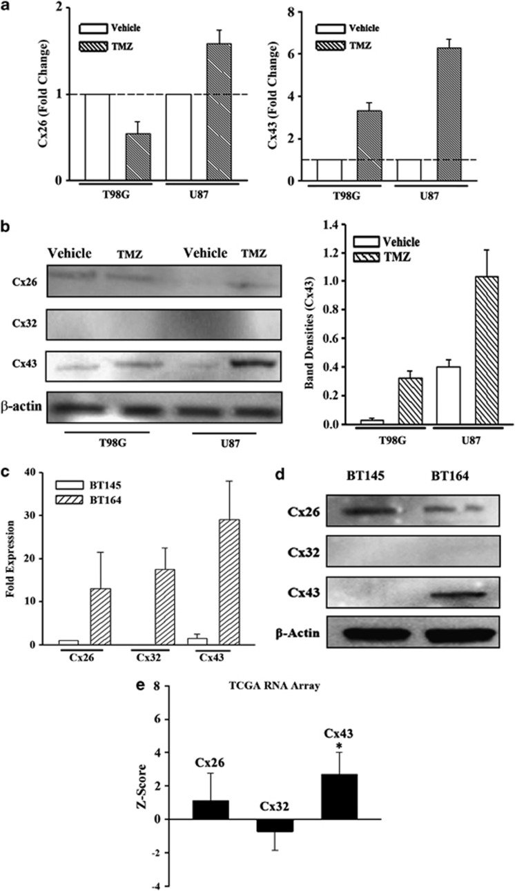
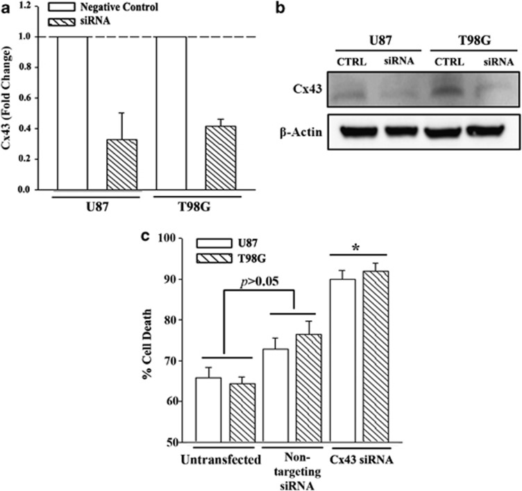
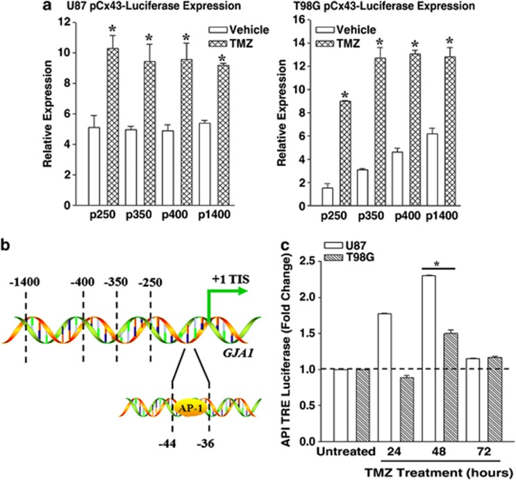
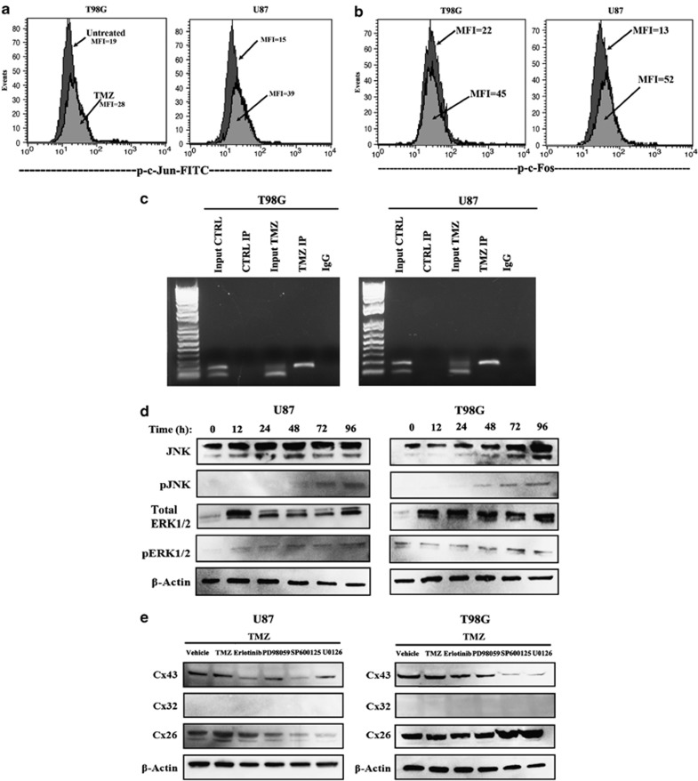
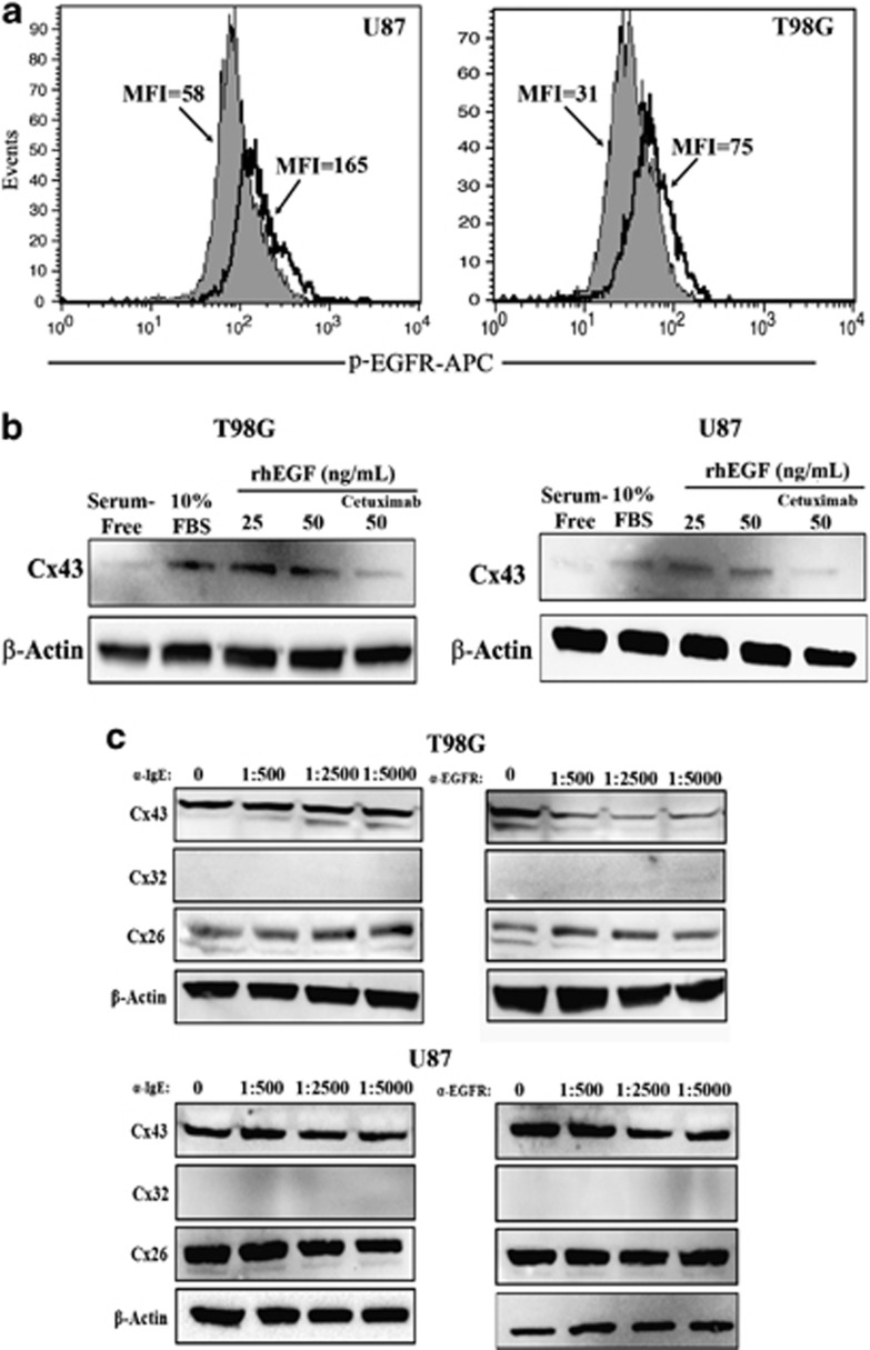
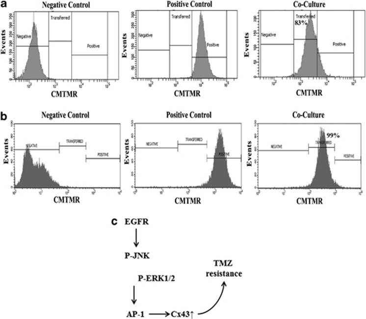
Glioblastoma Multiforme (GBM) is an aggressive adult primary brain tumor with poor prognosis. GBM patients develop resistance to the frontline chemotherapy, temozolomide (TMZ). As the connexins (Cx) have been shown to have a complex role in GBM, we investigated the role of Cx43 in TMZ resistance. Cx43 was increased in the TMZ-resistant low passage and cell lines. This correlated with the data in The Cancer Genome Atlas. Cx43 knockdown, reporter gene assays, chromatin immunoprecipitation assay, real-time PCR and western blots verified a role for Cx43 in TMZ resistance. This occurred by TMZ-resistant GBM cells being able to activate epidermal growth factor receptor (EGFR). In turn, EGFR activated the JNK-ERK1/2-AP-1 axis to induce Cx43. The increased Cx43 was functional as indicated by gap junctional intercellular communication among the resistant GBM cells. Cell therapy could be a potential method to deliver drugs, such as anti-EGF to tumor cells. Similar strategies could be used to reverse the expression of Cx43 to sensitize GBM cells to TMZ. The studies showed the potential for targeting EGF in immune therapy. These agents can be used in conjunction with stem cell therapy to treat GBM.


Figures

Figure 1
Figure 1
Increased Cx43 in TMZ-treated GBM cells. (a) U87 and T98G cells were treated with 200 μM TMZ. After 72 h, real-time PCR was performed with primers specific for Cx26 (left) and Cx43 (right). (b) Whole-cell extracts were prepared from U87 and T98G cells treated with TMZ for 72 h, and then analyzed by western blots for Cx26, Cx32 and Cx43 (left). The normalized band densities for Cx43 protein is shown at right. (c) RNA from low-passage cell lines from a patient with recurrent GBM (TMZ resistance BT164) and a naive patient (TMZ-sensitive BT145) were studied by real-time PCR for Cx26, Cx32 and Cx43 mRNA. The data are shown for the mean relative fold expression±S.D., n=4. (d) Whole-cell extracts from BT145 and BT164 were analyzed by western blot for Cx43. (e) TCGA level 3 with >500 GBM tissues was used to verify the expressions of Cx26, Cx32 and Cx43 expressions. The results are shown as the mean±S.D. for all samples within the database.* P<0.05 versus Cx26 or Cx32
Figure 2
Figure 2
Cell viability of TMZ-treated Cx43 knockdown GBM cells. U87 and T98G were transfected with Cx43-targeted siRNA or a non-targeting siRNA (Negative Control, Neg Ctrl). The cells were analyzed by real-time PCR for Cx43 mRNA (a) and whole-cell extracts in western blot with anti-Cx43 (b). The knockdown cells, Neg Ctrl and untransfected cells were treated with 200 μM TMZ. After 72 h, the cells were studied for viability using the CytoTox 96 LDH release. The results are presented as mean±S.D. of triplicates from each of four independent experiments (n=12).*P<0.05 versus Neg Ctrl and untransfected cells
Figure 3
Figure 3
Effect of TMZ on the 5′ regulatory region of GJA1 (Cx43). T98G and U87 cells were transfected with luciferase reporter vectors with various inserts, upstream of the transcriptional initiation site of the GJA1 5′ regulatory region. The transfectants were treated with vehicle or 200 μM TMZ for 72 h. Whole-cell lysates from the viable cells were analyzed for luciferase activity. The relative expression in luciferase activities are presented as the mean±S.D. The data represent triplicates from four independent experiments, n=12 (a). Shown is the AP-1 site within the 5′ regulatory region Cx43 (GJA1) (b). GBM cell lines were transfected with a reporter vector containing AP-1 Tandem Repeats. The transfectants were treated with 200 μM TMZ. After 72 h, whole-cell extracts were studied for luciferase levels. The results are presented as mean±S.D. of triplicates in four independent experiments, n=12 (c). *P<0.05 versus vehicle
Figure 4
Figure 4
EGFR-mediated activation of AP-1 in TMZ-treated GBM cells. (b) U87 and T98G cells were treated with 200 μM TMZ. After 72 h, the viable cells were analyzed by flow cytometry for phospho (p)-c-Jun (a) and p-c-Fos activation. The dark histograms represent untreated cells and the gray histograms, TMZ treatment. The MFI of each histogram is depicted with an arrow. (c) ChIP analyses were performed for AP-1 binding to endogenous Cx43 using TMZ-resistant GBM cells. The complex was precipitated with anti-c-Jun. Shown are the PCR of the precipitated gDNA with primers spanning the AP-1 site. (d) Western blots were performed for the upstream activators of AP-1 activation, pERK and p-JNK using whole-cell extracts from untreated (Time 0) and timeline treatment of U87 and T98G with 200 μM TMZ. The timeline studies occurred at 12 h intervals up to 96 h. (e) GBM cells were pretreated different pharmacological agents along the EGFR signaling pathway. After 24 h, the cells were washed and then exposed to 200 μM TMZ for 72 h. Whole-cell extracts were analyzed by western blots for Cx26, Cx32 and Cx43
Figure 5
Figure 5
Effects of EGFR signaling on Cx43 expression. (a) Flow cytometry was performed for cell surface expression for EGFR with untreated and TMZ-resistant GBM cells. Resistance was established with 200 μM TMZ for 72 h. The solid histogram represents untreated cells and the open histogram represents treated cells. The MFI of each histogram is shown with an arrow. (b) GBM cells were treated in sera-free media with 25 and 50 ng/ml of rhEGF. In parallel, the 50 ng/ml point also contained Cetuximab at 1/500 dilution. In parallel, the cells were stimulated with 10% FBS alone and with serum-free medium alone. At 24 h, whole-cell lysates were analyzed by western blots for Cx43. (c) TMZ-resistant U87 and T98G cells were established with 200 μM TMZ, in the presence or absence of Cetuximab (α-EGFR) or Omalizumab (α-IGE). Each antibody was used at the following final dilutions: 1 : 500, 1 : 2500 and 1 : 5000. After 72 h, whole-cell lysates were analyzed by western blot for Cx26, Cx32 and Cx43
Figure 6
Figure 6
GBM cells form functional gap junctions. (a) Ten unlabeled U87 or T98G cells were cocultured for 72 h with ten CMTMR-labeled (Cell Tracker Orange) cells (center). Dye transfer was evaluated by flow cytometry. (b) A diagram shows TMZ-resistant GBM cells with activated EGFR, which phosphorylated JNK and ERK1/2 to activate AP-1 for Cx43 expression

References

    1. Shahar T, Nossek E, Steinberg DM, Rozovski U, Blumenthal DT, Bokstein F, et al. The impact of enrollment in clinical trials on survival of patients with glioblastoma. J Clin Neurosci. 2012;19:1530–1534. - PubMed
    1. Park CK, Lee SH, Kim T, Choi S, Park SH, Heo D, et al. The value of temozolomide in combination with radiotherapy during standard treatment for newly diagnosed glioblastoma. J Neurooncol. 2013;112:277–283. - PubMed
    1. Zhang J, Stevens MF, Bradshaw TD. Temozolomide: mechanisms of action, repair and resistance. Curr Mol Pharmacol. 2012;5:102–114. - PubMed
    1. Johannessen TCA, Bjerkvig R. Molecular mechanisms of temozolomide resistance in glioblastoma multiforme. Expert Rev Anticancer Ther. 2012;12:635–642. - PubMed
    1. Happold C, Roth P, Wick W, Schmidt N, Florea AM, Silginer M, et al. Distinct molecular mechanisms of acquired resistance to temozolomide in glioblastoma cells. J Neurochem. 2012;122:444–455. - PubMed

Publication types

MeSH terms