. 2014 Mar 27;10(3):e1003507.
doi: 10.1371/journal.pcbi.1003507. eCollection 2014 Mar.

Investigation of inflammation and tissue patterning in the gut using a Spatially Explicit General-purpose Model of Enteric Tissue (SEGMEnT)

Affiliations

Investigation of inflammation and tissue patterning in the gut using a Spatially Explicit General-purpose Model of Enteric Tissue (SEGMEnT)

Chase Cockrell et al. PLoS Comput Biol. .

Abstract

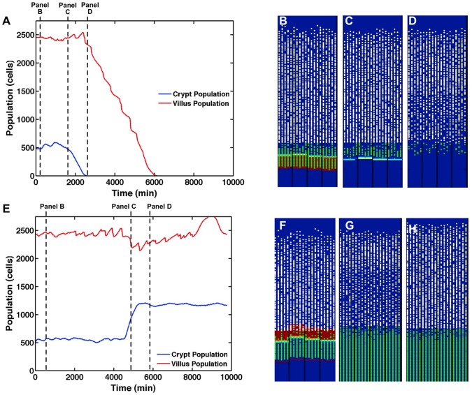
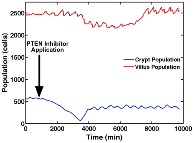
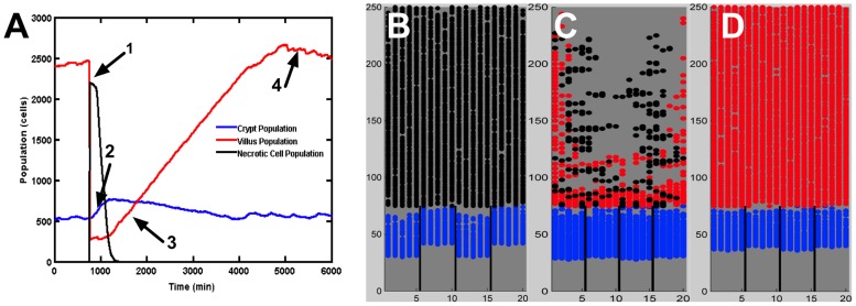
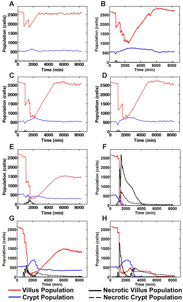
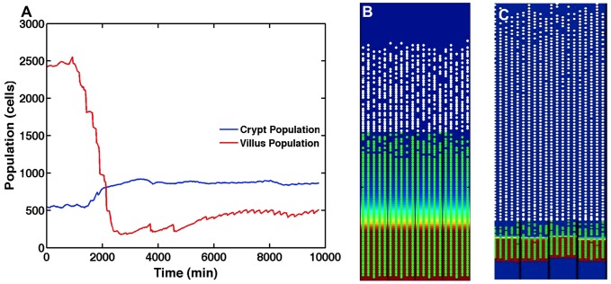
The mucosa of the intestinal tract represents a finely tuned system where tissue structure strongly influences, and is turn influenced by, its function as both an absorptive surface and a defensive barrier. Mucosal architecture and histology plays a key role in the diagnosis, characterization and pathophysiology of a host of gastrointestinal diseases. Inflammation is a significant factor in the pathogenesis in many gastrointestinal diseases, and is perhaps the most clinically significant control factor governing the maintenance of the mucosal architecture by morphogenic pathways. We propose that appropriate characterization of the role of inflammation as a controller of enteric mucosal tissue patterning requires understanding the underlying cellular and molecular dynamics that determine the epithelial crypt-villus architecture across a range of conditions from health to disease. Towards this end we have developed the Spatially Explicit General-purpose Model of Enteric Tissue (SEGMEnT) to dynamically represent existing knowledge of the behavior of enteric epithelial tissue as influenced by inflammation with the ability to generate a variety of pathophysiological processes within a common platform and from a common knowledge base. In addition to reproducing healthy ileal mucosal dynamics as well as a series of morphogen knock-out/inhibition experiments, SEGMEnT provides insight into a range of clinically relevant cellular-molecular mechanisms, such as a putative role for Phosphotase and tensin homolog/phosphoinositide 3-kinase (PTEN/PI3K) as a key point of crosstalk between inflammation and morphogenesis, the protective role of enterocyte sloughing in enteric ischemia-reperfusion and chronic low level inflammation as a driver for colonic metaplasia. These results suggest that SEGMEnT can serve as an integrating platform for the study of inflammation in gastrointestinal disease.


Conflict of interest statement

The authors have declared that no competing interests exist.

Figures

Figure 1
Figure 1. Modular control structure for SEGMEnT.
A simple representation of SEGMEnT's modular control structure is shown in Panel A: morphogenesis cell signaling determines cell behavior, the aggregate of which defines the tissue morphology. An inflammatory module acts as a controller on the morphogenesis processes. Because of the structure/function relationship in gut epithelial tissue the tissue morphology further modulates the morphogenesis cell signaling. Panel B demonstrates future planned control modules to be added to SEGMEnT in order to expand its functional representation (boxes and connectors with dashed lines).
Figure 2
Figure 2. SEGMEnT topology.
Panel A contains a histology cross section of ileal tissue (top) and scanning electron microscopy of the mucosal surface of ileum (bottom). These images are juxtaposed with Panel B, which is the topology used by SEGMEnT where crypts and villi are represented with a matrix of rectangular prisms. Each individual crypt or villus is then “unwrapped” onto a 2-dimensional grid (Panel C), on which signaling interactions, morphogen diffusion and physical cellular actions take place.
Figure 3
Figure 3. Overall schematic of SEGMEnT.
Letter A depicts the cellular processes linked to Gut Epithelial Cell (GEC) type (Letter B), their spatial location in terms of the crypt-villus architecture (Letter C), and expected gradients of different morphogens (Letter D). Biologically realistic gradients for Wingless-related integration site (Wnt), Bone Morphogenetic Protein (BMP), Sonic Hedgehog Homolog (Hh), β-catenin, and Akt are shown.
Figure 4
Figure 4. Baseline healthy ileal tissue dynamics.
Panel A demonstrates stable cell populations in the crypt and villus over time with complete renewal of the epithelial tissue every 5 days of simulated time. Panel B displays a SEGMEnT screenshot at homeostasis. Note that this 2d projection is the unwrapped 3d topology seen in Figure 2. All subsequent SEGMEnT screenshots are presented in this fashion to aid in the interpretation of its behavior, but are simulated in a 3d model. Differentiating GECs in the crypts are seen as green circles; white circles represent differentiated GECs on the villi. The red shading in the background represents areas of Wingless-related integration site (Wnt) activity; the absence of Wnt is seen as a blue background. Note that at the crypt-villus junction there is a region of undifferentiated GECs with no Wnt. Since activated Wnt signaling is identified and localized by the accumulation of β-catenin, the absence of Wnt in this zone denotes a region after the activation of β-catenin destruction complex and before all the β-catenin has been destroyed, at which point differentiation is triggered and occurs. This pattern of β-catenin activity is consistent with patterns seen in published histological data seen in Figures 3a and 3c in Ref .
Figure 5
Figure 5. Morphogen knockout/inhibition simulations.
Panel A displays average crypt and villus GEC populations in SEGMEnT when exposed to Wingless-related integration site (Wnt) inhibition. Panels B–D displays three frames from a SEGMEnT simulation of Wnt inhibition. Panel B represents baseline homeostasis prior to Wnt application. Panel C was captured at t = 1755 min, shortly after Wnt application. Cells then quickly differentiate as the β-catenin destruction complex is activated; the increased Hh signal from the newly differentiated cells combined with the Wnt inhibitor quickly eliminate all Wnt activity. Panel D was captured at t = 2160 min, at which point with the elimination of Wnt, the crypt no longer exists, there are few proliferating cells, and the villi have lost significant cellularity. Panel E displays average crypt and villus GEC populations in SEGMEnT when exposed to Sonic Hedgehog Homolog (Hh) inhibition. Panel F represents the system at baseline homeostasis prior the application of Hh inhibition. Panel G was captured at 5010 min, shortly after application of Hh inhibition. After application of the Hh inhibitor, the crypt expands rapidly. Initially, there is slight villus shrinkage as differentiation is temporarily halted by the now uninhibited Wingless-related integration site (Wnt) gradient. Panel H was captured at t = 6000 min. As the system adjusts to the lack of Hh, homeostasis is reached and persistent crypt hyperplasia exists throughout the system (Panel H).
Figure 6
Figure 6. PTEN inhibition.
Average crypt and villus populations are presented when exposed to Phosphotase and tensin homolog (PTEN) inhibition. Immediately subsequent to the PTEN inhibition (Arrow), levels of Sonic Hedgehog Homolog (Hh) spike, resulting in an immediate shrinkage in the crypt. With Wingless-related integration site (Wnt) inhibited by the higher levels of Hh, cell proliferation slows, resulting in slight villus atrophy. The system then reaches a new equilibrium absent PTEN via these redundant pathways, resulting in essentially the baseline tissue architecture. It is notable that the compensatory dynamics seen in the simulation experiments occur in a period not captured by the sampling interval in .
Figure 7
Figure 7. Local injury to the gut mucosa and subsequent reconstitution of crypt-villus architecture.
Panel A displays populations of living gut epithelial cells (GECs) on the crypt and villus, and necrotic cells upon local tissue injury and subsequent healing. Injury is induced by causing necrosis of an entire villus, resulting in a rapid drop of villus GEC population and spike in necrotic cell population (Arrow 1). In the time period directly subsequent to villus death the crypt grows rapidly; this is due to the sudden loss of Sonic Hedgehog Homolog (Hh) signaling as most of the differentiated cells on the villus have died. The death of the villi cells reduces the Wnt inhibition of the surviving crypt GECs, resulting in a growth spike in the crypt population (Arrow 2) that precedes the reconstitution of the villus population (Arrow 3). All during this process the inflammatory response is clearing the necrotic cells, allowing the regulatory functions of the morphogen pathways to normalize, leading to regrowth of the villus back to the homeostatic state (Arrow 4). Panels B–D display three screenshots from a simulation of localized epithelial damage/injury. Undifferentiated transit amplifying (TA) cells are shaded in blue. Differentiated enterocytes are shaded in red. Necrotic cells are shaded in black. Panel B, captured at t = 750 min, demonstrates that the epithelial insult results in necrotic cell death throughout the majority of the villus. Panel C, captured at t = 2250 min, shows that the necrotic cells continue to damage surrounding tissue until clearance by macrophages and neutrophils (not explicitly represented in these screenshots). Panel D, captured at t = 4500 min, demonstrates the recovered tissue architecture after regrowth of the epithelial cell populations.
Figure 8
Figure 8. Normal and aberrant tissue response ischemia/reperfusion across a range of ischemic times.
With 30 minutes of ischemia (Panel A) there is minimal necrosis and the system returns to normal by ∼24 hrs. Increasing amounts of ischemia at 2 hrs (Panel B), 3 hrs (Panel C) and 4 hrs (Panel D) all demonstrate recoverable tissue architecture. With 5 hrs ischemia (Panel E) there is a persistent alteration in the tissue composition. Application of 6 hrs ischemia (Panel F) results in a non-recoverable injury. Panels G and H show corresponding behavior in the absence of sloughing (Panel G = 30 min ischemia, Panel H = 3 hrs ischemia). The lack of sloughing increases the number of necrotic cells in place, leading to increased inflammatory stimuli and the propagation of inflammation-mediated damage, converting readily recoverable ischemia at 30 minutes and 3 hrs to correspondingly persistent effects on the tissue architecture at 30 minutes ischemia (Panel G) and tissue necrosis with 3 hrs of ischemia (Panel H).
Figure 9
Figure 9. Colonic metaplasia in the ileal pouch.
Panel A displays average crypt and villus gut epithelial cell (GEC) populations after exposure to sustained low-level toll-like receptor (TLR4) stimulation and signaling (an abstraction of fecal stasis). This low-level up-regulation of inflammation communicates via our hypothesized Phosphotase and tensin homolog (PTEN) mechanisms, leading to increased apoptosis, shortening the villus, as well as an inhibition of the Sonic Hedgehog homolog (Hh) pathway, which increases the size of the proliferative compartment (i.e. crypt). Panel B displays a screenshot from SEGMEnT when simulating conditions leading to colonic metaplasia. Crypt hyperplasia and villus atrophy are clearly evident (compare with normal homeostatic condition in Panel C, and as seen in Figure 4C), along with a shift in the villus to crypt height ratio that matches the alterations seen in colonic metaplasia as reported in Ref .

References

    1. Turner JR (2004) The Gastrointestinal Tract. In: Vinay Kumar AKA, Jon C. Aster, Nelson Fausto, Robbins and Cotran: Pathological Basis of Disease, 8th Edition. Philadelphia, PA: Saunders-Elsevier.
    1. Korpe PS, Petri WA Jr (2012) Environmental enteropathy: critical implications of a poorly understood condition. Trends Mol Med 18: 328–336. - PMC - PubMed
    1. De Plaen IG (2013) Inflammatory signaling in necrotizing enterocolitis. Clin Perinatol 40: 109–124. - PMC - PubMed
    1. Latella G, Papi C (2012) Crucial steps in the natural history of inflammatory bowel disease. World J Gastroenterol 18: 3790–3799. - PMC - PubMed
    1. Seal JB, Morowitz M, Zaborina O, An G, Alverdy JC (2010) The molecular Koch's postulates and surgical infection: a view forward. Surgery 147: 757–765. - PubMed

Publication types

Substances