. 2014 May;6(5):564-76.
doi: 10.1039/c4ib00015c.

Microfluidic source-sink model reveals effects of biophysically distinct CXCL12 isoforms in breast cancer chemotaxis

Affiliations

Microfluidic source-sink model reveals effects of biophysically distinct CXCL12 isoforms in breast cancer chemotaxis

S P Cavnar et al. Integr Biol (Camb). 2014 May.

Abstract

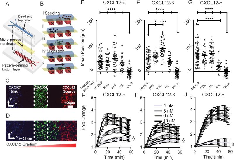
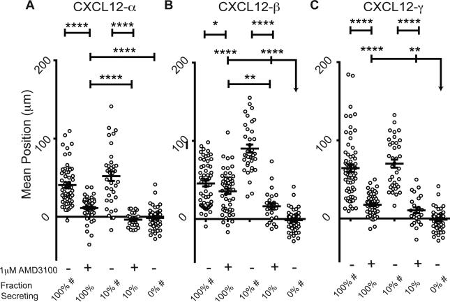
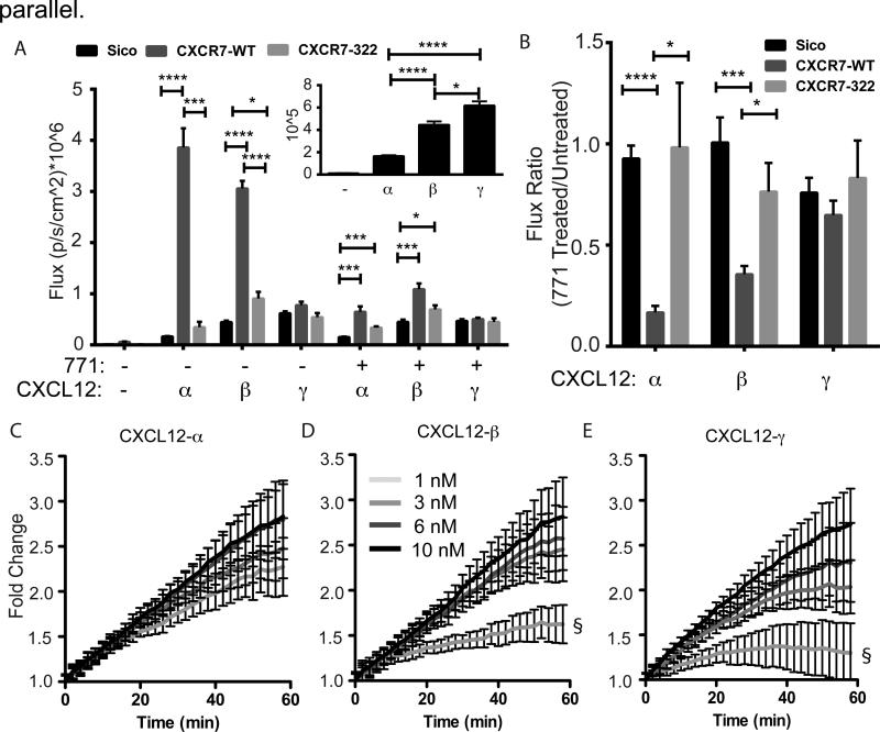
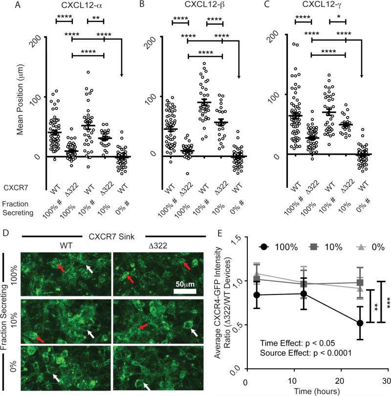
Chemokines critically regulate chemotaxis in normal and pathologic states, but there is limited understanding of how multicellular interactions generate gradients needed for cell migration. Previous studies of chemotaxis of CXCR4+ cells toward chemokine CXCL12 suggest the requirement of cells expressing scavenger receptor CXCR7 in a source-sink system. We leveraged an established microfluidic device to discover that chemotaxis of CXCR4 cells toward distinct isoforms of CXCL12 required CXCR7 scavenging only under conditions with higher than optimal levels of CXCL12. Chemotaxis toward CXCL12-β and -γ isoforms, which have greater binding to extracellular molecules and have been largely overlooked, was less dependent on CXCR7 than the more commonly studied CXCL12-α. Chemotaxis of CXCR4+ cells toward even low levels of CXCL12-γ and CXCL12-β still occurred during treatment with a FDA-approved inhibitor of CXCR4. We also detected CXCL12-γ only in breast cancers from patients with advanced disease. Physiological gradient formation within the device facilitated interrogation of key differences in chemotaxis among CXCL12 isoforms and suggests CXCL12-γ as a biomarker for metastatic cancer.


Figures

Figure 1
Figure 1. Microfluidic source-sink-migration device
(A, B) Schematic multilayered microfluidic device fabrication and representation of patterned cell seeding. Controlled flow through bottom channels results in 200 µm wide cell patterns with 200 µm gaps. Parts i, ii, and iii depict stepwise addition of CXCL12-secreting cells, CXCR7+ cells, and CXCR4+ cells, respectively. Parts iv and v represent conditions before and after migration. (C, D) Representative confocal images of patterned cells in the device at t=0 (C corresponds to B iv) and after 24 hours (D corresponds to B v). The CXCL12 secreting cells co-express FP650 (red); CXCR4+ cells express NLS-AcGFP (green); and CXCR7+ cells are stained with Hoescht 33342 (blue). The dashed white line denotes channel boundaries that define the starting position. The graded red triangle below (D) denotes the gradient direction. (E-G) Average position of CXCR4+ cells after 24 hours of migration toward CXCL12-α, -β, or -γ. Each point represents the mean position of~300±50 cells from 1 of 6 view fields from 10-11 devices. Fraction of secreting cells denotes the relative dilution of CXCL12-isoform secreting cells patterned with non-secreting cells. Data are shown as mean values ± S.E.M. (n=6 view fields for 4-11 devices per condition). The bars represent statistical comparison between pairs of conditions. The arrow denotes multiple paired comparisons to the same condition (*p<0.05, ***p<0.005, ****p<0.0001). Data for 100% and 0% secreting cells are marked (#) to designate the same data plotted for comparison in multiple figures. Matched conditions were performed in parallel. (H-J) Cells expressing a luciferase complementation reporter for association of CXCR4 and β-arrestin 2 were incubated with increasing equimolar concentrations of synthetic CXCL12-α, β, or γ. Data were graphed as mean values ± S.E.M. (n=4 measurements) from one of two representative experiments. Fold change in bioluminescence is relative to untreated cells at corresponding time points. The symbol § demarcates statistical differences by Tukey post hoc test between concentrations for the final time point. For CXCL12-α, 1nM is different from 6nM (p<0.01) and 10 nM (p<0.01). For CXCL12-β, 1nM is different from 6nM (p<0.05) and 10 nM (p<0.01). For CXCL12-γ, 1nM is different from all other concentrations (p<0.0001). Comparisons between isoforms are in supplemental information (Fig. S6).
Figure 2
Figure 2. AMD3100 limits migration of CXCR4+ cells toward CXCL12 isoforms
(A-C) Positions of CXCR4+ cells within migration devices were determined after 24 hours of migration in the absence or presence of 1 µM AMD3100. Data are plotted as average positions ± S.E.M. (n=6 view fields each for 4-11 devices per condition, similar to previous figures). Fraction of secreting cells denotes the relative percent of CXCL12-isoform secreting cells relative to control cells patterned in the source position. The bar represents the statistical comparison between pairs of conditions. The arrow denotes multiple paired comparisons to the same condition (*p<0.05, **p<0.01, ****p<0.0001). Data for 100%, 10%, and 0% secreting cells are marked (#) to designate the same data plotted for comparison in multiple figures. Matched conditions were performed in
Figure 3
Figure 3. CXCR7-dependent scavenging of CXCL12-isoforms
(A) 231 cells expressing CXCR7-GFP-WT, CXCR7-Δ322-GFP or no CXCR7 were incubated for 1 hour with equal levels (based on Gaussia luciferase activity) of cell-secreted CXCL12- α, β, or γ. Following incubation and acid wash to remove extracellular CXCL12, we measured internal Gaussia luciferase activity to quantify internalization of CXCL12. Photon flux values are reported as mean ± S.E.M. (n=4 measurements) from one of three representative experiments. The inset highlights only CXCL12-isoforms binding to CXCR7-negative 231-Sico cells. (B) Ratio of internalized bioluminescence signal (A) between cells incubated with inhibitor of CXCL12 binding to CXCR7 (771) relative to untreated cells. Statistical demarcations compare data between bars (* p< 0.05, ***p<0.005, **** p<0.0001). (C-E) Cells expressing a luciferase complementation reporter for association of CXCR7 and β-arrestin 2 were incubated with increasing equimolar concentrations of synthetic CXCL12-α, β, or γ. Data were graphed as mean values ± S.E.M. (n=4 measurements) from one of two representative experiments. Gray-scale code for concentrations indicated in panel D is the same for all isoforms. Fold change in bioluminescence is relative to untreated cells at corresponding time points. The symbol § demarcates statistical differences by Tukey post hoc test between concentrations for the final time point. There are no statistical differences between concentrations of CXCL12-α (C). For CXCL12-β, 1nM is different from 3nM (p<0.01), 6nM (p<0.01), and 10 nM (p<0.0001). For CXCL12-γ, 1nM is different from 3nM (p<0.05), 6nM (p<0.01), and 10 nM (p<0.0001). Comparisons between isoforms are in supplemental information (Fig. S5).
Figure 4
Figure 4. CXCR7 scavenging is necessary for chemotaxis of CXCR4+ cells in response to higher levels of CXCL12
(A-C) Migration of CXCR4+ cells toward various fractions of cells secreting different isoforms of CXCL12 in the presence of cells expressing either CXCR7-WT or a mutant lacking the carboxy terminus of the receptor (CXCR7-Δ322). Data are graphed as average position ± S.E.M. of migrating CXCR4+ cells after 24 hours (n=6 view fields each for 4-11 devices per condition, similar to previous figures). Fraction of secreting cells denotes the relative dilution of CXCL12-isoform secreting cells. The bar represents the statistical comparison between pairs of conditions. The arrow denotes multiple paired comparisons to the same condition (*p<0.05, **p<0.01, ****p<0.0001). Data for 100%, 10%, and 0% secreting cells are marked (#) to designate the same data plotted for comparison in multiple figures. Matched conditions were performed in parallel. (D) Representative Z-stack compressions of confocal images of CXCR4-GFP+ cells after 24 hours patterned in the context of dilutions of CXCL12-β (0, 10, and 100%) and with WT and CXCR7-Δ322 cells. Red arrows highlight intracellular CXCR4-GFP vesicles. White arrows denote cell membrane CXCR4-GFP. (E) Time course quantification of CXCR4-GFP intensity in devices patterned in the context of dilutions of CXCL12-β source cells (0, 10, and 100%). The plot depicts the mean ± SEM of the ratio between average CXCR4-GFP fluorescence intensity for devices with Δ322-CXCR7 relative to WT-CXCR7 (n=5 images from one of two representative experiments). Two-way ANOVA reveals significant time and source effects without significant interactions. The bars represent statistical significance by the Tukey post hoc test only for the 100% source at the 24 hour time point (** p<0.01, ***, p<0.005).

References

    1. Raman D, Sobolik-Delmaire T, Richmond A. Chemokines in health and disease. Experimental Cell Research. 2011;317(5):575–589. - PMC - PubMed
    2. Burkhardt AM, Homey B, Zlotnik A. Homeostatic chemokine receptors and organ-specific metastasis. Nature Reviews Immunology. 2011;11(9):597+. - PubMed
    3. Balkwill FR. The chemokine system and cancer. The Journal of Pathology. 2012;226(2):148–157. - PubMed
    4. Zhang GX, Baker CM, Kolson DL, Rostami AM. Chemokines and chemokine receptors in the pathogenesis of multiple sclerosis. Multiple Sclerosis. 2000;6(1):3–13. - PubMed
    5. Zernecke A, Shagdarsuren E, Weber C. Chemokines in atherosclerosis: an update. Arterioscler Throm Vasc Biol. 2008;28(11):1897–1908. - PubMed
    6. Krumbholz M, Theil D, Cepok S, Hemmer B, Kivisäkk P, Ransohoff RM, Hofbauer M, Farina C, Derfuss T, Hartle C, Newcombe J, Hohlfeld R, Meinl E. Chemokines in multiple sclerosis: CXCL12 and CXCL13 up-regulation is differentially linked to CNS immune cell recruitment. Brain. 2006;129(1):200–211. - PubMed
    1. Wu Y, Zhao R. The Role of Chemokines in Mesenchymal Stem Cell Homing to Myocardium. Stem Cell Reviews and Reports. 2012;8(1):243–250. - PubMed
    2. Fukuda S, Pelus LM. Chemokine-mobilized adult stem cells; defining a better hematopoietic graft. Leukemia. 2008;22(3):466+. - PMC - PubMed
    1. Yu SR, Burkhardt M, Nowak M, Ries J, Petrasek Z, Scholpp S, Schwille P, Brand M. Fgf8 morphogen gradient forms by a source-sink mechanism with freely diffusing molecules. Nature. 2009;461(7263):533–6. doi: 10.1038/nature08391. Epub 2009 Sep 9. - PubMed
    2. Boldajipour B, Mahabaleshwar S, Kardash E, Reichman-Fried M, Blaser H, Minina S, Wilson D, Xu Q, Raz E. Control of chemokine-guided cell migration by ligand sequestration. Cell. 2008;132(3):463–473. - PubMed
    3. Torisawa Y, Mosadegh B, Bersano-Begey T, Steele J, Luker K, Luker G, Takayama S. Microfluidic platform for chemotaxis in gradients formed by CXCL12 source-sink cells. Integr Biol (Camb) 2010;2(11-12):680–686. - PMC - PubMed
    1. Scholpp S, Brand M. Endocytosis controls spreading and effective signaling range of Fgf8 protein. Current biology. 2004;14(20):1834–1841. - PubMed
    2. Naumann U, Cameroni E, Pruenster M, Mahabaleshwar S, Raz E, Zerwes H, Rot A, Thelen M. CXCR7 functions as a scavenger for CXCL12 and CXCL11. PLoS One. 2010;5(2):e9175. - PMC - PubMed
    3. Luker K, Steele J, Mihalko L, Luker G. Constitutive and chemokine-dependent internalization and recycling of CXCR7 in breast cancer cells to degrade chemokine ligands. Oncogene. 2010;29:4599–4610. - PMC - PubMed
    4. Luker K, Lewin S, Mihalko L, Schmidt B, Winkler J, Coggins N, Thomas D, Luker G. Scavenging of CXCL12 by CXCR7 promotes tumor growth and metastasis of CXCR4-positive breast cancer cells. Oncogene. 2012 doi: 10.1038/onc.2011.633. [Epub ahead of print] - PMC - PubMed
    5. Ray P, Mihalko L, Coggins N, Moudgil P, Ehrlich A, Luker K, Luker G. Carboxy-terminus of CXCR7 regulates receptor localization and function. Int J Biochem Cell Biol. 2012;44(4):669–678. - PMC - PubMed
    6. Fredericks Z, Pitcher J, Lefkowitz R. Identification of the G protein-coupled receptor kinase phosphorylation sites in the human beta2-adrenergic receptor. J Biol Chem. 1996;271(23):13796–13803. - PubMed
    7. Sanchez-Alcaniz J, Haege S, Mueller W, Pla R, Mackay F, Schulz S, Lopez-Bendito G, Stumm R, Marin O. Cxcr7 controls neuronal migration by regulating chemokine responsiveness. Neuron. 2011;69(1):77–90. - PubMed
    8. Wang Y, Li G, Stanco A, Long J, Crawford D, Potter G, Pleasure S, Behrens T, Rubenstein J. CXCR4 and CXCR7 have distinct functions in regulating interneuron migration. Neuron. 2011;69(1):61–76. - PMC - PubMed
    1. Weber M, Hauschild R, Schwarz J, Moussion C, de Vries I, Legler DF, Luther SA, Bollenbach T, Sixt M. Interstitial Dendritic Cell Guidance by Haptotactic Chemokine Gradients. Science. 2013;339(6117):328–332. - PubMed

Publication types

MeSH terms