

doi: 10.1126/science.1249410.
Structure of the yeast mitochondrial large ribosomal subunit
Affiliations
- PMID: 24675956
- PMCID: PMC4046073
- DOI: 10.1126/science.1249410
Item in Clipboard
Structure of the yeast mitochondrial large ribosomal subunit
Science.
.
Abstract
Mitochondria have specialized ribosomes that have diverged from their bacterial and cytoplasmic counterparts. We have solved the structure of the yeast mitoribosomal large subunit using single-particle cryo-electron microscopy. The resolution of 3.2 angstroms enabled a nearly complete atomic model to be built de novo and refined, including 39 proteins, 13 of which are unique to mitochondria, as well as expansion segments of mitoribosomal RNA. The structure reveals a new exit tunnel path and architecture, unique elements of the E site, and a putative membrane docking site.
Figures
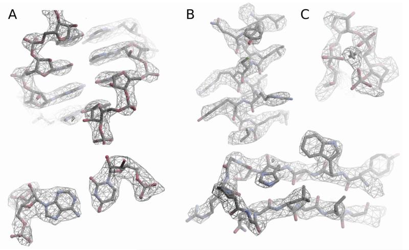
Examples of the density in cryo-EM maps of the 54S subunit. A. RNA, showing a double helix (top) and a base pair (bottom) B. Alpha helix and a beta turn in proteins showing side chains C. Mg ion that coordinates a tight turn in RNA.
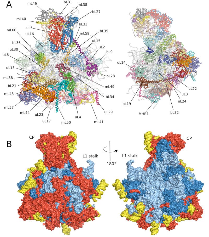
Overview of the yeast 54S ribosomal subunit. A. Cytoplasmic and side views of final model with 21S rRNA colored in gray. B. Cytoplasmic and interface views colored showing regions conserved with bacteria (blue), found in both yeast and mammalian mitochondria (red) and present only in yeast mitochondria (yellow).
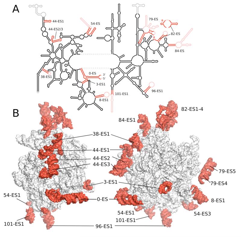
A. Schematic diagram of the secondary structure of 21S rRNA. B. Two views of tertiary structure showing mitochondria-specific ESs in red labeled after the conserved helix according to standard numbering (see text).
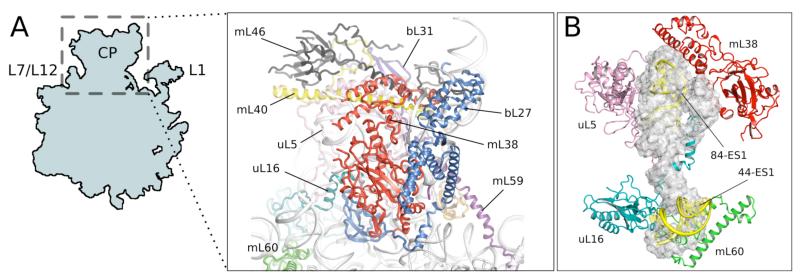
Central protuberance (CP) of the 54S subunit. A. Proteins involved in the C P. B. Elements that replace 5S rRNA (gray) in the CP
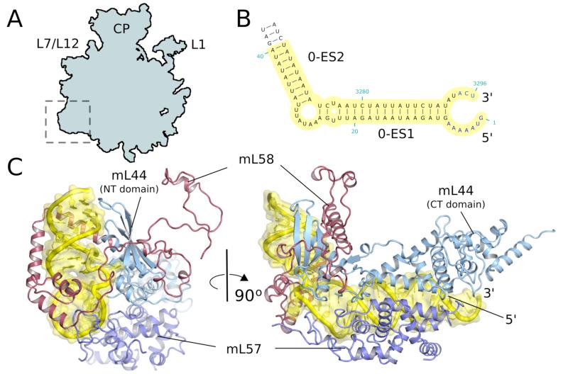
A membrane facing protuberance. A. Site of expansion of the 54S relative to bacteria. B. ES0 of 21S rRNA serves as an anchor for the protuberance. C. Two views of the protuberance showing ho w mitochondria-specific proteins bind to the expansion segment.
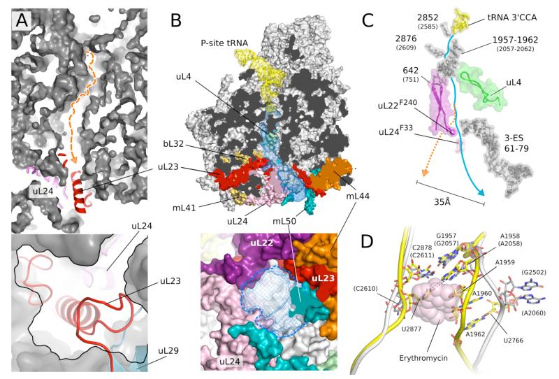
The putative exit tunnel in the 54S subunit. A. The bacterial tunnel (top, path in orange) is blocked by a mitochondria-specific extension to uL23 (detail in bottom panel). B. The 54S tunnel (blue mesh) has been extensively remodeled by mitochondria-specific protein elements (colored) (top) resulting in a tunnel exit (bottom) that is substantially different from that in bacteria. In A and B the lower panels are 90 ° rotations of the upper panels. C. Conservation of elements that make up the entrance and top part of the tunnel. The constriction at the top is narrower, and two aromatic amino acids that line the tunnel make a close approach. The path of the bacterial tunnel (orange) would exit 35 Å away. D. Narrowing of the 54S tunnel entrance (yellow) due to an AU base pair not found in bacteria (white) that would cause a steric clash with macrolides such as erythromycin (pink).
Comment in
-
Biochemistry. The resolution revolution.Science. 2014 Mar 28;343(6178):1443-4. doi: 10.1126/science.1251652. Science. 2014. PMID: 24675944 No abstract available.
References
-
- Ott M, Herrmann JM. Co-translational membrane insertion of mitochondrially encoded proteins. Biochim Biophys Acta. 2010;1803:767. - PubMed
-
- Jones CN, Miller C, Tenenbaum A, Spremulli LL, Saada A. Antibiotic effects on mitochondrial translation and in patients with mitochondrial translational defects. Mitochondrion. 2009;9:429. - PubMed
Publication types
MeSH terms
Substances
Associated data
- Actions
Grants and funding
LinkOut - more resources
Full Text Sources
Other Literature Sources
Molecular Biology Databases
