Review
. 2014 Apr 22;20(1):191-201.
doi: 10.2119/molmed.2013.00165.

Molecular mechanisms of pulmonary arterial remodeling

Affiliations
Review

Molecular mechanisms of pulmonary arterial remodeling

Patrick Crosswhite et al. Mol Med. .

Abstract

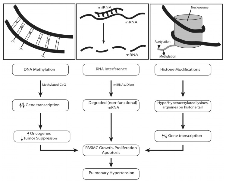
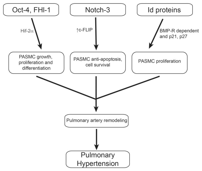
Pulmonary arterial hypertension (PAH) is characterized by a persistent elevation of pulmonary arterial pressure and pulmonary arterial remodeling with unknown etiology. Current therapeutics available for PAH are primarily directed at reducing the pulmonary blood pressure through their effects on the endothelium. It is well accepted that pulmonary arterial remodeling is primarily due to excessive pulmonary arterial smooth muscle cell (PASMC) proliferation that leads to narrowing or occlusion of the pulmonary vessels. Future effective therapeutics will be successful in reversing the vascular remodeling in the pulmonary arteries and arterioles. The purpose of this review is to provide updated information on molecular mechanisms involved in pulmonary arterial remodeling with a focus on growth factors, transcription factors, and epigenetic pathways in PASMC proliferation. In addition, this review will highlight novel therapeutic strategies for potentially reversing PASMC proliferation.


Figures

Figure 1
Figure 1
Summary of molecular mechanisms involved in pulmonary remodeling. In pulmonary artery smooth muscle cells, inhibition of the potassium channels leads to cell depolarization and calcium entry, stimulating excess calcium release from the sarcoplasmic reticulum leading to calcium-calmodulin–regulated cell proliferation. When growth factors (PDGF, VEGF, TGF-β) and mitogens (ET-1) bind to their receptors, various signaling pathways are activated which increase cytosolic calcium levels and activate the MAPK signaling pathway–stimulating transcription factors (c-fos, c-jun, c-myc, and so on) and increase cell proliferation. Prostacyclin’s antagonistic effects act by increasing cAMP concentration, which simultaneously inhibits MAPK signaling and activates the PKA/MLCK growth-suppressive pathway. The vasodilator nitric oxide diffuses into the cell and stimulates cGMP production via guanylyl cyclase that results in MLCK dephosphorylation that inhibits cell growth and relaxes the SMC, leading to vasodilation. Finally, depolarization of the mitochondrial membrane by the mitoKATP channels (see mitoKATP above) delays apoptosis by inhibiting cytochrome c release and generates hydrogen peroxide (H2O2), stimulating transcription factors that drive cell proliferation. Cyto-C, cytochrome c; CaM: calcium-calmodulin complex; S.R, sarcoplasmic reticulum; RyR, ryanodine receptor; PKA, protein kinase A; AC, adenylyl cyclase; GC, guanylyl cyclase; N.O., nitric oxide. Lightning bolt indicates hyperpolarization.
Figure 2
Figure 2
Epigenetics and pulmonary hypertension. Methylation of CpG islands in the promoter region of genes can lead to either hypo- or hyperacetylated promoters. This, in turn, can lead to increased or decreased gene transcription. While the pathways are not clearly defined, it is expected that an increase in oncogenes and/or a decrease in tumor suppressor genes can promote the abnormal cell proliferation of PASMCs. RNA interference is a well-described mechanism where miRNAs target specific mRNA and lead to degradation, preventing translation into mature protein. Lastly, histone modifications (either acetylated or methylated lysines and arginines) can promote or prevent transcription factor-mediated gene transcription and lead to increased cell proliferation or apoptosis resistance. These epigenetic mechanisms provide an alternative route for PH development besides mutations associated with the DNA sequence itself. ↑, Increase or upregulation; ↓ decrease or downregulation.
Figure 3
Figure 3
Novel transcription factors and pulmonary hypertension. Oct-4, normally associated with embryonic stem cell differentiation, has been suggested to play a role in hypoxia-mediated proliferation of PASMCs through the ability of Hif-2α to bind the Oct-4 promoter and trigger cell growth. Similar to Oct-4, Fhl-1 also was demonstrated to promote PASMC growth and proliferation through activation of the Hif-1/2 factors in response to hypoxia. Increased levels of Notch-3 were shown to induce c-FLIP, an antiapoptotic mediator, in PASMCs, and results in increased apoptosis resistance. Notch-3 also may interact with upstream regulators of the MAPK signaling pathway (including Src and Ras) to promote cell proliferation. Lastly, the Id protein family may act cooperatively to regulate BMP-R-dependent PASMC proliferation via their effects on the cyclin-dependent kinases and have been shown to be affected by prostacyclin therapy, a vital treatment resource for PH patients.

References

    1. Anderson JR, Nawarskas JJ. Pharmacotherapeutic management of pulmonary arterial hypertension. Cardiol Rev. 2010;18:148–62. - PubMed
    1. Morrell NW, et al. Cellular and molecular basis of pulmonary arterial hypertension. J Am Coll Cardiol. 2009;54:S20–31. - PMC - PubMed
    1. Giaid A, Saleh D. Reduced expression of endothelial nitric oxide synthase in the lungs of patients with pulmonary hypertension. N Engl J Med. 1995;333:214–21. - PubMed
    1. Szczeklik J, et al. Effects of prostaglandin E1 on pulmonary circulation in patients with pulmonary hypertension. Br Heart J. 1978;40:1397–401. - PMC - PubMed
    1. Christman BW, et al. An imbalance between the excretion of thromboxane and prostacyclin metabolites in pulmonary hypertension. N Engl J Med. 1992;327:70–5. - PubMed

Publication types

MeSH terms

Substances