Review
. 2014 Mar 27;10(3):e1003894.
doi: 10.1371/journal.ppat.1003894. eCollection 2014 Mar.

Viral OTU deubiquitinases: a structural and functional comparison

Affiliations
Review

Viral OTU deubiquitinases: a structural and functional comparison

Ben A Bailey-Elkin et al. PLoS Pathog. .

Abstract

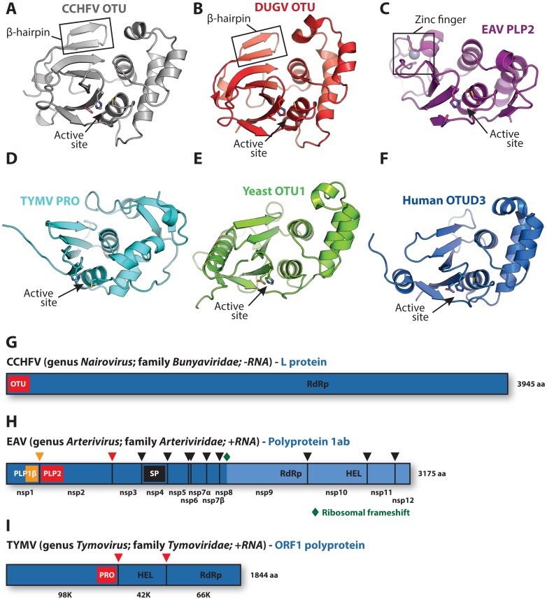
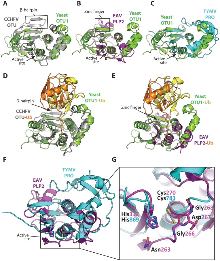
Recent studies have revealed that proteases encoded by three very diverse RNA virus groups share structural similarity with enzymes of the Ovarian Tumor (OTU) superfamily of deubiquitinases (DUBs). The publication of the latest of these reports in quick succession prevented proper recognition and discussion of the shared features of these viral enzymes. Here we provide a brief structural and functional comparison of these virus-encoded OTU DUBs. Interestingly, although their shared structural features and substrate specificity tentatively place them within the same protease superfamily, they also show interesting differences that trigger speculation as to their origins.


Conflict of interest statement

The authors have declared that no competing interests exist.

Figures

Figure 1
Figure 1. Viral and eukaryotic OTU domain structures and viral protein context.
Crystal structures of (A) CCHFV OTU (3PT2) , (B) DUGV OTU (4HXD) , (C) EAV PLP2 (4IUM) , (D) TYMV PRO (4A5U) , , (E) yeast OTU1 (3BY4) , and (F) human OTUD3 (4BOU) . The β-hairpin motifs of CCHFV OTU and DUGV OTU are indicated in boxes in panels A and B, respectively, and the zinc-finger motif of EAV PLP2 is boxed in panel C. Active sites are indicated with arrows. The CCHFV OTU, DUGV OTU, EAV PLP2, and yeast OTU1 domains were crystallized in complex with Ub, which has been removed for clarity. Structure images were generated using PyMol . (G) Schematic representation of the CCHFV large (L) protein , . A similar organization is found in the DUGV L protein, but is not depicted. The OTU domain resides in the N-terminal region of this protein and is not involved in autoproteolytic cleavage events . (H) Schematic representation of the EAV polyprotein 1ab . PLP2 resides in nonstructural protein 2 (nsp2) and is responsible for the cleavage between nsp2 and nsp3 . (I) Schematic representation of the TYMV ORF1 polyprotein . PRO resides in the N-terminal product of this polyprotein and is responsible for two internal cleavages , . Key replicative enzymes are indicated in G, H, and I. Colored arrowheads denote cleavage sites for the indicated protease domains. HEL, helicase; PLP, papain-like protease; RdRp, RNA-dependent RNA polymerase; SP, serine protease.
Figure 2
Figure 2. Superpositions of the viral OTU proteases with yeast OTU1 and one another.
Superpositions of yeast OTU1 (3BY4) with (A) CCHFV OTU (3PT2) , RMSD: 1.8 Å over 112 residues, (B) EAV PLP2 (4IUM) , RMSD: 2.8 Å over 69 residues, and (C) TYMV PRO (4A5U) , , RMSD: 1.4 Å over 76 residues. Superpositions of the yeast OTU1-Ub complex with (D) the CCHFV OTU-Ub complex and (E) the EAV PLP2-Ub complex, highlighting the difference in the orientation of Ub between the two viral OTU domains versus the eukaryotic yeast OTU1 domain. The Ub that is complexed with yeast OTU1 is depicted in yellow, while the Ub complexed with CCHFV OTU or EAV PLP2 is depicted in orange. (F) Superposition of EAV PLP2 and TYMV PRO, RMSD: 2.5 Å over 53 residues. (G) Close-up of the active site region (boxed) of the superposition depicted in F. Side chains of the catalytic cysteine (Cys270 and Cys783 for EAV PLP2 and TYMV PRO, respectively) and histidine (His332 and His869 for EAV PLP2 and TYMV PRO, respectively) residues are shown as sticks, as well as the active site Asn263 for EAV PLP2. The backbone amide group of Asp267 likely contributes to the formation of the oxyanion hole in the active site of EAV PLP2, yet a functionally equivalent residue is absent in TYMV PRO. The Gly266 and Gly268 residues flanking Asp267 in EAV PLP2 are depicted as sticks as well, for clarity. Note the alternative orientation of the active site cysteine residue of TYMV PRO which, unlike EAV PLP2, was not determined in covalent complex with an Ub suicide substrate. All alignments were generated using the PDBeFOLD server , and thus the reported RMSD values differ from those reported in Table 1, in which the DALI server was used. The yeast OTU1, CCHFV OTU, and EAV PLP2 domains were all crystallized in complex with Ub, which has been removed in panels A, B, C, F, and G for clarity. All images were generated using PyMol . RMSD, root-mean-square deviation.

References

    1. Komander D, Rape M (2012) The ubiquitin code. Annu Rev Biochem 81: 203–229. - PubMed
    1. Clague MJ, Urbe S (2010) Ubiquitin: same molecule, different degradation pathways. Cell 143: 682–685. - PubMed
    1. Jiang X, Chen ZJ (2012) The role of ubiquitylation in immune defence and pathogen evasion. Nat Rev Immunol 12: 35–48. - PMC - PubMed
    1. Oudshoorn D, Versteeg GA, Kikkert M (2012) Regulation of the innate immune system by ubiquitin and ubiquitin-like modifiers. Cytokine Growth Factor Rev 23: 273–282. - PMC - PubMed
    1. Huang TT, D'Andrea AD (2006) Regulation of DNA repair by ubiquitylation. Nat Rev Mol Cell Biol 7: 323–334. - PubMed

MeSH terms

Substances

LinkOut - more resources