. 2014 Jun;44(6):1831-42.
doi: 10.3892/ijo.2014.2354. Epub 2014 Mar 21.

All trans-retinoic acid (ATRA) induces re-differentiation of early transformed breast epithelial cells

Affiliations

All trans-retinoic acid (ATRA) induces re-differentiation of early transformed breast epithelial cells

Maria F Arisi et al. Int J Oncol. 2014 Jun.

Abstract

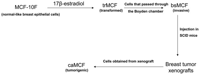
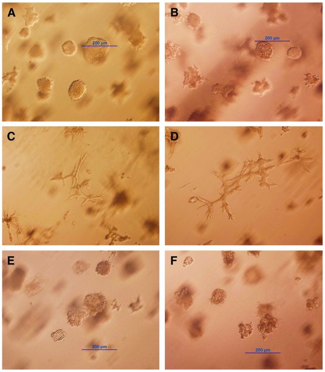
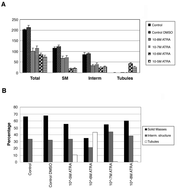
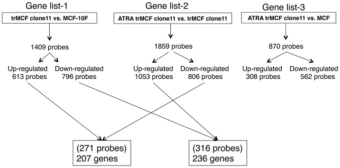
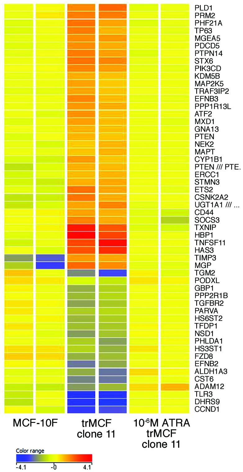
Retinoids have been used as potential chemotherapeutic or chemopreventive agents because of their differentiative, anti-proliferative, pro-apoptotic and antioxidant properties. We investigated the effect of all trans-retinoic acid (ATRA) at different stages of the neoplastic transformation using an in vitro model of breast cancer progression. This model was previously developed by treating the MCF-10F human normal breast epithelial cells with high dose of estradiol and consists of four cell lines which show a progressive neoplastic transformation: MCF-10F, normal stage; trMCF, transformed MCF-10F; bsMCF, invasive stage; and caMCF, tumorigenic stage. In 3D cultures, MCF-10F cells form tubules resembling the structures in the normal mammary gland. After treatment with estradiol, these cells formed tubules and spherical masses which are indicative of transformation. Cells that only formed spherical masses in collagen were isolated (trMCF clone 11) and treated with ATRA. After treatment with 10 or 1 µM ATRA, the trMCF clone 11 cells showed tubules in collagen; 10 and 43% of the structures were tubules in cells treated with 10 and 1 µM ATRA, respectively. Gene expression studies showed that 207 genes upregulated in transformed trMCF clone 11 cells were downregulated after 1 µM ATRA treatment to levels comparable to those found in the normal breast epithelial cells MCF-10F. Furthermore, 236 genes that were downregulated in trMCF clone 11 were upregulated after 1 µM ATRA treatment to similar levels shown in normal epithelial cells. These 443 genes defined a signature of the ATRA re-programming effect. Our results showed that 1 µM ATRA was able to re-differentiate transformed cells at early stages of the neoplastic process and antagonistically regulate breast cancer associated genes. The invasive and tumorigenic cells did not show any changes in morphology after ATRA treatment. These results suggest that ATRA could be used as a chemopreventive agent to inhibit the progression of premalignant lesions of the breast.


Figures

Figure 1.
Figure 1.
In vitro-in vivo model of cell transformation. The human normal-like MCF-10F cells were treated with high dose of estradiol and named early transformed breast epithelial cells (trMCF). The trMCF cells were seeded on a Boyden chamber and the cells that invaded, bsMCF, were selected and expanded. The bsMCF cells were injected in the fat mammary pad of SCID mice producing breast tumor xenografts. These xenografts were surgically removed and tumor cells were expanded giving origin to caMCF. The trMCF cells did not produced tumors when injected in SCID mice.
Figure 2.
Figure 2.
All-trans retinoic acid (ATRA) induces morphological changes in transformed cells trMCF clone 11. The trMCF clone 11 cells were plated in collagen matrix (3-D cultures) after being treated continuously for 26 days with different concentrations of ATRA. The cells were photograph after 8 days in collagen. (A) trMCF clone 11 cells (control); (B) trMCF clone 11 cells treated with 0.1% DMSO (vehicle, control); (C) trMCF clone 11 after being treated with 10−5 M (10 μM) ATRA; (D) trMCF clone 11 cells after being treated with 10−6 M (1 μM) ATRA; (E) trMCF clone 11 cells after being treated with 10−7 M ATRA; (F) trMCF clone 11 cells after being treated with 10−8 M ATRA.
Figure 3.
Figure 3.
Spherical masses, tubules and intermediate structures formed in collagen by trMCF clone 11 before and after ATRA treatments. The trMCF clone 11 cells form spherical masses in collagen and some intermediate structures. The trMCF clone 11 cells were treated with different concentrations of all-trans retinoic acid (ATRA) for 26 days; after ATRA treatments, the cells were plated in collagen. (A) Total number of different structures in collagen of trMCF clone 11 cells before and after ATRA treatment. Total number of spherical masses (SM), tubules and intermediate structures (spherical masses with prolongations) per well are shown. (B) Different structures on collagen matrix of trMCF clone 11 cells after treatment with different concetrations of ATRA. Percentage of different structures in collagen.
Figure 4.
Figure 4.
Invasion assay of trMCF clone 11 before and after ATRA treatments. The cell index of Matrigel-coated wells (invasion) at different time points are shown. The invasion capacity of the trMCF clone 11 did not show significant differences after 10−5 M ATRA (▲) or 10−6 M ATRA (■) compared with control after 16 h.
Figure 5.
Figure 5.
Unsupervised sample classification by principle coordinate analysis (PCoA). Two arrays were performed for each cell line and treatment: MCF-10F (MCF_CTR1 and MCF_CTR2), trMCF clone 11 (E2_CTR1 and E2_CTR2), 10−5 M ATRA trMCF clone 11 (E2_Hi1 and E2_Hi2) and 10−6 M ATRA trMCF clone 11 (E2_Lo1 and E2_Lo2). The trMCF clone 11 cells treated with 10−5 M ATRA (E2_Hi) or 10−6 M ATRA (E2_Lo) shown a major difference with trMCF clone 11 cells (E2_CTR) and minor differences with MCF-10F cells (MCF_CTR).
Figure 6.
Figure 6.
Representation of the gene expression studies showing number of dysregulated genes. Expression studies were performed in the early transformed trMCF clone 11 cells before and after treatment with 10−6 M ATRA (ATRA trMCF clone 11) and normal human breast epithelial MCF-10F cells. A total of 207 genes upregulated in the transformed trMCF clone 11 (compared to the normal MCF-10F) were downregulated after treatment with 10−6 M ATRA and, 236 genes that were downregulated in trMCF clone 11 (compared to MCF-10F) were upregulated by 10−6 M ATRA treatment. These 443 genes defined a gene signature programming the reverse-transformation effect by ATRA.
Figure 7.
Figure 7.
Heat map of selected genes in normal breast epithelial cells and early transformed cells before and after 10−6 M ATRA treatment. The expressions of genes involved in cell morphology are shown; also some genes from the aryl hydrocarbon and RAR pathways and genes involved in chromatin modification are shown. The genes that were dysregulated in the early transformed breast epithelial cells (trMCF clone 11) reached normal levels, similar to the normal breast epithelial cells MCF-10F, after treatment with 10−6 M ATRA. Red, yellow or blue colors represent expression levels above, at or below the mean level across the samples.

Similar articles

Cited by

References

    1. Theodosiou M, Laudet V, Schubert M. From carrot to clinic: an overview of the retinoic acid signaling pathway. Cell Mol Life Sci. 2010;67:1423–1445. - PMC - PubMed
    1. Duester G. Retinoic acid synthesis and signaling during early organogenesis. Cell. 2008;134:921–931. - PMC - PubMed
    1. Mongan NP, Gudas LJ. Diverse actions of retinoid receptors in cancer prevention and treatment. Differentiation. 2007;75:853–870. - PubMed
    1. Connolly R, Nguyen NK, Sukumar S. Molecular pathways: current role and future directions of the retinoic acid pathway in cancer prevention and treatment. Clin Cancer Res. 2013;19:1651–1659. - PMC - PubMed
    1. Bushue N, Wan YJ. Retinoid pathway and cancer therapeutics. Adv Drug Deliv Rev. 2010;62:1285–1298. - PMC - PubMed

Publication types

MeSH terms