

Inflammation as well as angiogenesis may participate in the pathophysiology of brain radiation necrosis
- PMID: 24676944
- PMCID: PMC4100008
- DOI: 10.1093/jrr/rru017
Inflammation as well as angiogenesis may participate in the pathophysiology of brain radiation necrosis
Abstract
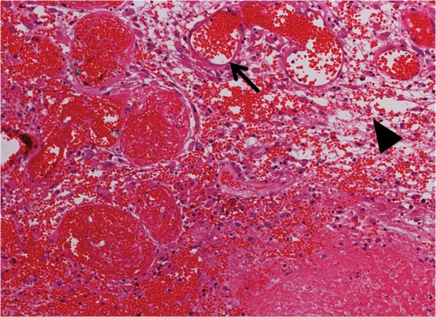
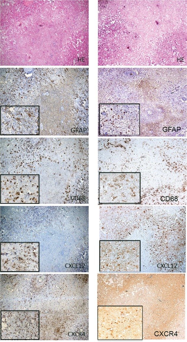
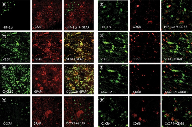
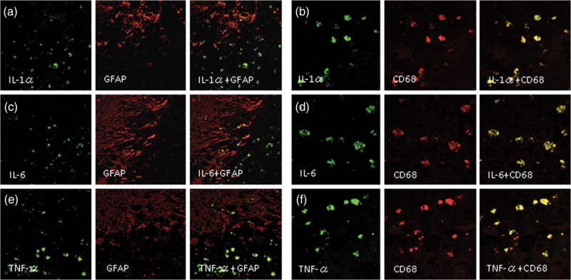
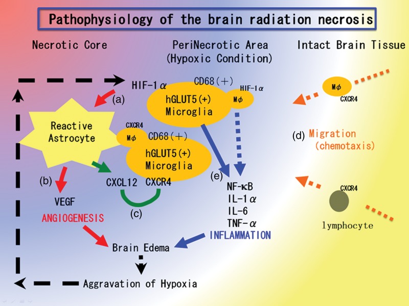
Radiation necrosis (RN) after intensive radiation therapy is a serious problem. Using human RN specimens, we recently proved that leaky angiogenesis is a major cause of brain edema in RN. In the present study, we investigated the same specimens to speculate on inflammation's effect on the pathophysiology of RN. Surgical specimens of symptomatic RN in the brain were retrospectively reviewed by histological and immunohistochemical analyses using hematoxylin and eosin (H&E) staining as well as immunohistochemical staining for VEGF, HIF-1α, CXCL12, CXCR4, GFAP, CD68, hGLUT5, CD45, IL-1α, IL-6 TNF-α and NF-kB. H&E staining demonstrated marked angiogenesis and cell infiltration in the perinecrotic area. The most prominent vasculature was identified as thin-walled leaky angiogenesis, i.e. telangiectasis surrounded by prominent interstitial edema. Two major cell phenotypes infiltrated the perinecrotic area: GFAP-positive reactive astrocytes and CD68/hGLUT5-positive cells (mainly microglias). Immunohistochemistry revealed that CD68/hGLUT5-positive cells and GFAP-positive cells expressed HIF-1α and VEGF, respectively. GFAP-positive cells expressed chemokine CXCL12, and CD68/hGLUT5-positive cells expressed receptor CXCR4. The CD68/hGLUT5-positive cells expressed pro-inflammatory cytokines IL-1α, IL-6 and TNF-α in the perinecrotic area. VEGF caused leaky angiogenesis followed by perilesional edema in RN. GFAP-positive cells expressing CXCL12 might attract CXCR4-expressing CD68/hGLUT5-positive cells into the perinecrotic area. These accumulated CD68/hGLUT5-positive cells expressing pro-inflammatory cytokines seemed to aggravate the RN edema. Both angiogenesis and inflammation might be caused by the regulation of HIF-1α, which is well known as a transactivator of VEGF and of the CXCL12/CXCR4 chemokine axis.
Keywords: CXCL12/CXCR4 chemokine axis; brain radiation necrosis; inflammation; microglia; pro-inflammatory cytokine.
© The Author 2014. Published by Oxford University Press on behalf of The Japan Radiation Research Society and Japanese Society for Radiation Oncology.
Figures
References
-
- Furuse M, Kawabata S, Kuroiwa T, et al. Repeated treatments with bevacizumab for recurrent radiation necrosis in patients with malignant brain tumors: a report of 2 cases. J Neurooncol. 2011;102:471–5. - PubMed
-
- Gonzalez J, Kumar AJ, Conrad CA, et al. Effect of bevacizumab on radiation necrosis of the brain. Int J Radiat Oncol Biol Phys. 2007;67:323–6. - PubMed
-
- Nonoguchi N, Miyatake S, Fukumoto M, et al. The distribution of vascular endothelial growth factor-producing cells in clinical radiation necrosis of the brain: pathological consideration of their potential roles. J Neurooncol. 2011;105:423–31. - PubMed
-
- Miyatake S, Kuroiwa T, Kajimoto Y, et al. Fluorescence of non-neoplastic, magnetic resonance imaging-enhancing tissue by 5-aminolevulinic acid: case report. Neurosurgery. 2007;61:E1101–3. discussion E1103–4. - PubMed
Publication types
MeSH terms
Substances
LinkOut - more resources
Full Text Sources
Other Literature Sources
Medical
Molecular Biology Databases
Research Materials
Miscellaneous
