. 2014 Nov;229(11):1822-30.
doi: 10.1002/jcp.24636.

Probiotic L. reuteri treatment prevents bone loss in a menopausal ovariectomized mouse model

Affiliations

Probiotic L. reuteri treatment prevents bone loss in a menopausal ovariectomized mouse model

Robert A Britton et al. J Cell Physiol. 2014 Nov.

Abstract

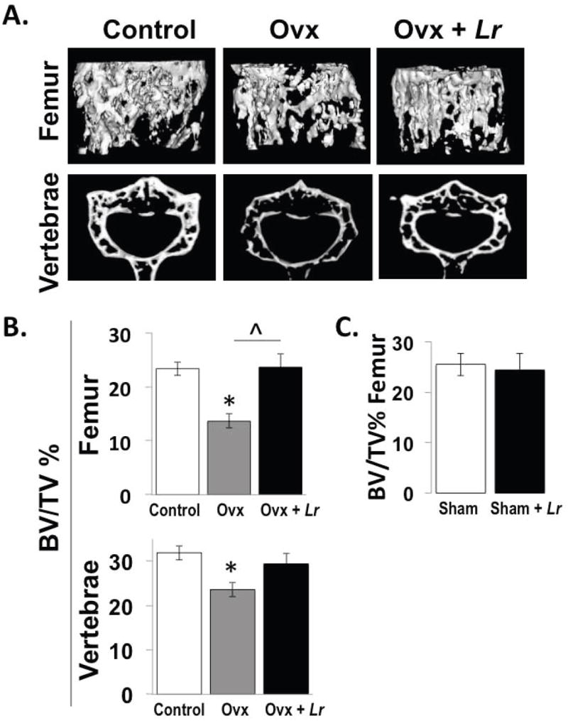
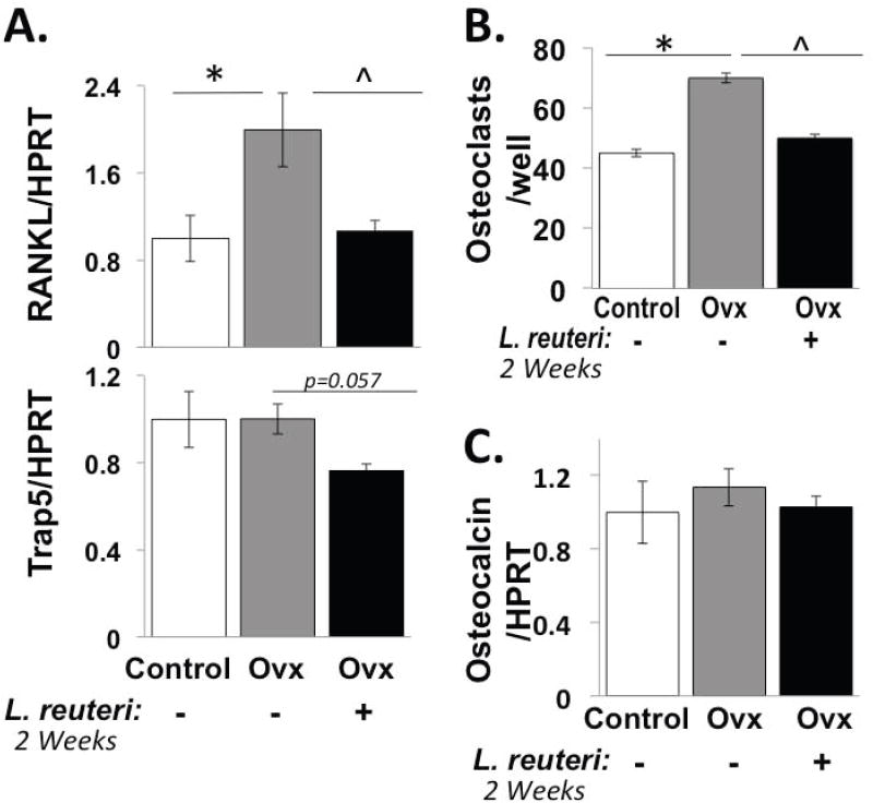
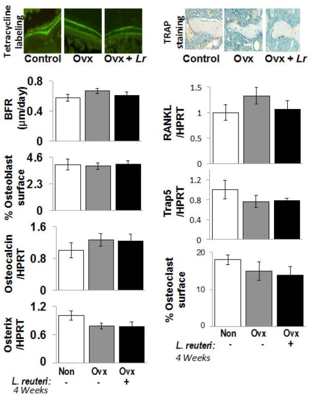
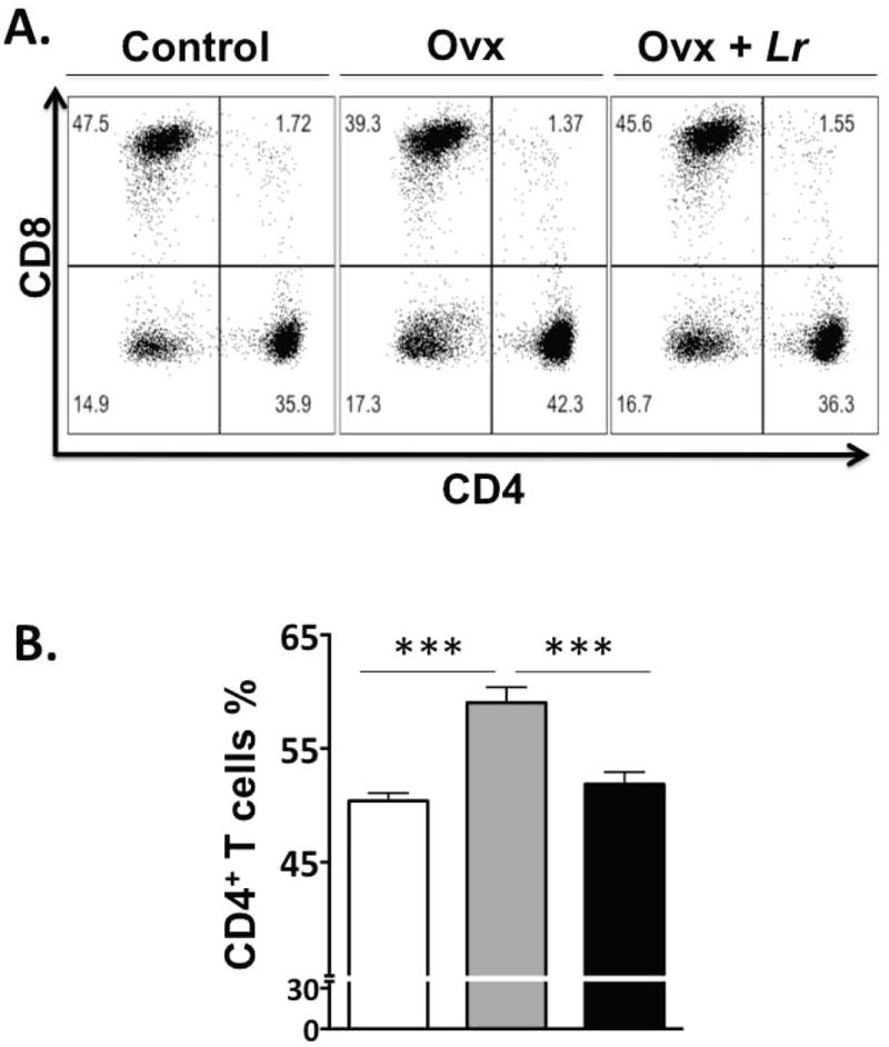
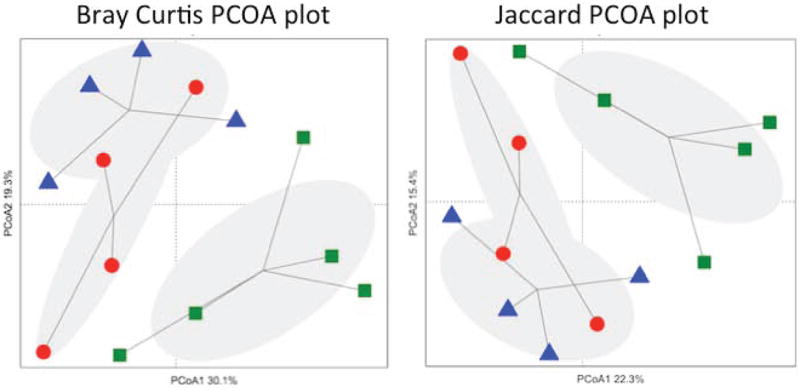
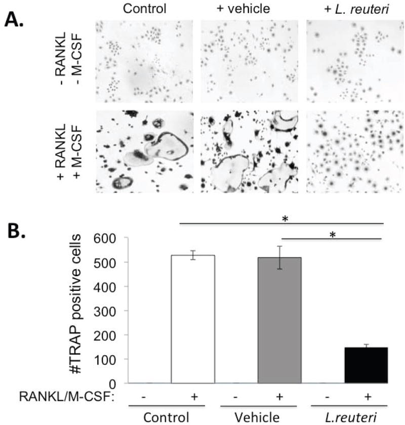
Estrogen deficiency is a major risk factor for osteoporosis that is associated with bone inflammation and resorption. Half of women over the age of 50 will experience an osteoporosis related fracture in their lifetime, thus novel therapies are needed to combat post-menopausal bone loss. Recent studies suggest an important role for gut-bone signaling pathways and the microbiota in regulating bone health. Given that the bacterium Lactobacillus reuteri ATCC PTA 6475 (L. reuteri) secretes beneficial immunomodulatory factors, we examined if this candidate probiotic could reduce bone loss associated with estrogen deficiency in an ovariectomized (Ovx) mouse menopausal model. Strikingly, L. reuteri treatment significantly protected Ovx mice from bone loss. Osteoclast bone resorption markers and activators (Trap5 and RANKL) as well as osteoclastogenesis are significantly decreased in L. reuteri-treated mice. Consistent with this, L. reuteri suppressed Ovx-induced increases in bone marrow CD4+ T-lymphocytes (which promote osteoclastogenesis) and directly suppressed osteoclastogenesis in vitro. We also identified that L. reuteri treatment modifies microbial communities in the Ovx mouse gut. Together, our studies demonstrate that L. reuteri treatment suppresses bone resorption and loss associated with estrogen deficiency. Thus, L. reuteri treatment may be a straightforward and cost-effective approach to reduce post-menopausal bone loss.


Conflict of interest statement

All authors state that they have no conflict of interest.

Figures

Figure 1
Figure 1. Ovariectomy (Ovx) mice treated with L. reuteri for 4 weeks display femur and vertebral trabecular bone volumes that are similar to ovary intact control mice
Representative micro-computed tomography isosurface images (A) and percent bone volume fraction (BV/TV) quantitative pooled data (B, C) obtained from the trabecular bone region of the distal femur and L3 vertebrae of non-Ovx control (white bar), Ovx (gray bar), and Ovx+L. reuteri treated (black bar) mice (A, B) and from sham versus sham plus L. reuteri treatment (C). L. reuteri was given by gavage only as described in the methods. Values are averages ± standard error, n=8 per group, * p<0.05 compared to Non-Ovx, ˆ p<0.05 compared to Ovx as determined by one-way ANOVA followed by Tukey HSD.
Figure 2
Figure 2. L. reuteri treatment prevents Ovx-induced increases in RANKL and bone marrow osteoclastogenesis after 2 weeks of probiotic treatment
Conditions are non-Ovx, intact mice (white bar); Ovx, ovariectomized mice (gray bar); Ovx + Lr, Ovx mice gavaged with L. reuteri for 2 weeks (black bar). (A) Levels of TRAP5 and RANKL RNA in whole tibia bone. Levels were expressed relative to HPRT, a non-modulated house-keeping gene. (B) Harvested mouse bone marrow cells from the mice above were cultured in vitro with RANKL and M-CSF and the number of osteoclasts counted per well. (C) Osteocalcin mRNA levels were measured in whole tibia bone and expressed relative to HPRT. Data values are average ± standard error, n ≥ 8 per group, * p<0.05 to Non-Ovx, ˆ p<0.05 to Ovx by one-way ANOVA followed by Tukey HSD.
Figure 3
Figure 3. Osteoclast and osteoblast markers don’t show significant changes in response to Ovx +/- L. reuteri treatment at 4 weeks
Top left: Representative fluorescent microscope photographs depicting the two pulses of calcein incorporation. The space between the two bands represents the mineral apposition rate (MAR). Top right: Representative microscope photographs of TRAP stained femur metaphyseal sections. Osteoclasts stain purple. Left Bar Graphs: Bone formation rate (BFR), osteoblast surface/total trabecular surface, and tibial RNA levels of osteocalcin and osterix (relative to HPRT an unaltered housekeeping gene). Right Bar Graphs: Tibial RNA levels of RANKL and Trap5 relative to HPRT, as well as osteoclast surface/total trabecular surface. Conditions are non-Ovx (white); Ovx, ovariectomized mice (gray); and Ovx + Lr, Ovx mice gavaged with Lactobacillus reuteri for 4 weeks (black). Values are averages ± standard error, n=8 per group, * p<0.05 to Non-Ovx by one-way ANOVA followed by Tukey HSD.
Fig 4
Fig 4. FACS analysis of bone marrow cells
Control, OVX and OVX+LR mice were treated as described and femurs collected during euthanasia at 2 weeks after treatment. Femurs were flushed and bone marrow cells were collected, processed and stained for cell surface markers for identifying the various immune cells as described in the methods. CD4+ T-lymphocytes were identified as CD3+CD4+ T-cells; Dot plots are shown at the top and cumulative data presented in graph in the bottom. In the dot plots, the numbers in the quadrants indicate the percentage of cells. N=8 for each condition; ***P<0.001 as determined by ANOVA followed by Bonferroni.
Figure 5
Figure 5. Two-dimensional projection of the principal coordinate analysis (PCoA) of bacterial 16S sequences from mouse jejunum samples along the first two principal axes
Left. PCoA was calculated using the Bray-Curtis similarity measures (coordinate axis 1 percent variation explained=30.1%, coordinate axis 2=19.3%). Right. PCoA was calculated using the Jaccard similarity measure (coordinate axis 1 percent variation explained=22.3%, coordinate axis 2=15.4%). Non-OVX mice (▲), OVX mice (●), L.reuteri-treated OVX mice (■).
Figure 6
Figure 6. Effect of L. reuteri secreted factors on osteoclast differentiation in Raw 264.7 cells
(A) Representative images of TRAP+ multinuclear giant cells at 7 days after RANKL and M-CSF treatment. (a) Cells only (Negative control) (b) RANKL and M-CSF (Positive control) (c) MEM-α (d) MEM-α plus RANKL and M-CSF (e) L. reuteri conditioned MEM-α (LR CM) (f) LR CM plus RANKL and M-CSF. (B) Quantification of TRAP+ multinuclear (>3 nuclei/cell) giant cells at 7 days after treatment with RANKL (100ng/ml) and M-CSF (10ng/ml) per visual field under light microscopy. Values are averages ± standard deviations. Conditions were done in triplicate and the entire experiment repeated 4 times. * p < 0.05 by Students t-test.

Similar articles

Cited by

References

    1. ESHRE Capri Workshop Group. Bone fractures after menopause. Hum Reprod Update. 2010;16(6):761–773. - PubMed
    1. Abrams SA, Griffin IJ, Hawthorne KM, Liang L, Gunn SK, Darlington G, Ellis KJ. A combination of prebiotic short- and long-chain inulin-type fructans enhances calcium absorption and bone mineralization in young adolescents. The American journal of clinical nutrition. 2005;82(2):471–476. - PubMed
    1. Atarashi K, Tanoue T, Shima T, Imaoka A, Kuwahara T, Momose Y, Cheng G, Yamasaki S, Saito T, Ohba Y, Taniguchi T, Takeda K, Hori S, Ivanov II, Umesaki Y, Itoh K, Honda K. Induction of colonic regulatory T cells by indigenous Clostridium species. Science. 2011;331(6015):337–341. - PMC - PubMed
    1. Backhed F, Fraser CM, Ringel Y, Sanders ME, Sartor RB, Sherman PM, Versalovic J, Young V, Finlay BB. Defining a healthy human gut microbiome: current concepts, future directions, and clinical applications. Cell host & microbe. 2012;12(5):611–622. - PubMed
    1. Bouxsein ML, Myers KS, Shultz KL, Donahue LR, Rosen CJ, Beamer WG. Ovariectomy-induced bone loss varies among inbred strains of mice. J Bone Miner Res. 2005;20(7):1085–1092. - PubMed

Publication types

MeSH terms