. 2014 May 6;55(5):3012-21.
doi: 10.1167/iovs.13-13554.

Retinal pigment epithelial cell death by the alternative complement cascade: role of membrane regulatory proteins, calcium, PKC, and oxidative stress

Affiliations

Retinal pigment epithelial cell death by the alternative complement cascade: role of membrane regulatory proteins, calcium, PKC, and oxidative stress

Ping Yang et al. Invest Ophthalmol Vis Sci. .

Abstract

Purpose: Retinal pigment epithelial (RPE) cell death is an important feature of the advanced forms of AMD. Complement alternative pathway (AP) activation is associated with RPE cell death in AMD. In this study, we developed a new model to initiate AP activation on RPE cells and investigated the cellular mechanisms modulating AP activation-mediated RPE cell death.

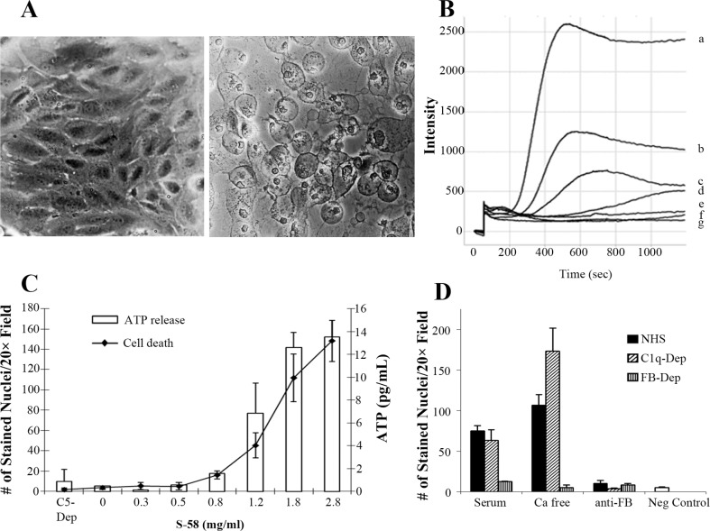
Methods: An anti-RPE antibody was developed. A spontaneously arising human RPE cell line (ARPE-19) and donor RPE cells were primed with this antibody followed by stimulation with 6% C1q-depleted human serum (C1q-Dep) to activate AP. Complement activation was evaluated by flow cytometry and immunofluorescent staining. Cellular response to complement activation was examined by measurement of intracellular calcium and adenosine triphosphate (ATP) release. Cell viability was assessed by Sytox orange, tetrazolium salt, and lactate dehydrogenase release assays.

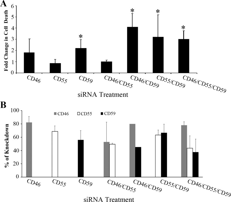
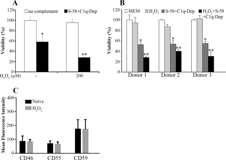
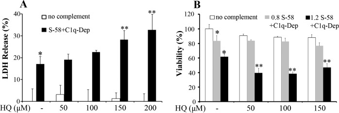
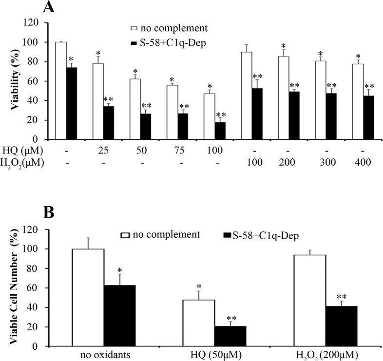
Results: Alternative pathway complement-mediated RPE cell death was associated with membrane attack complex formation and a rapid rise in intracellular calcium followed by release of ATP. Downregulation of membrane complement regulatory proteins and protein kinase C (PKC) inhibition increased cell susceptibility to complement attack. Pretreatment of RPE cells with either hydrogen peroxide or hydroquinone enhanced cell death. Chronic repetitive treatment of RPE cells with low levels of oxidants also enhanced complement-mediated cell death.

Conclusions: Activation of complement through the alternative pathway induces sublytic and lytic phases of complement attack on RPE cells, leading to cell death modulated by extracellular calcium, membrane complement regulatory proteins, and intracellular signaling mechanisms. Single-dose oxidant exposure and low-dose repetitive oxidant exposure rendered RPE cells more susceptible to complement-mediated death.

Keywords: AMD; PKC; RPE; calcium; complement; oxidative stress.


Figures

Figure 1
Figure 1
S-58 primed ARPE-19 cells in the presence of C1q-Dep form functional MAC, which kills cells through the AP but not the CP. (A) ARPE-19 cells were primed with S-58 (1.2 mg/mL) and then incubated with either 6% C1q-Dep or 6% C5-Dep. Complement attack on primed ARPE-19 cells leads to cell swelling (right panel) as compared with primed cells treated with C5-Dep (left panel). (B) Cells were primed with varying S-58 concentrations (a = 2.8 mg/mL, b = 1.8 mg/mL, c = 1.2 mg/mL, d = 0.8 mg/mL, e = 0.6 mg/mL, and f = 2.8 mg/mL in the absence of serum) or g = serum alone, with Fluo-4AM (2 μM) washed twice, and then incubated with 6% C1q-Dep or 6% C5-Dep. Intracellular calcium was determined by Fluo-4AM fluorescence intensity measurement. (C) Cells were primed with S-58 at the indicated concentrations and then incubated for 3 hours with either 6% C1q-Dep or 6% C5-Dep. The number of Sytox Orange-stained nuclei of three nonoverlapping fields per well using a ×20 objective was quantified by MetaMorph image analysis software, and is an indicator of cell death. Adenosine triphosphate release was quantified with an ATP Bioluminescence Assay Kit HS II. (D) Cells primed with S-58 (1.2 mg/mL) were washed with Ca2+-free HBSA containing EGTA and then washed in Ca2+-free HBSA without EGTA and then challenged with 6% NHS, 6% C1q-Dep, or 6% FB-Dep, in the presence or absence of exogenous Ca2+, or in the presence of an inhibitory anti-FB monoclonal antibody (20 μg/mL). Cell death was determined as described in (C). Negative control: absence of S-58.
Figure 2
Figure 2
Effects on cell survival of siRNA-mediated knockdown of ARPE-19 cell mCRPs. (A) ARPE-19 cells were transfected for 6 hours with either nontargeting control siRNA or the indicated siRNAs (2 nM) followed by media replacement. Transfected cells were cultured for 72 hours, replated in 96-well plates and cultured for an additional 24 hours. Cells were then incubated with S-58 (1.2 mg/mL) and then treated with 6% C1q-Dep. Cell death was determined by penetration of membrane impermeant dye Sytox Orange as described in the Methods. *P < 0.05 versus control siRNA-treated cells. (B) ARPE-19 cells were transfected, replated, and cultured as described in (A). Cells were then harvested and processed for FACS analysis. Individual mCRP knockdown experiments were repeated three or more times (CD46: n = 3, CD55: n = 5, CD59: n = 3) and combination mCRP knockdowns (excluding CD46/CD59) were repeated twice. The average knockdown percentage was calculated based on mean fluorescence intensity as described in figure S6. Error bars correspond to SDs of replicate experiments.
Figure 3
Figure 3
Calcium depletion and PKC or MEK1 inhibition modulate the response of ARPE-19 cells to complement attack. (A) To obtain extracellular calcium-free conditions, S-58-primed cells were washed with Ca2+-free HBSA containing EGTA (HBSS –Ca2+/–Mg2+ containing 0.02% MgCl2, 1% BSA, 0.5 mM EGTA) and then washed in Ca2+-free HBSA with no EGTA. Cells were then incubated with media supplemented with 6% C1q-Dep in Ca2+-free HBSA without EGTA in the presence of 1.5 mM Ca2+or in the absence of Ca2+. (B) Extracellular calcium-free conditions were obtained in S-58 primed cells as described in (A). Media supplemented with 6% C1q-Dep in Ca2+-free HBSA without EGTA were then added to the cells in the presence or absence of calcium at the indicated levels. The 0 mM calcium refers to the conditions before serum was added. (C, D). Cells were primed for 30 minutes with S-58 (1.2 mg/mL), washed and incubated for 10 minutes with chelerythrine (C) and PD98059 (D) at the indicated concentrations. Pre-incubation solution was removed, and then 6% C1q-Dep diluted in HBSS with the corresponding inhibitors was added. Cell death was determined as described in Figure 1C.
Figure 4
Figure 4
Oxidative stress synergizes with complement to enhance cell death. (A) The ARPE-19 cells were pretreated with the indicated concentrations of H2O2 for 1.5 hours, primed with S-58 (1.2 mg/mL), and then incubated with 6% C1q-Dep. Cell viability was determined by WST-1 assay. *(P < 0.05) versus medium alone or oxidant alone. **(P < 0.05) versus complement alone or oxidant alone. (B) Retinal pigment epithelial cells from three donors were stimulated with H2O2 (200 μM) for 1.5 hours, primed with S-58 (1.2 mg/mL), and then incubated with 6% C1q-Dep. Cell viability was examined by WST-1 assay. *(P < 0.05) versus MEM alone or oxidant alone. **(P < 0.05) versus complement alone or oxidant alone. (C) The ARPE-19 cells were treated for 1.5 hours with H2O2 (0.5 mM). Cell surface mCRP levels were determined by FACS.
Figure 5
Figure 5
Hydroquinone sensitizes donor RPE cells to complement-mediated cell injury. (A, B). Retinal pigment epithelial cells were pretreated with the indicated concentrations of HQ for 1.5 hours, incubated with S-58 Ab (1.2 mg/mL in [A], 0.8 mg/mL and 1.2 mg/mL denoted “0.8” and “1.2” in [B]), and then incubated with 6% C1q-Dep. Cell permeability and cell viability were determined by LDH assay (A) and WST-1 assay (B), respectively, in RPE cells from a 51-year-old donor. *(P < 0.05) versus MEM alone and **(P < 0.05) versus complement alone or oxidant alone.
Figure 6
Figure 6
Different oxidants generate different ROS levels. Retinal pigment epithelial cells from a 62-year-old donor were loaded with 20 μM CM-H2DCFDA for 30 minutes and washed twice. (A) Cells were treated with or without various oxidant concentrations, and ROS production was measured by relative fluorescence units (RFU) at the indicated times using a fluorescence plate reader (490-nm excitation, 522-nm emission). *P < 0.05 versus DMEM, HQ and atRal. (B) Cells assayed in (A) were washed and replaced with serum-free medium for 2 hours. Reactive oxygen species was measured followed by cell viability determination by WST assay. *P < 0.05 versus medium. **P < 0.05 versus HQ.
Figure 7
Figure 7
Repetitive HQ and H2O2 exposures sensitize donor RPE cells to complement-mediated cell injury. (A) and (B) Retinal pigment epithelial cells were treated with the indicated concentrations of HQ and H2O2 in phenol free MEM containing 1% FBS every other day for 1 week (four exposures), primed with S-58 (1.2 mg/mL) for 30 minutes, washed once, and then incubated with 6% C1q-Dep for determination of cell viability (A) and viable cell number (B) in RPE cells from a 62-year-old donor. *(P < 0.05) versus MEM alone and **(P < 0.05) versus complement alone or oxidant alone.

References

    1. Van der Schaft TL, Mooy CM, de Bruijn WC, de Jong PT. Early stages of age-related macular degeneration: an immunofluorescence and electron microscopy study. Br J Ophthalmol. 1993; 77: 657–661. - PMC - PubMed
    1. Johnson LV, Leitner WP, Staples MK, Anderson DH. Complement activation and inflammatory processes in Drusen formation and age related macular degeneration. Exp Eye Res. 2001; 73: 887– 896. - PubMed
    1. Miller RA, Bookstein F, Van der Meulen J, et al. Candidate biomarkers of aging: age-sensitive indices of immune and muscle function covary in genetically heterogeneous mice. J Gerontol A Biol Sci Med Sci. 1997; 52: B39– B47. - PMC - PubMed
    1. Mullins RF, Russell SR, Anderson DH, Hageman GS. Drusen associated with aging and age-related macular degeneration contain proteins common to extracellular deposits associated with atherosclerosis, elastosis, amyloidosis, and dense deposit disease. FASEB J. 2000; 14: 835– 846. - PubMed
    1. Edwards AO, Ritter R III, Abel KJ, Manning A, Panhuysen C, Farrer LA. Complement factor H polymorphism and age-related macular degeneration. Science. 2005; 308: 421– 424. - PubMed

Publication types

MeSH terms

LinkOut - more resources