

Thiol isomerases in thrombus formation
- PMID: 24677236
- PMCID: PMC4067134
- DOI: 10.1161/CIRCRESAHA.114.301808
Thiol isomerases in thrombus formation
Abstract
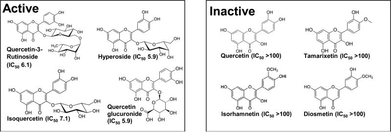
Protein disulfide isomerase (PDI), ERp5, and ERp57, among perhaps other thiol isomerases, are important for the initiation of thrombus formation. Using the laser injury thrombosis model in mice to induce in vivo arterial thrombus formation, it was shown that thrombus formation is associated with PDI secretion by platelets, that inhibition of PDI blocked platelet thrombus formation and fibrin generation, and that endothelial cell activation leads to PDI secretion. Similar results using this and other thrombosis models in mice have demonstrated the importance of ERp5 and ERp57 in the initiation of thrombus formation. The integrins, αIIbβ3 and αVβ3, play a key role in this process and interact directly with PDI, ERp5, and ERp57. The mechanism by which thiol isomerases participate in thrombus generation is being evaluated using trapping mutant forms to identify substrates of thiol isomerases that participate in the network pathways linking thiol isomerases, platelet receptor activation, and fibrin generation. PDI as an antithrombotic target is being explored using isoquercetin and quercetin 3-rutinoside, inhibitors of PDI identified by high throughput screening. Regulation of thiol isomerase expression, analysis of the storage, and secretion of thiol isomerases and determination of the electron transfer pathway are key issues to understanding this newly discovered mechanism of regulation of the initiation of thrombus formation.
Keywords: antiplatelet agents; antithrombotic agents; blood platelets; platelet aggregation inhibitor; thrombosis.
Figures
References
-
- Givol D, Goldberger RF, Anfinsen CB. Oxidation and disulfide interchange in the reactivation of reduced ribonuclease. The Journal of biological chemistry. 1964;239:PC3114–3116. - PubMed
-
- De Lorenzo F, Goldberger RF, Steers E, Jr., Givol D, Anfinsen B. Purification and properties of an enzyme from beef liver which catalyzes sulfhydryl-disulfide interchange in proteins. The Journal of biological chemistry. 1966;241:1562–1567. - PubMed
-
- Vaux D, Tooze J, Fuller S. Identification by anti-idiotype antibodies of an intracellular membrane protein that recognizes a mammalian endoplasmic reticulum retention signal. Nature. 1990;345:495–502. - PubMed
-
- Chen K, Lin Y, Detwiler TC. Protein disulfide isomerase activity is released by activated platelets. Blood. 1992;79:2226–2228. - PubMed
Publication types
MeSH terms
Substances
Grants and funding
LinkOut - more resources
Full Text Sources
Other Literature Sources
Medical
Molecular Biology Databases
Miscellaneous
