. 2014 Jul;29(7):1531-1540.
doi: 10.1002/jbmr.2200.

METTL21C is a potential pleiotropic gene for osteoporosis and sarcopenia acting through the modulation of the NF-κB signaling pathway

Affiliations

METTL21C is a potential pleiotropic gene for osteoporosis and sarcopenia acting through the modulation of the NF-κB signaling pathway

Jian Huang et al. J Bone Miner Res. 2014 Jul.

Abstract

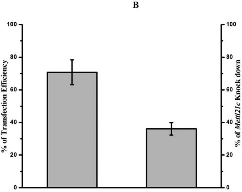
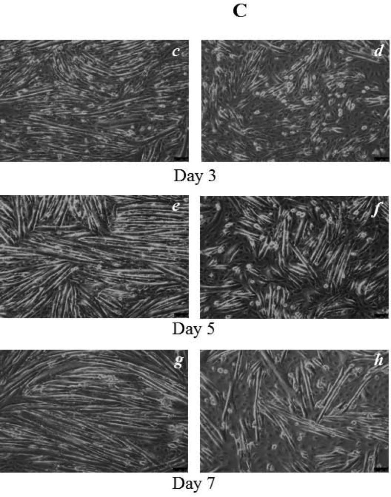
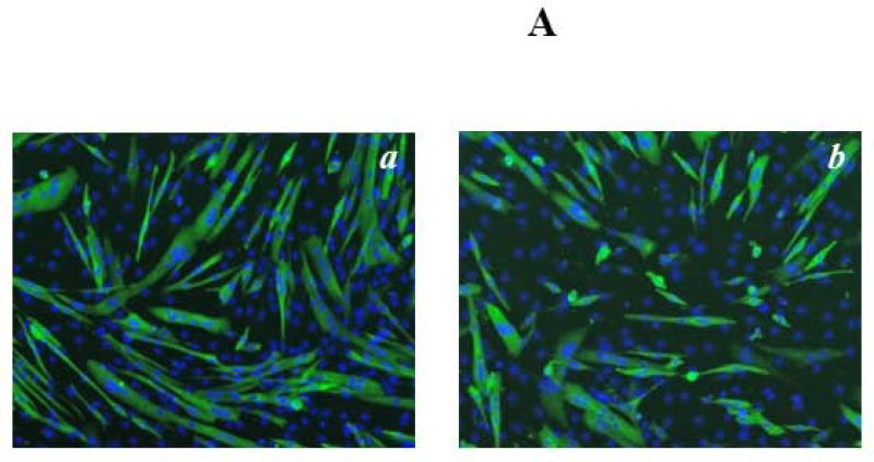
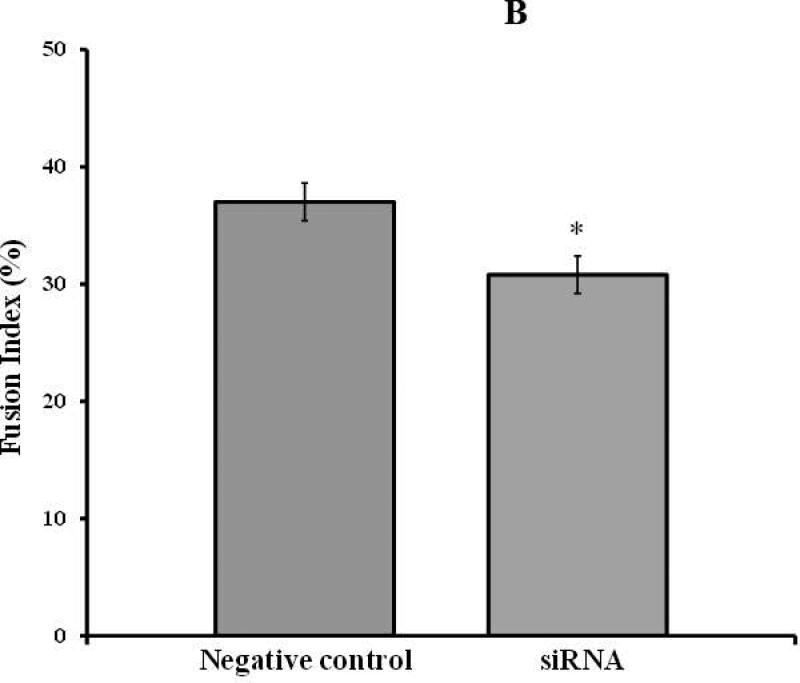
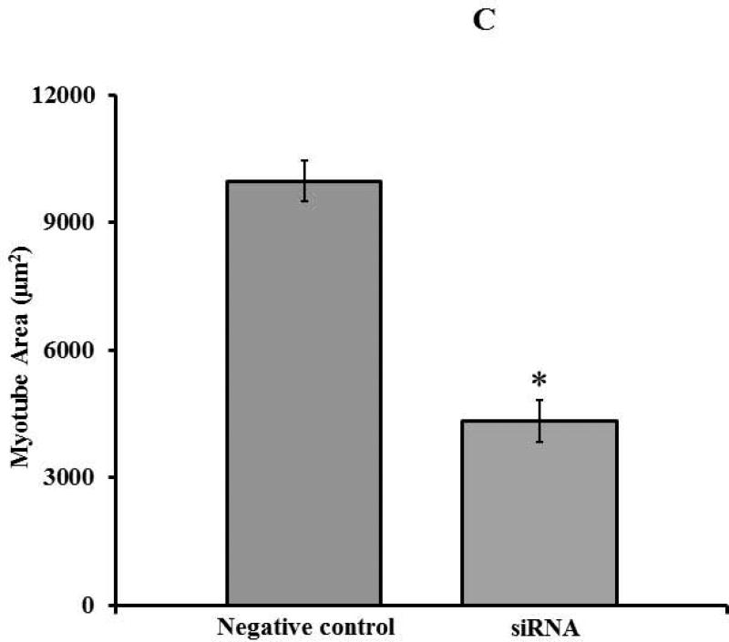
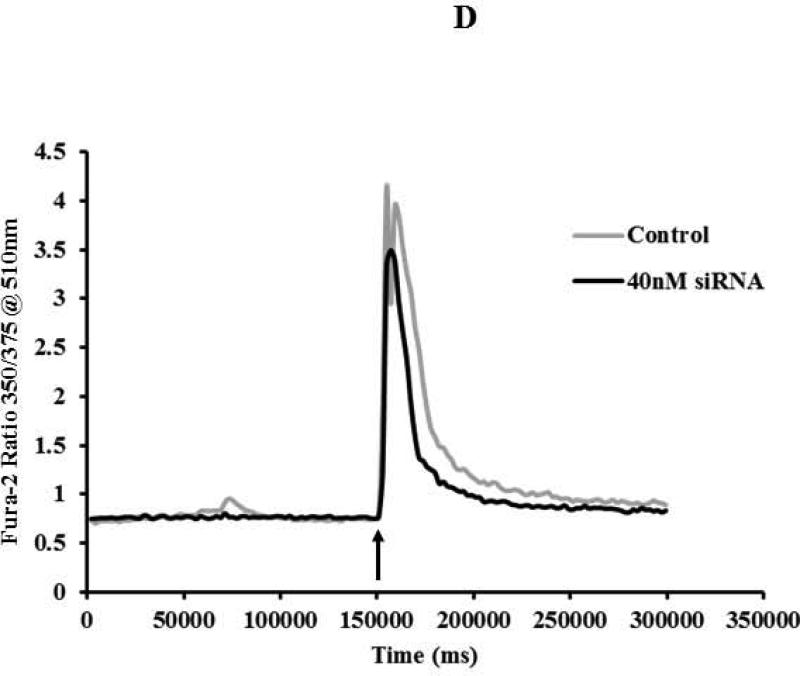
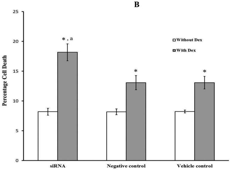
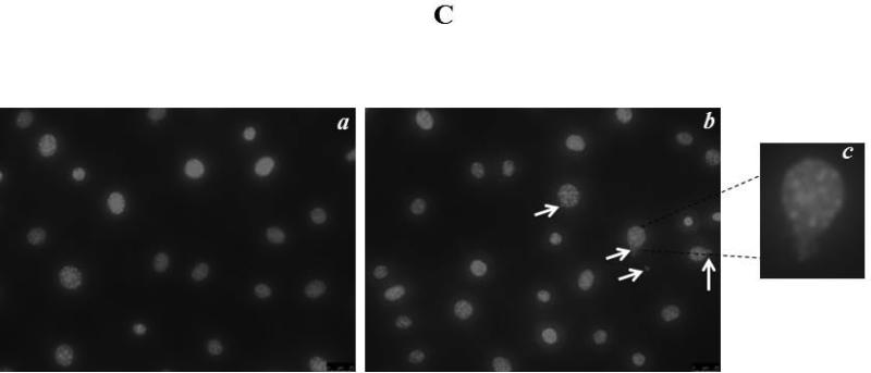
Sarcopenia and osteoporosis are important public health problems that occur concurrently. A bivariate genome-wide association study (GWAS) identified METTL21c as a suggestive pleiotropic gene for both bone and muscle. The METTL21 family of proteins methylates chaperones involved in the etiology of both myopathy and inclusion body myositis with Paget's disease. To validate these GWAS results, Mettl21c mRNA expression was reduced with siRNA in a mouse myogenic C2C12 cell line and the mouse osteocyte-like cell line MLO-Y4. At day 3, as C2C12 myoblasts start to differentiate into myotubes, a significant reduction in the number of myocytes aligning/organizing for fusion was observed in the siRNA-treated cells. At day 5, both fewer and smaller myotubes were observed in the siRNA-treated cells as confirmed by histomorphometric analyses and immunostaining with myosin heavy chain (MHC) antibody, which only stains myocytes/myotubes but not myoblasts. Intracellular calcium (Ca(2+)) measurements of the siRNA-treated myotubes showed a decrease in maximal amplitude peak response to caffeine, suggesting that less Ca(2+) is available for release due to the partial silencing of Mettl21c, correlating with impaired myogenesis. In siRNA-treated MLO-Y4 cells, 48 hours after treatment with dexamethasone there was a significant increase in cell death, suggesting a role of Mettl21c in osteocyte survival. To investigate the molecular signaling machinery induced by the partial silencing of Mettl21c, we used a real-time PCR gene array to monitor the activity of 10 signaling pathways. We discovered that Mettl21c knockdown modulated only the NF-κB signaling pathway (ie, Birc3, Ccl5, and Tnf). These results suggest that Mettl21c might exert its bone-muscle pleiotropic function via the regulation of the NF-κB signaling pathway, which is critical for bone and muscle homeostasis. These studies also provide rationale for cellular and molecular validation of GWAS, and warrant additional in vitro and in vivo studies to advance our understanding of role of METTL21C in musculoskeletal biology.

Keywords: BONE-MUSCLE INTERACTIONS; GENETIC RESEARCH; HUMAN ASSOCIATION STUDIES; OSTEOCYTES; SKELETAL MUSCLE.


Figures

Fig 1
Fig 1. Partial Knockdown of Mettl21C Alters the Morphological Phenotype of C2C12 Myoblasts during Myogenic Differentiation Indicating Impaired Myogenesis
A) Representative image of C2C12 myoblasts 24 hours after transfection by All Star negative control siRNA (40nM). a) Phase contrast image, b) Fluorescent image, both images taken from the same representative area. For quantification, 4 different areas (~400 cells) were used from 5 different experiments. B) Under these experimental conditions (see Methods for details) transfection efficiency equaled 70.8 ± 7.6%, leading to a decrease of 36.1 ± 4.0% in Mettl21c expression as detected by RT-qPCR. C) Each pair of images (c-d, e-f, g-h) shows representative high resolution 14-BIT CCD camera phase contrast images of the negative control and Mettl21csiRNA-treated C2C12 cells, at 3, 5, and 7 days of differentiation, respectively. Significantly less in number and smaller myotubes are observed in Mettl21c-siRNA-treated C2C12 cells during differentiation. Vehicle control results were essentially identical to the negative control (See Table 1). Calibration bar = 100μm.
Fig 2
Fig 2. Partial Silencing of Mettl21c Reduces Fusion Index, Myotube Cell area, and Calcium Release from the Sarcoplasmic Reticulum
A) Representative fluorescence images of DAPI-stained nuclei (blue) and myosin heavy chain antibody (MHC, green) of C2C12 at 3 days of differentiation after transfection. a) Negative control and b) Mettl21c-siRNA-treated C2C12 cells. B) Summary data show that the Fusion Index in Mettl21c-siRNA-treated C2C12 cells decreased significantly compared to negative control (p<0.05). C) Summary data show that myotube cell area in Mettl21c-siRNA-treated C2C12 cells drastically decreased compared to negative control (p<0.0001). D) Representative calcium transients induced by 20mM caffeine (arrow) on C2C12 myotubes loaded with Fura-2/AM. In Mettl21c-siRNA-treated C2C12 myotubes at day 5 of differentiation, compared to negative control, the amplitude peak calcium response to caffeine was significantly decreased and the relaxation phase of the transient was shorter (p<0.01).
Fig 3
Fig 3. Partial Knockdown of Mettl21c in MLO-Y4 osteocytes increased cell death induced by dexamethasone
A) Representative image of MLO-Y4 osteocytes 24 hours after transfection of All Star negative control siRNA (200nM). a) Phase contract image, b) Fluorescent image of tagged siRNA; Both images were taken from the same area; c) Merging of a) and b). B) Summary data of cell death in MLO-Y4 cells transfected with Mettl21C siRNA, with and without dexamethasone (Dex) treatment. Cell death was detected 48h after treatment by trypan blue exclusion assay (*Dex increased cell death under all conditions; and aMettl21c knockdown induced cell death levels higher than Dex alone, p < 0.05). C) The Nuclear fragmentation assay shows the a) absence of nuclear blebbing in control MLO-Y4 cells as compared with the b) enhanced blebbing in the siRNA-treated MLO-Y4 osteocytes; c) The enlarged image clearly shows the blebbing process in siRNA-treated MLO-Y4 osteocytes.

Similar articles

Cited by

References

    1. Bijlsma AY, Meskers CG, Westendorp RG, Maier AB. Chronology of age-related disease definitions: osteoporosis and sarcopenia. Ageing Res Rev. 2012;11(2):320–4. - PubMed
    1. Buford TW, Anton SD, Judge AR, Marzetti E, Wohlgemuth SE, Carter CS, et al. Models of accelerated sarcopenia: critical pieces for solving the puzzle of age-related muscle atrophy. Ageing Res Rev. 2010;9(4):369–83. - PMC - PubMed
    1. Brotto M, Abreu EL. Sarcopenia: pharmacology of today and tomorrow. J Pharmacol Exp Ther. 2012;343(3):540–6. - PMC - PubMed
    1. Janssen I, Shepard DS, Katzmarzyk PT, Roubenoff R. The healthcare costs of sarcopenia in the United States. J Am Geriatr Soc. 2004;52(1):80–5. - PubMed
    1. Karsenty G, Ferron M. The contribution of bone to whole-organism physiology. Nature. 2012;481(7381):314–20. - PMC - PubMed

Publication types

MeSH terms