. 2014 May;23(5):627-38.
doi: 10.1002/pro.2445. Epub 2014 Mar 11.

Completing the structural family portrait of the human EphB tyrosine kinase domains

Affiliations

Completing the structural family portrait of the human EphB tyrosine kinase domains

Ross C Overman et al. Protein Sci. 2014 May.

Abstract

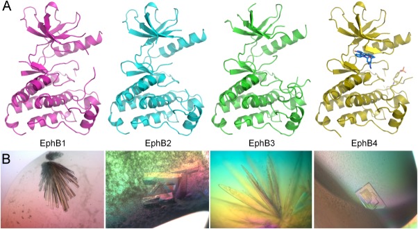
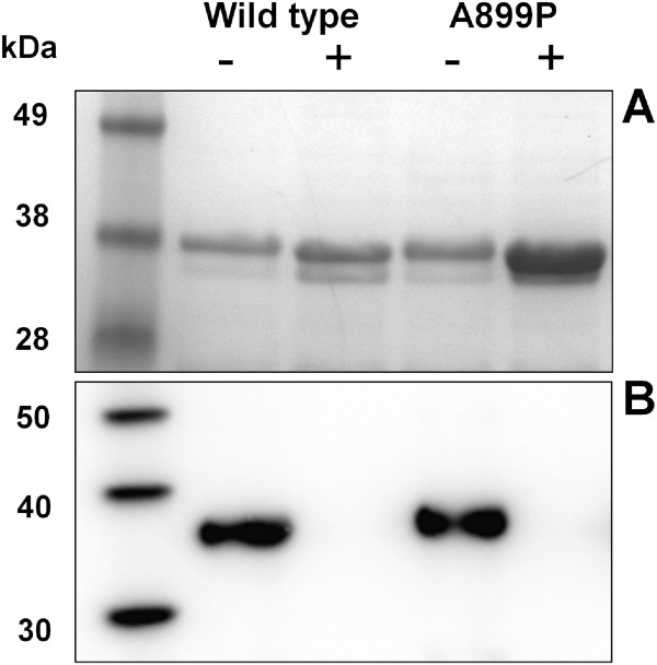
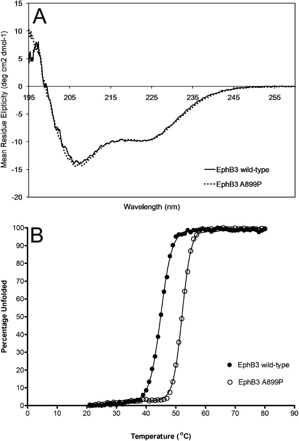
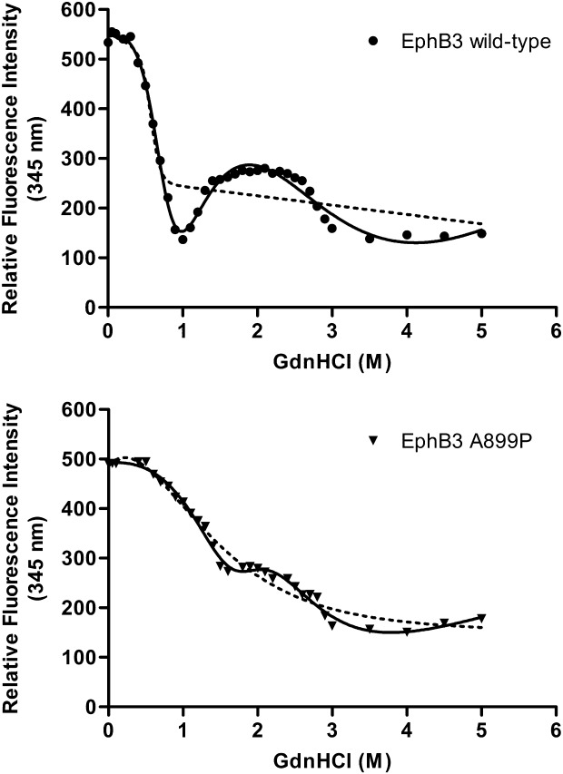
The EphB receptors have key roles in cell morphology, adhesion, migration and invasion, and their aberrant action has been linked with the development and progression of many different tumor types. Their conflicting expression patterns in cancer tissues, combined with their high sequence and structural identity, present interesting challenges to those seeking to develop selective therapeutic molecules targeting this large receptor family. Here, we present the first structure of the EphB1 tyrosine kinase domain determined by X-ray crystallography to 2.5Å. Our comparative crystalisation analysis of the human EphB family kinases has also yielded new crystal forms of the human EphB2 and EphB4 catalytic domains. Unable to crystallize the wild-type EphB3 kinase domain, we used rational engineering (based on our new structures of EphB1, EphB2, and EphB4) to identify a single point mutation which facilitated its crystallization and structure determination to 2.2 Å. This mutation also improved the soluble recombinant yield of this kinase within Escherichia coli, and increased both its intrinsic stability and catalytic turnover, without affecting its ligand-binding profile. The partial ordering of the activation loop in the EphB3 structure alludes to a potential cis-phosphorylation mechanism for the EphB kinases. With the kinase domain structures of all four catalytically competent human EphB receptors now determined, a picture begins to emerge of possible opportunities to produce EphB isozyme-selective kinase inhibitors for mechanistic studies and therapeutic applications.

Keywords: EphB1; EphB2; EphB3; EphB4; activation mechanism; crystallography; ligand-binding sites; protein engineering.


Figures

Figure 1
Figure 1
Structural gallery of the EphB kinase domains. (A) Backbone ribbon representations of the kinase domain structures of EphB1 (pink), EphB2 (cyan), EphB3 A899P (green), and EphB4 plus staurosporine (yellow). (B) Corresponding light microscope images of diffraction-quality crystals of EphB kinase domains. PyMol was used to prepare all structure images (http://www.pymol.org).
Figure 2
Figure 2
E. coli expression results of EphB3 kinase domains. (A) shows an SDS PAGE gel analysis of hexahistidine affinity-purified samples from soluble fraction of E. coli cell lysates; 0.5 ml of culture equivalent loaded per lane,-/+ refers to co-expression with PTP1B. (B) Shows an anti-phosphotyrosine western blot analysis of the same affinity purified samples; 0.5 µg of kinase loaded per lane.
Figure 3
Figure 3
Thermal unfolding of EphB3 kinases. (A) shows a molar concentration adjusted 260 to 195 nm CD wavelength scan of each of EphB3 and EphB3 A899P kinase domains to demonstrate their secondary-structure profiles, n = 3—the web server K2d was used to estimate the percentages of protein secondary structure from these circular dichroism spectra; EphB3 wild-type: 32% alpha helical, 15% beta strand, 52% random coil, EphB3 A899P: 36% alpha helical, 16% beta strand, 48% random coil. (B) Shows averaged thermal-unfolding transitions obtained from CD experiments monitored at 222 nm (alpha-helical response). Unfolding transition data were fitted to a six-parameter unfolding equation, using Prism software to generate comparative melting temperatures (Tm) and van't Hoff enthalpies of unfolding (ΔUH(Tm)).
Figure 4
Figure 4
Chaotropic unfolding of EphB3 kinases. Unfolding of EphB3 wild-type and A899P kinase domains with increasing GdnHCl was monitored by following the progressive quenching of internal tryptophan fluorescence (Ex 295 nm, Em 345 nm). Units shown on the y-axis are relative fluorescence units. Curves were fitted to both two-state (dotted line) and three-state (unbroken line) models. n = 3 for all.
Figure 5
Figure 5
EphB3 structure observations. (A) Backbone representation of EphB catalytic domain structures [color scheme follows Fig. 1(A)], after superposition on the C-terminal lobes. (B) A ribbon overlay of the active sites of EphB4 (black) with CMPD1 (grey) (PDB: 2VWU), and EphB3 (green) highlighting the key difference at Gly699 (Cys717 in EphB3) and Ala700 (Ser706 in EphB2), the surface shown is that of EphB3; (C) surface representation of the EphB3 substrate-binding groove with the partially ordered EphB3 activation loop and HRD motif (green sticks), overlaid with part of the EPHOPT peptide (orange sticks) from the EphA3 complex structure (3FXX).

References

    1. Pasquale EB. Eph-ephrin bidirectional signaling in physiology and disease. Cell. 2008;133:38–52. - PubMed
    1. Kullander K, Klein R. Mechanisms and functions of Eph and ephrin signalling. Nat Rev Mol Cell Biol. 2002;3:475–486. - PubMed
    1. Pasquale EB. Eph receptor signalling casts a wide net on cell behaviour. Nat Rev Mol Cell Biol. 2005;6:462–475. - PubMed
    1. Pasquale EB. Eph receptors and ephrins in cancer: bidirectional signalling and beyond. Nat Rev Cancer. 2010;10:165–180. - PMC - PubMed
    1. Choi Y, Syeda F, Walker JR, Finerty PJ, Jr, Cuerrier D, Wojciechowski A, Liu Q, Dhe-Paganon S, Gray NS. Discovery and structural analysis of Eph receptor tyrosine kinase inhibitors. Bioorg Med Chem Lett. 2009;19:4467–4470. - PMC - PubMed

Publication types