. 2014 Jun;243(6):753-64.
doi: 10.1002/dvdy.24118. Epub 2014 Mar 12.

The minus-end actin capping protein, UNC-94/tropomodulin, regulates development of the Caenorhabditis elegans intestine

Affiliations

The minus-end actin capping protein, UNC-94/tropomodulin, regulates development of the Caenorhabditis elegans intestine

Elisabeth Cox-Paulson et al. Dev Dyn. 2014 Jun.

Abstract

Background: Tropomodulins are actin-capping proteins that regulate the stability of the slow-growing, minus-ends of actin filaments. The C. elegans tropomodulin homolog, UNC-94, has sequence and functional similarity to vertebrate tropomodulins. We investigated the role of UNC-94 in C. elegans intestinal morphogenesis.

Results: In the embryonic C. elegans intestine, UNC-94 localizes to the terminal web, an actin- and intermediate filament-rich structure that underlies the apical membrane. Loss of UNC-94 function results in areas of flattened intestinal lumen. In worms homozygous for the strong loss-of-function allele, unc-94(tm724), the terminal web is thinner and the amount of F-actin is reduced, pointing to a role for UNC-94 in regulating the structure of the terminal web. The non-muscle myosin, NMY-1, also localizes to the terminal web, and we present evidence that increasing actomyosin contractility by depleting the myosin phosphatase regulatory subunit, mel-11, can rescue the flattened lumen phenotype of unc-94 mutants.

Conclusions: The data support a model in which minus-end actin capping by UNC-94 promotes proper F-actin structure and contraction in the terminal web, yielding proper shape of the intestinal lumen. This establishes a new role for a tropomodulin in regulating lumen shape during tubulogenesis.

Keywords: actomyosin contractility; terminal web; tubulogenesis.


Figures

Figure 1
Figure 1
Tropomodulin structure and UNC-94 localization in the embryonic C. elegans intestine. A: Structure of Human Tmod1, C. elegans UNC-94 and TMD-2 and position of mutations that were studied. Tropomodulins have two actin capping regions: a tropomyosin-dependent actin binding domain at the N-terminus (TM-CAP) and an actin binding domain at the C-terminus that contains leucine rich repeats (LRR-CAP). The percentage similarity of these domains to those in human Tmod1 is indicated below the domain (Yamashiro et al., 2012). The unc-94 gene encodes two isoforms that differ in their first exon and 5′ UTR. The protein structure of UNC-94a is shown, and lines indicate which regions of the gene encode the protein domains. The tmd-2 gene also encodes two isoforms, with tmd-2b encoding a C-terminal extension that is not present in tmd-2a. The protein structure of TMD-2b is shown, and lines indicate which regions of the gene encode the protein domains. The location of mutations that were studied is shown below the gene structures. B: Embryo co-stained for UNC-94 (via antibody staining) and F-actin (via phalloidin staining). Arrow points to co-localization at the intestinal terminal web. In all images, the anterior of the intestine is positioned to the left.
Figure 2
Figure 2
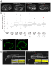
UNC-94 is required for proper intestinal shape in C. elegans embryos and adults. A: Embryos stained with phalloidin (3 - 3.5 fold stage). Red arrows indicate regions of abnormally wide intestinal lumen diameter. These are representative images and do not show the most extreme phenotypes observed. Bar = 10 microns. B: Box plots showing distribution of maximum lumen diameters for 3 - 3.5 fold C. elegans embryos of various genotypes. Black asterisks: statistically different from wild-type (p < 0.001 in a two-sided Student’s t-test). Blue asterisk: statistically different from wild-type; erm-1::gfp control (p < 0.001 in a Mann-Whitney U test). Red asterisk: statistically different from tmd-2(ok3417);L4440(RNAi) (p < 0.001 in a Mann-Whitney U test) but not statistically different from unc-94(RNAi). L4440 is the empty feeding vector used as a negative control. C: 3D reconstructions of 3-fold stage wild-type and unc-94(tm724) embryos expressing erm-1::gfp. Bar = 10 microns. Movies available in Supplemental Material. D: 3D reconstructions of adult wild-type (lateral view) and unc-94(tm724) (transverse view) worms expressing erm-1::gfp. Insets show 3D models of the intestinal surface in the boxed regions and are magnified. The anteroposterior (AP), dorsal-ventral (DV), and transverse (T) axes are indicated. White arrows point to the excretory cell body and red arrows point to cystic excretory canals. Bar = 50 microns.
Figure 3
Figure 3
UNC-94 regulates F-actin levels in the intestinal terminal web, but does not regulate levels of the intermediate filament protein, IFB-2. A: Images of phalloidin stained 3 - 3.5 fold embryos and graph showing quantification of fluorescence intensity. Images on the left show a wild-type embryo (average fluorescence intensity = 1155 on a scale of 0–4095) and a unc-94(tm724) embryo (average fluorescent intensity = 816). B: Images of 3 - 3.5 fold embryos immunostained for IFB-2 with the monoclonal MH33 antibody and graph showing quantification of fluorescence intensity. Images on the left are from 3D reconstructions, and the arrowhead points to small cytoplasmic accumulations of IFB-2. In A and B, bars indicate the standard error of the mean. Asterisk indicates a statistically significance difference in comparison to the wild-type control (p< 0.001 in a two-sided Student’s t-test).
Figure 4
Figure 4
Loss of UNC-94 function does not compromise cell junctions. A: Top images show 2-fold embryos stained for HMR-1/cadherin to mark adherens junctions (green), and IFB-2 (blue) to mark the terminal web. Bottom images are of 2-fold embryos stained for AJM-1, a basal component of apical junctions in C. elegans. In the unc-94(tm724) embryo shown, there is greater distance between some of the junctions within the same intestinal ring, indicating that the lumen is wider in these regions. Asterisks mark the first intestinal ring. Bar = 10 microns. Enlargements of the HMR-1 and AJM-1 staining in the boxed regions are provided to the right of the originals. B: TEM images of adult C. elegans worms. Top images show electron dense apical junctions (AJ), and bottom images show microvilli (M) and the terminal web (TW).
Figure 5
Figure 5
Increasing actomyosin contraction partially rescues the unc-94(tm724) mutant phenotype. A: Wild-type and unc-94(tm724) C. elegans embryos immunostained for NMY-1 and IFB-2 (to mark the terminal web). B: Quantification of fluorescence intensity in wild-type and unc-94(tm724) C. elegans embryos immunostained for NMY-1. Bars show standard error of the mean. C: C. elegans embryo expressing MEL-11::GFP. D: Graph showing maximum lumen diameters of wild-type and unc-94(tm724) mutants treated with mel-11(RNAi) or with the empty feeding vector (L4440). Widths were measured at the widest part of the lumen; and the percentage of 3 - 3.5 fold embryos with the designated lumen widths is shown. E: 3D reconstruction of let-502(ca201) heterozygous embryo stained for F-actin. Red arrow points to an area where the intestinal lumen is expanded, but not flattened. Movie available in Supplemental Material. Bars = 10 microns.
Figure 6
Figure 6
UNC-94 does not nucleate F-actin in vitro. Pyrene labeled G-actin at 4 μM was pre-incubated with buffer only (black), or buffer with 0.4 μM UNC-94 (red) or 0.8 μM UNC-94 (green), and was polymerized at time zero by adding polymerization buffer. Kinetics of actin polymerization was monitored by pyrene fluorescence, which is increased upon polymerization.

References

    1. Bernadskaya YY, Patel FB, Hsu HT, Soto MC. Arp2/3 promotes junction formation and maintenance in the Caenorhabditis elegans intestine by regulating membrane association of apical proteins. Mol Biol Cell. 2011;22:2886–2899. - PMC - PubMed
    1. Bretscher A, Weber K. Localization of actin and microfilament-associated proteins in the microvilli and terminal web of the intestinal brush border by immunofluorescence microscopy. J Cell Biol. 1978;79:839–845. - PMC - PubMed
    1. Buechner M. Tubes and the single C. elegans excretory cell. Trends Cell Biol. 2002;10:479–84. - PubMed
    1. Burgess DR. Reactivation of intestinal epithelial cell brush border motility: ATP-dependent contraction via a terminal web contractile ring. J Cell Biol. 1982;95:853–863. - PMC - PubMed
    1. Carberry K, Wiesenfahrt T, Geisler F, Stocker S, Gerhardus H, Uberbach D, Davis W, Jorgensen E, Leube RE, Bossinger O. The novel intestinal filament organizer IFO-1 contributes to epithelial integrity in concert with ERM-1 and DLG-1. Development. 2012;139:1851–1862. - PMC - PubMed

Publication types

MeSH terms

LinkOut - more resources