Review
. 2014 May;23(5):551-65.
doi: 10.1002/pro.2446. Epub 2014 Mar 19.

Structure and function of LGR5: an enigmatic G-protein coupled receptor marking stem cells

Affiliations
Review

Structure and function of LGR5: an enigmatic G-protein coupled receptor marking stem cells

Kaavya Krishna Kumar et al. Protein Sci. 2014 May.

Abstract

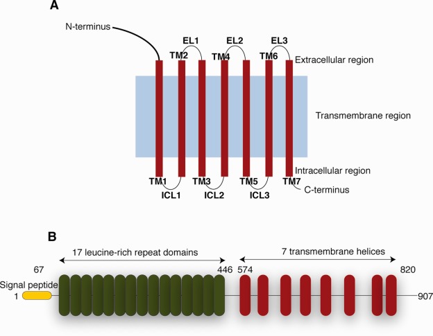
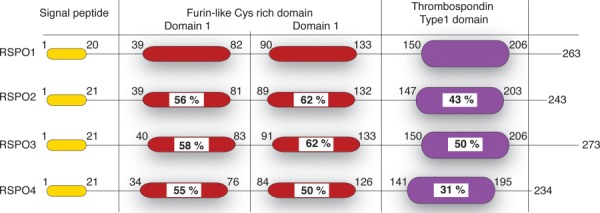
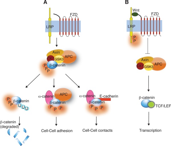
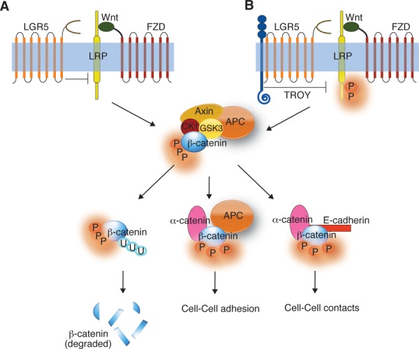
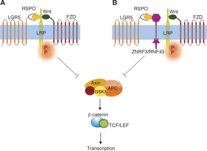
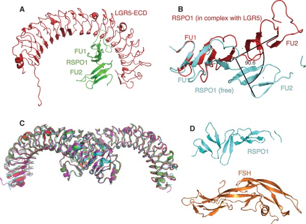
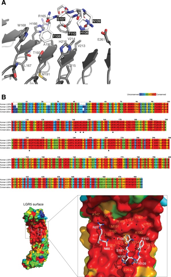
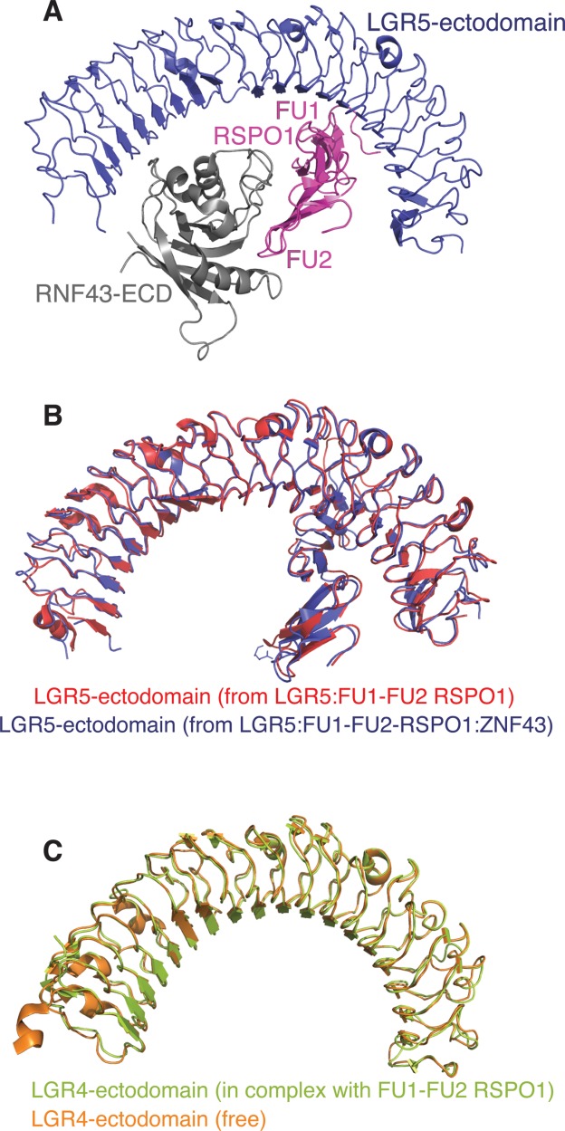
G-protein coupled receptors (GPCRs) are an important class of membrane protein that transmit extracellular signals invoked by sensing molecules such as hormones and neurotransmitters. GPCR dysfunction is implicated in many diseases and hence these proteins are of great interest to academia and the pharmaceutical industry. Leucine-rich repeat-containing GPCRs contain a characteristic extracellular domain that is an important modulator of intracellular signaling. One member of this class is the leucine-rich repeat-containing G-protein-coupled receptor 5 (LGR5), a stem cell marker in intestinal crypts, and mammary glands. LGR5 modulates Wnt signaling in the presence of the ligand R-spondin (RSPO). The mechanism of activation of LGR5 by RSPO is not understood, nor is the intracellular signaling mechanism known. Recently reported structures of the extracellular domain of LGR5 bound to RSPO reveal a horseshoe-shaped architecture made up of consecutive leucine-rich repeats, with RSPO bound on the concave surface. This review discusses the discovery of LGR5 and the impact it is having on our understanding of stem cell and cancer biology of the colon. In addition, it covers functional relationships suggested by sequence homology and structural analyses, as well as some intriguing conundrums with respect to the involvement of LGR5 in Wnt signaling.

Keywords: GPCR; LGR5; RSPO; Wnt signaling; colon cancer; stem cells.


Figures

Figure 1
Figure 1
Schematic presentation of the general structure of GPCRs and LGR5. (A) General architecture of GPCRs. (B) LGR5 contains a signal peptide (yellow) followed by 17 leucine-rich repeat (LRR) domains (red). It contains a linker region between the last LRR and the first TM domain, followed by a seven helical TM domain homologs to rhodopsin-like GPCR.
Figure 2
Figure 2
Schematic representation of the domain architecture of RSPO. RSPOs contain a signal peptide followed by two furin-like Cys-rich repeats (red). It contains a thrombospondin type1 domain (violet) and a C-terminal tail of varying lengths. Numbers represent the amino-acid numbers for RSPO. Sequence identity compared to RSPO1 is written as % within the domains.
Figure 3
Figure 3
Wnt signaling pathways. (A) In the absence of Wnt, the “destruction complex” (formed by Axin, GSK3, CK1, and APC) phosphorylates β-catenin targeting for ubiquitination and subsequent degradation. In addition, phospho-β-catenin is involved in cell-cell adhesion (with α-catenin and APC) and in cell–cell contacts (with α-catenin and E-cadherin). (B) When Wnt is present, it binds to FZD and LRP forming a ternary complex. This complex inhibits the phosphorylation of β-catenin by the “destruction complex” resulting in translocation of β-catenin into the nucleus. In the nucleus β-catenin binds TCF/LEF resulting in gene transcription.
Figure 4
Figure 4
Effect of LGR5 overexpression on Wnt signaling. (A) Overexpression of LGR5 might antagonize Wnt signaling by sequestering LRP5/6, resulting in β-catenin degradation. (B) LGR5 might downregulate Wnt signaling by recruiting TROY that might, in turn, inhibit LRP5/6 leading to the degradation of β-catenin. Scenarios (A) and (B) results in an increase in cell-cell adhesion and cell-cell contacts.
Figure 5
Figure 5
Effect of RSPO:LGR5 complex on Wnt signaling. (A) LGR5:RSPO interacts with FZD, LRP, and Wnt to form a “potentiating complex” that inhibits the phosphorylation of β-catenin by the “destruction complex.” This results in gene transcription (enhance Wnt signaling). (B) The LGR5:RSPO complex might interact with the negative Wnt regulator, ZNRF3/RNF43 to enhance Wnt signaling.
Figure 6
Figure 6
Crystal structures of LGR5-ectodomain:RSPO1 complexes. (A) X-ray crystal structure of the LGR5-ECD (red) in complex with the two furin-like domains (FU1-FU2) of RSPO1 (green) (PDB code: 4BSS). (B) The structures of the FU1-FU2 domains from free RSPO1 (cyan, PDB code: 4BSO) and RSPO1 in complex with LGR5 (red, PDB code: 4BSS) show a 90.5° change in orientation relative to each other. (C) Overlay (Cα over 482 residues LGR5:RSPO complex) of the four crystal forms of LGR5:RSPO complex. P61224 (green, PDB code:4BST), C2 (cyan, PDB code: 4BSU), P22121 (magenta, PDB code: 4BSR), P21 (red, PDB code: 4BSS). (D) Structure of RSPO1 (cyan; PDB code: 4BSO) as compared to FSH structure (orange; PDB code: 1FL7).
Figure 7
Figure 7
LGR5:RSPO interface. (A) Residues R165 to W168 on LGR5 (gray) make close contacts with residues F106 to F110 on RSPO1 (white). (B) Sequence alignment of human LGR4–6. Residues are colored according to conservation (Highly conserved (Red) to poorly conserved (Blue). Residues that make a H-bond with RSPO1 are marked with a dotted-line (black) (Top). The surface representation of LGR5 colored according to the sequence conservation with RSPO residues in stick representation (white) (bottom). Residues 106–110 in RSPO1 (stick representation; white) are lined by residues in LRR5 (R165, H166, L167, and W168), LRR6 (A190, M191, T192, and L193) and LRR7 (V213, V214, L215, and H216) of LGR5 (surface representation).
Figure 8
Figure 8
Structures of LGR5/4-ectodomain:RSPO1 complexes. (A) Structure of LGR5-ECD (blue) in a ternary complex with FU1-FU2 domains of RSPO1 (magenta) and RNF43-ECD (gray) (PDB code: 4KNG). (B) Overlay of LGR5-ectodomain:RSPO1 (PDB code: 4BSS) and LGR5-ectodomain:RSPO1:RNF43-ectodomain (PDB code: 4KNG) (Cα 543). (C) The structures of free LGR4 (orange, PDB code: 4LI1) and LGR4 in complex with FU1-FU2 domains of RSPO1 (light green, PDB code: 4LI2) overlay with a RMSD of 0.6 Å (Cα 452).

References

    1. Fredriksson R, Lagerstrom MC, Lundin LG, Schioth HB. The G-protein-coupled receptors in the human genome form five main families. Phylogenetic analysis, paralogon groups, and fingerprints. Mol Pharmacol. 2003;63:1256–1272. - PubMed
    1. Ji TH, Grossmann M, Ji I. G protein-coupled receptors. I. Diversity of receptor-ligand interactions. J Biol Chem. 1998;273:17299–17302. - PubMed
    1. Lundstrom K. Latest development in drug discovery on G protein-coupled receptors. Curr Protein Pept Sci. 2006;7:465–470. - PubMed
    1. Chun E, Thompson AA, Liu W, Roth CB, Griffith MT, Katritch V, Kunken J, Xu F, Cherezov V, Hanson MA, Stevens RC. Fusion partner toolchest for the stabilization and crystallization of G protein-coupled receptors. Structure. 2012;20:967–976. - PMC - PubMed
    1. Haga K, Kruse AC, Asada H, Yurugi-Kobayashi T, Shiroishi M, Zhang C, Weis WI, Okada T, Kobilka BK, Haga T, Kobayashi T. Structure of the human M2 muscarinic acetylcholine receptor bound to an antagonist. Nature. 2012;482:547–551. - PMC - PubMed

Publication types