. 2014 Jun;32(6):1503-14.
doi: 10.1002/stem.1677.

Erythropoiesis from human embryonic stem cells through erythropoietin-independent AKT signaling

Affiliations

Erythropoiesis from human embryonic stem cells through erythropoietin-independent AKT signaling

William S Kim et al. Stem Cells. 2014 Jun.

Abstract

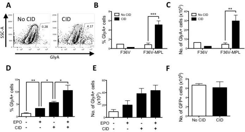
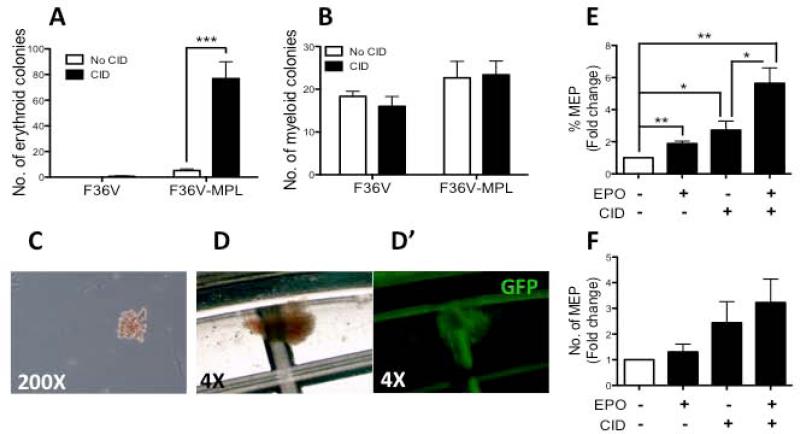
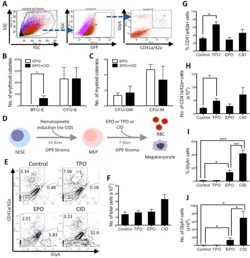
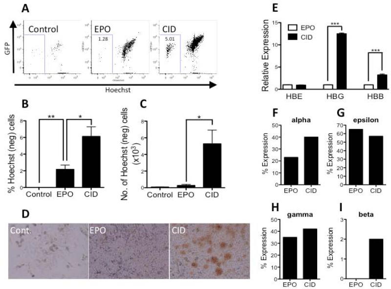
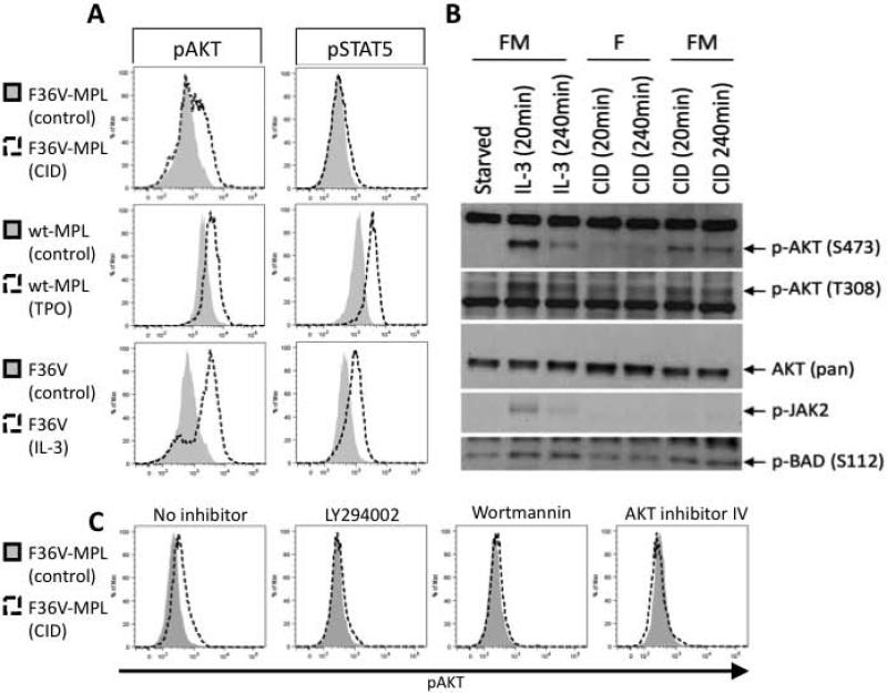
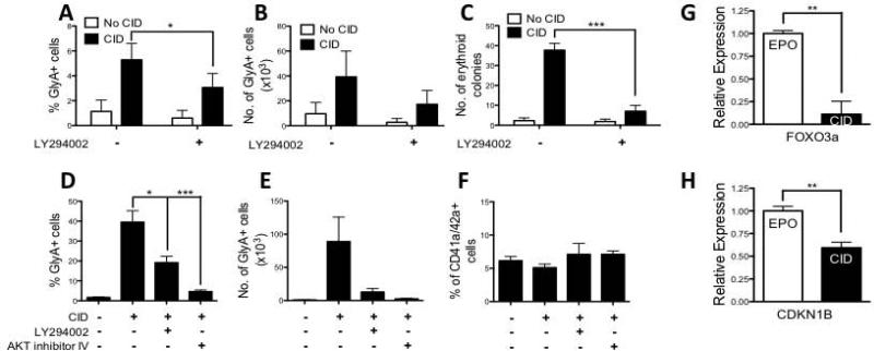
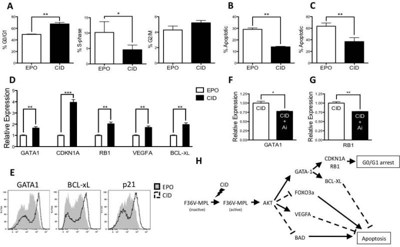
Unlimited self renewal capacity and differentiation potential make human pluripotent stem cells (PSC) a promising source for the ex vivo manufacture of red blood cells (RBCs) for safe transfusion. Current methods to induce erythropoiesis from PSC suffer from low yields of RBCs, most of which are immature and contain embryonic and fetal rather than adult hemoglobins. We have previously shown that homodimerization of the intracellular component of MPL (ic-MPL) induces erythropoiesis from human cord blood progenitors. The goal of this study was to investigate the potential of ic-MPL dimerization to induce erythropoiesis from human embryonic stem cells (hESCs) and to identify the signaling pathways activated by this strategy. We present here the evidence that ic-MPL dimerization induces erythropoietin (EPO)-independent erythroid differentiation from hESC by inducing the generation of erythroid progenitors and by promoting more efficient erythroid maturation with increased RBC enucleation as well as increased gamma:epsilon globin ratio and production of beta-globin protein. ic-MPL dimerization is significantly more potent than EPO in inducing erythropoiesis, and its effect is additive to EPO. Signaling studies show that dimerization of ic-MPL, unlike stimulation of the wild type MPL receptor, activates AKT in the absence of JAK2/STAT5 signaling. AKT activation upregulates GATA-1 and FOXO3 transcriptional pathways with resulting inhibition of apoptosis, modulation of cell cycle, and enhanced maturation of erythroid cells. These findings open up potential new targets for the generation of therapeutically relevant RBC products from hPSC.

Keywords: AKT; Erythropoiesis; Erythropoietin; GATA-1; Human embryonic stem cells; MPL.


Figures

Figure 1
Figure 1. Dimerization of ic-MPL induces erythropoiesis from hESC
H1 hESCs were transduced with F36V-MPL or F36V (control vector) and cultured on murine OP9 stroma for 14 days with or without CID or EPO (no other cytokines added) (schema Supplementary Figure 2A). After 14 days of culture, the cells were counted and analyzed by flow cytometry for generation of erythroid (GlyA+) cells. A) Representative immunophenotype of F36V-MPL transduced cells cultured +/− CID (day 14, gated on GFP+ human [murine CD29-] cells). B-F) Summary of FACS data from Day 14 cultures (n=5 experiments). D-F) FACS data is shown from F36V-MPL expressing cells only. *P<0.05, **P<0.01, ***P<0.001. GlyA+ cell number was calculated by multiplying the total cell count with the total GlyA+ percentage represented in each experiment. F) Total number of DAPI-mCD29-GFP+ cells from Day 14 culture (n=3 experiments).
Figure 2
Figure 2. Dimerization of ic-MPL induces the generation of erythroid progenitors
H1 hESCs were transduced with F36V-MPL or F36V (control vector) and cultured on OP9 stroma for 14 days with or without CID and/or EPO. After 14 days of culture, the cells were counted and (A-D’) replated in methylcellulose (with no CID) to measure CFU or (E,F) analyzed by FACS. A) number of erythroid colonies (BFU-E and CFU-E) and B) myeloid colonies (CFU-GM and CFU-M) per 150,000 cells (n=15 experiments;). C) CFU-E and D) BFU-E were both scored as ‘erythroid colonies’ in A. D’) shows GFP expression of BFU-E shown in D. E, F) Summary of FACS data (n=3 experiments), shown as E) frequency of MEP (%GlyA+CD42a/CD42a+) and F) number of MEP; Fold increase is relative to no agent control. *P<0.05, **P<0.01, ***P<0.001.
Figure 3
Figure 3. Dimerization of ic-MPL in MEP induces robust erythroid differentiation
Hematopoietic differentiation was induced from F36V-MPL transduced hESC on OP9 stroma Day 0-2 in BMP4, VEGF, and FGF and then Day 3-14 in SCF, Flt-3L, IL-3, TPO, and EPO (schema Supplementary Figure 2B). A) GFP+ MEP were isolated by FACS on day 14 and cultured in B,C) CFU assay containing EPO ± CID for 14 days or D-J) on OP9 stroma for a further 7 days (i.e. total 21 days of culture) in the presence of either TPO, EPO, CID or no agent (Control) and then analyzed by FACS. A) Representative FACS sort gating for isolation of Day 14 MEP (GFP+ MEP are shown as green events in back-gating to FSC/SSC panel). B) Number of erythroid colonies (BFU-E and CFU-E) and C) myeloid colonies per dish per 10,000 cells plated are shown (n=3 experiments; *P<0.05). D) Schema of experimental setup for E-J. E) Representative FACS analysis Day 21. F) Total number of DAPI-GFP+ cells from Day 21 culture (n=3 experiments). G-J) Summary of FACS data at Day 21 shown as: G,H) % and number of megakaryocytes (GlyA-CD41a/CD42a+); I,J) % and number of erythroid (GlyA+CD41a/42a-) cells (n=3); *P<0.05, **P<0.01, ***P<0.001.
Figure 4
Figure 4. Dimerization of ic-MPL induces more efficient erythroid maturation than erythropoietin
A-C) Erythroid enucleation, D) hemoglobinization, and E-I) Globin expression were analyzed after differentiation of isolated MEP in either EPO, CID or neither (Control). MEP were initially generated after 14 days without CID and then cultured on OP9 stroma in the three conditions shown for a further 7 days (see schema Fig 3D). A) Representative FACS analysis of Hoechst and GFP expression to assess enucleation of erythroid cells (gated from GlyA+ gated cells). B,C) Summary of FACS data measuring enucleated erythroid cells (% Hoechstneg cells within GlyA+ gate) (n=4). D) Light microscopy of Day 21 stromal co-culture showing large, hemoglobinized clones only in CID culture. E) Relative mRNA of globins HBE (embryonic), HBG (fetal), and HBB (adult) by q-PCR. β-actin was used as a housekeeping gene (n=3). HPLC results shown as F) % alpha-globin expression relative to total globin (i.e. beta, alpha, gamma, and epsilon) and G-I) % expression from beta-locus (beta, gamma, epsilon). *P<0.05, **P<0.01, ***P<0.001
Figure 5
Figure 5. Dimerization of ic-MPL activates AKT but not JAK2/STAT5 signaling
Signal transduction analysis of Ba/F3 cells expressing F36V-MPL (FM), wild-type (full length) human MPL (wt-MPL), or F36V (F) transgenes. Cells were cytokine-starved overnight prior to stimulation with IL-3, TPO, CID or no agent (control), after which the cells were processed for A,C) phosphoflow and B) western blot analysis. A) Representative FACS analysis of pAKT (S473) and pSTAT5. The time point of maximal activation is shown for each agent: CID 60min, TPO 60min, and IL-3 30min. B) Representative Western Blot analysis of pAKT (S473 and T308 residues), pan-AKT (control), pJAK2, and pBAD (S112). F36V-MPL (FM); F36V (F). C) Cells were stimulated +/− CID for 60min with or without inhibitors as shown. Representative FACS analysis for pAKT (S473) is shown.
Figure 6
Figure 6. Induction of erythropoiesis from hESC by ic-MPL dimerization requires PI3K/AKT activation
A-C) F36V-MPL transduced hESC were cultured on OP9 stroma in hematopoietic induction medium from Day 0-14 with or without CID (no cytokines added). From Day 7-14, cells were also treated ± PI3K inhibitor (LY294002). Day 14 cells were processed for A,B) FACS analysis and C) CFU assay. A,B) %GlyA+ and number GlyA+ cells, respectively (n=5). C) Total number of erythroid colonies per 150,000 cells plated (n=6). D-F) Day 14 MEP generated from F36V-MPL transduced hESC were isolated by FACS for a further 7 days of culture (i.e. total 21 days of culture) in the presence of SCF, Flt-3L, IL-3, TPO, ± CID along with either LY294002 or AKT inhibitor IV and then analyzed at Day 21 by FACS (n=4). D) % and E) number of erythroid cells (GlyA+CD41a/CD42a-); F) % megakaryocytes (GlyA-CD41a/42a+). G,H) F36V-MPL transduced hESC were cultured on OP9 stroma in hematopoietic induction medium Day 0-14 without morphogens or cytokines, and then in liquid expansion culture Day 14-28 (see Supplementary Figure 2C). Relative gene expression in total Day 28 cultures shown by q-PCR in CID normalized to EPO. β-actin was used as a housekeeping gene. *P<0.05, **P<0.01. ***P<0.001.
Figure 7
Figure 7. ic-MPL induced erythropoiesis is mediated through modulation of cell cycle and survival
A-E) F36V-MPL transduced hESC were cultured on OP9 stroma in hematopoietic induction medium Day 0-14 without morphogens or cytokines, and then in liquid expansion culture Day 14-28 (see Supplementary Figure 2C), and analyzed at Day 28. A) Cell cycle analysis: G0/G1 (BrdUneg7AADdim), S-phase (BrdUpos7AADdim), and G2/M cells (BrdUneg7AADhi). B) % Apoptosis defined as BrdUneg7AADneg. C) % Apoptosis based on Annexin V and PI (n=4). D) Relative expression of genes shown by q-PCR in CID normalized to EPO. β-actin was used as a housekeeping gene. E) Representative intracellular flow analysis of GATA1, BCL-xL, and p21 (CDKN1A) expression in EPO (gray) or CID (open). F,G) Day 28 cells were further cultured in the same medium (as in Day 14-28 culture) for 7 days with or without the addition of AKT inhibitor IV prior to q-PCR analysis. H) Proposed model of ic-MPL induced erythropoiesis mediated through AKT signaling. *P<0.05, **P<0.01 ***P<0.001.

Similar articles

Cited by

References

    1. Carson J, Grossman B, Kleinman S, et al. Red Blood Cell Transfusion: A Clinical Practice Guideline From the AABB. Annals of Internal Medicine. 2012;157:49–U95. - PubMed
    1. Olivier E, Qiu C, Velho M, et al. Large-scale production of embryonic red blood cells from human embryonic stem cells. Experimental Hematology. 2006;34:1635–1642. - PubMed
    1. Lapillonne H, Kobari L, Mazurier C, et al. Red blood cell generation from human induced pluripotent stem cells: perspectives for transfusion medicine. Haematologica-the Hematology Journal. 2010;95:1651–1659. - PMC - PubMed
    1. Kendall RG. Erythropoietin. Clin Lab Haematol. 2001;23:71–80. - PubMed
    1. Parekh C, Sahaghian A, Kim W, et al. Novel Pathways to Erythropoiesis Induced by Dimerization of Intracellular C-Mpl in Human Hematopoietic Progenitors. Stem Cells. 2012;30:697–708. - PMC - PubMed

Publication types

MeSH terms