Review
. 2014 Mar;102(1):101-12.
doi: 10.1002/bdrc.21056.

Tendon-to-bone attachment: from development to maturity

Affiliations
Review

Tendon-to-bone attachment: from development to maturity

Elazar Zelzer et al. Birth Defects Res C Embryo Today. 2014 Mar.

Abstract

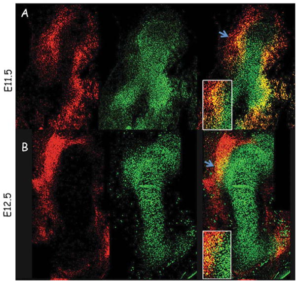
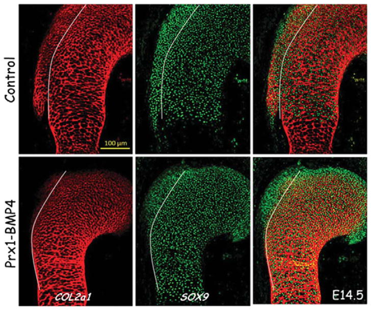
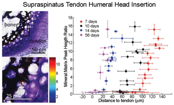
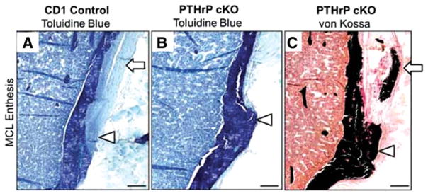
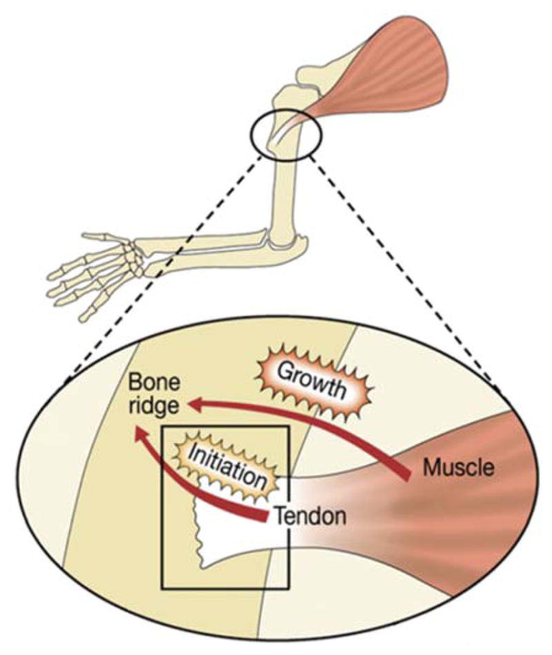
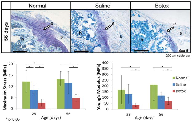
The attachment between tendon and bone occurs across a complex transitional tissue that minimizes stress concentrations and allows for load transfer between muscles and skeleton. This unique tissue cannot be reconstructed following injury, leading to high incidence of recurrent failure and stressing the need for new clinical approaches. This review describes the current understanding of the development and function of the attachment site between tendon and bone. The embryonic attachment unit, namely, the tip of the tendon and the bone eminence into which it is inserted, was recently shown to develop modularly from a unique population of Sox9- and Scx-positive cells, which are distinct from tendon fibroblasts and chondrocytes. The fate and differentiation of these cells is regulated by transforming growth factor beta and bone morphogenetic protein signaling, respectively. Muscle loads are then necessary for the tissue to mature and mineralize. Mineralization of the attachment unit, which occurs postnatally at most sites, is largely controlled by an Indian hedgehog/parathyroid hormone-related protein feedback loop. A number of fundamental questions regarding the development of this remarkable attachment system require further study. These relate to the signaling mechanism that facilitates the formation of an interface with a gradient of cellular and extracellular phenotypes, as well as to the interactions between tendon and bone at the point of attachment.

Keywords: BMP4; IHH; PTHrP; SCX; bone; bone eminence; cartilage; enthesis; musculoskeletal development; tendon insertion.


Conflict of interest statement

The authors have no conflicts of interest with the material reviewed in this article.

Figures

FIGURE 1
FIGURE 1
Bone eminence progenitors coexpress Sox9 and Scx. A (top row) and B (bottom row): Double fluorescence in situ hybridization of sagittal humerus sections from E11.5 to E12.5 wild-type mice, using antisense complementary RNA probes for Sox9 (green) and Scx (red). Blue arrows demarcate a field from which the deltoid and great tuberosities develop; box shows enlargement of eminence progenitors expressing both Sox9 and Scx (Blitz et al., 2013).
FIGURE 2
FIGURE 2
Schematic model for attachment unit formation in situ by segregation of a common progenitor pool to tenocytes and chondrocytes. At the onset (I), the bone anlage comprises differentiated chondrocytes (gray), and the attachment unit domain contains Sox9/Scx-positive progenitors (green). Next (II and III), progenitor cells gradually differentiate to tendon cells from one side (purple) and cartilage cells on the other side (gray) and form the attachment unit (IV). Although specification of attachment unit is regulated by TGFβ signaling (I), their differentiation to chondrocytes is regulated by BMP4 signaling from tendon progenitor cells (Blitz et al., 2013).
FIGURE 3
FIGURE 3
Immunofluorescence staining of humeral sections using anticollagen II (COL2A1) is shown in red, and anti-SOX9 antibodies in green indicates the presence of eminence progenitors at the deltoid tuberosity of the humeral head. Sections from E14.5 control and Prx1-Bmp4 mutants show that conditional knockout of Bmp4 in limb mesenchyme blocked the differentiation of bone eminence progenitors to cartilage. White lines mark the progenitor pool from which the deltoid tuberosity develops (Blitz et al., 2013).
FIGURE 4
FIGURE 4
Spatial gradients in mineral (as determined using Raman spectroscopy) form between tendon and bone at the developing entheses from the onset of endochondral ossification (7 days in the mouse supraspinatus tendon enthesis, as shown in the Von Kossa/Toluidine Blue stained sections on the left). Reproduced with permission from Schwartz et al., 2012.
FIGURE 5
FIGURE 5
Conditional deletion of PTHrP from Scx-expressing cells led to defects in medial collateral ligament (MCL) enthesis mineralization. A normal MCL enthesis is shown in (A). Note the tuberosity and distortion in (B) and (C) and the mineralization within the tuberosity and tendon itself in (C). The MCL tendon is identified by arrows in (A) and (C), and the enthesis site by arrowheads in (A)–(C). Reproduced with permission from Wang et al., 2013.
FIGURE 6
FIGURE 6
Blitz et al. (2009) suggested a model for the contribution of both tendons and muscles to bone eminence formation. Through a biphasic process, tendons regulate bone eminence initiation, and muscles control its subsequent growth. Further research is necessary to determine the mechanism whereby muscle contraction regulates eminence development. Reproduced with permission from Blitz et al., 2009.
FIGURE 7
FIGURE 7
Muscle paralysis dramatically impaired the development of the supraspinatus tendon-to-bone enthesis in mice. Top: A mature, compositionally graded enthesis (“e”) is seen 56 days postnatally in normal mice (scale bar =200 μm). In contrast, the enthesis in paralyzed shoulders appears disorganized, without a graded fibrocartilaginous transition between the supraspinatus tendon (“s”) and the humeral head bone (“h”). Reproduced with permission from Thomopoulos et al., 2007. Bottom: Maximum stress and modulus were significantly lower in the paralyzed group when compared with the normal and saline groups. Reproduced with permission from Schwartz et al., 2013.

References

    1. Akiyama H, Chaboissier MC, Martin JF, et al. The transcription factor Sox9 has essential roles in successive steps of the chondrocyte differentiation pathway and is required for expression of Sox5 and Sox6. Genes Dev. 2002;16:2813–2828. - PMC - PubMed
    1. Akiyama H, Kim JE, Nakashima K, et al. Osteo-chondroprogenitor cells are derived from Sox9 expressing precursors. Proc Natl Acad Sci USA. 2005;102:14665–14670. - PMC - PubMed
    1. Benjamin M, Ralphs JR. Fibrocartilage in tendons and ligaments—an adaptation to compressive load. J Anat. 1998;193 (Part 4):481–494. - PMC - PubMed
    1. Benjamin M, Kumai T, Milz S, Boszczyk BM, et al. The skeletal attachment of tendons—tendon “enthuses”. Comp Biochem Physiol A Mol Integr Physiol. 2002;133:931–945. - PubMed
    1. Biewener AA, Fazzalari NL, Konieczynski DD, Baudinette RV. Adaptive changes in trabecular architecture in relation to functional strain patterns and disuse. Bone. 1996;19:1–8. - PubMed

Publication types