

Gender differences in birdshot chorioretinopathy and the white dot syndromes: do they exist?
- PMID: 24678412
- PMCID: PMC3941241
- DOI: 10.1155/2014/146768
Gender differences in birdshot chorioretinopathy and the white dot syndromes: do they exist?
Abstract
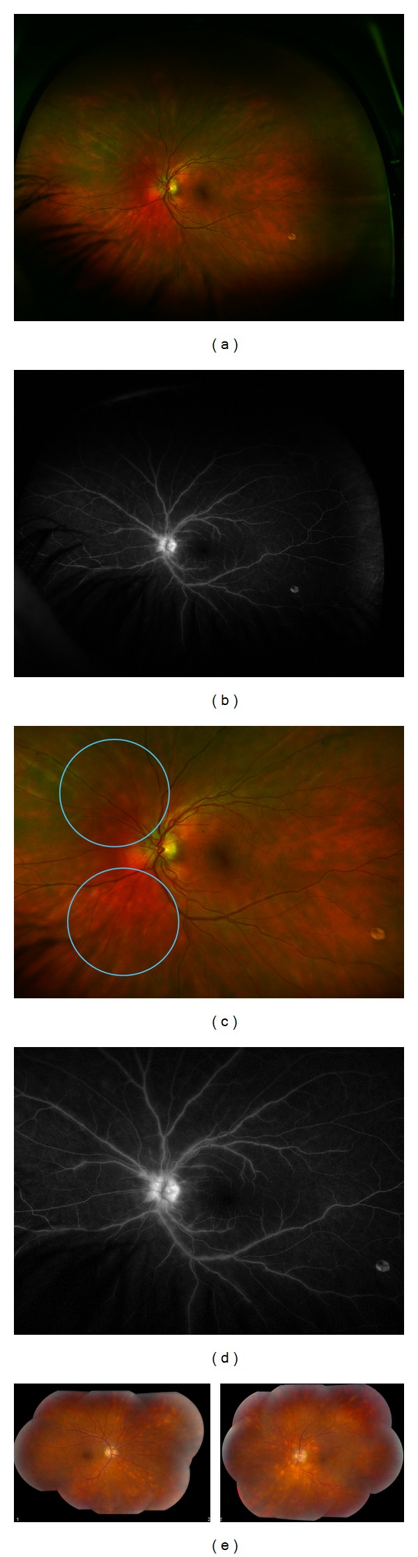
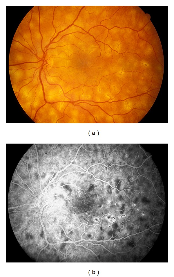
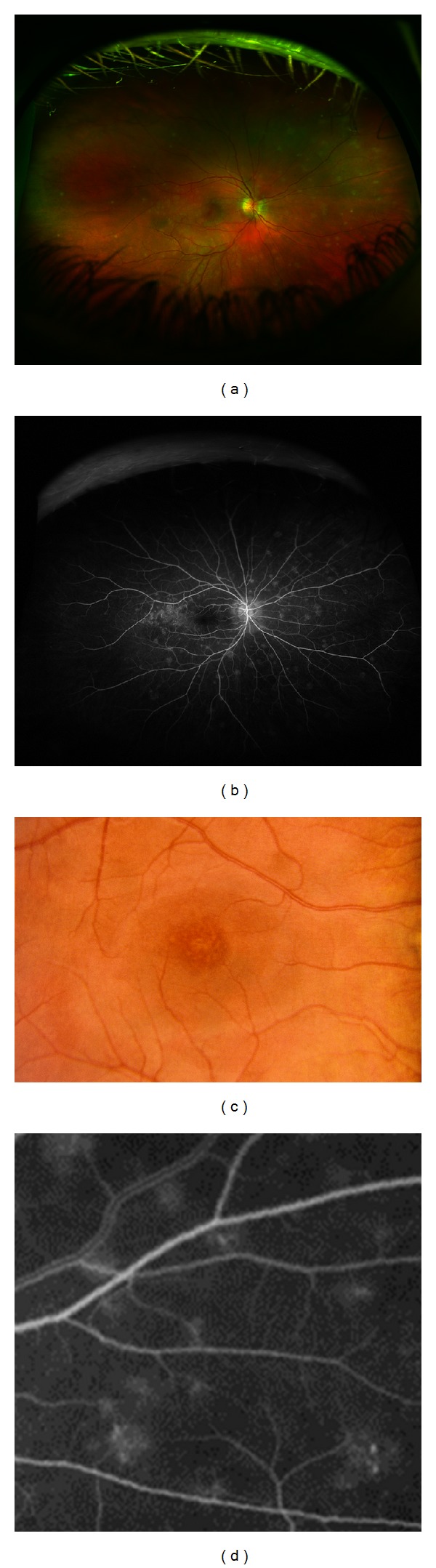
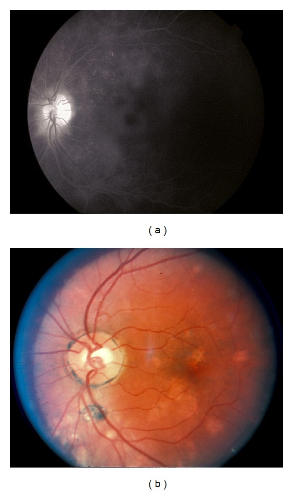
Inflammatory conditions that affect the posterior pole are diverse. Specifically, birdshot chorioretinopathy and the white dot syndromes present with multiple white dots in the fundus. These diseases appear to affect similar age groups but there is question as to whether or not a difference exists between the genders. This review summarizes the current studies on birdshot chorioretinopathy and the white dot syndromes as they are related to gender, exploring the differences, if any, which may exist between prevalence, clinical presentation, and treatment response for these diseases. Though the specific etiology of these diseases remains unclear, future treatments may be guided as to how these diseases affect the sexes differently.
Figures
Similar articles
-
Multi-modal imaging and anatomic classification of the white dot syndromes.Int J Retina Vitreous. 2017 Mar 20;3:12. doi: 10.1186/s40942-017-0069-8. eCollection 2017. Int J Retina Vitreous. 2017. PMID: 28331634 Free PMC article. Review.
-
A review of the inflammatory chorioretinopathies: the white dot syndromes.ISRN Inflamm. 2013 Oct 31;2013:783190. doi: 10.1155/2013/783190. eCollection 2013. ISRN Inflamm. 2013. PMID: 24294536 Free PMC article. Review.
-
The white dot syndromes.Compr Ophthalmol Update. 2007 Jul-Aug;8(4):179-200; discussion 203-4. Compr Ophthalmol Update. 2007. PMID: 17999832 Review.
-
The white dot syndromes.Am J Ophthalmol. 2004 Mar;137(3):538-50. doi: 10.1016/j.ajo.2004.01.053. Am J Ophthalmol. 2004. PMID: 15013878 Review.
-
Birdshot chorioretinopathy.Curr Opin Ophthalmol. 2006 Dec;17(6):545-50. doi: 10.1097/ICU.0b013e3280109479. Curr Opin Ophthalmol. 2006. PMID: 17065923 Review.
Cited by
-
Acute Posterior Multifocal Placoid Pigment Epitheliopathy (APMPPE): A Comprehensive Approach and Case Series: Systemic Corticosteroid Therapy Is Necessary in a Large Proportion of Cases.Medicina (Kaunas). 2022 Aug 8;58(8):1070. doi: 10.3390/medicina58081070. Medicina (Kaunas). 2022. PMID: 36013537 Free PMC article.
-
Optic Disc Edema Is an Under-Recognized Feature of Birdshot Chorioretinitis.J Neuroophthalmol. 2024 Dec 1;44(4):545-551. doi: 10.1097/WNO.0000000000002085. Epub 2024 Jan 25. J Neuroophthalmol. 2024. PMID: 38271082 Free PMC article.
-
Multi-modal imaging and anatomic classification of the white dot syndromes.Int J Retina Vitreous. 2017 Mar 20;3:12. doi: 10.1186/s40942-017-0069-8. eCollection 2017. Int J Retina Vitreous. 2017. PMID: 28331634 Free PMC article. Review.
-
The role of sex in uveitis and ocular inflammation.Int Ophthalmol Clin. 2015 Summer;55(3):111-31. doi: 10.1097/IIO.0000000000000072. Int Ophthalmol Clin. 2015. PMID: 26035764 Free PMC article. Review.
-
Birdshot chorioretinopathy: current knowledge and new concepts in pathophysiology, diagnosis, monitoring and treatment.Orphanet J Rare Dis. 2016 May 12;11(1):61. doi: 10.1186/s13023-016-0429-8. Orphanet J Rare Dis. 2016. PMID: 27175923 Free PMC article. Review.
References
-
- Franceschaetti A, Babel J. La choriorétinite en täche de bougie, manifestation de la maladie de Besnier-Boeck. Ophthalmologica. 1949;118:701–710. - PubMed
-
- Ryan SJ, Maumenee AE. Birdshot retinochoroidopathy. American Journal of Ophthalmology. 1980;89(1):31–45. - PubMed
-
- Henderly DE, Genstler AJ, Smith RE, Rao NA. Changing patterns of uveitis. American Journal of Ophthalmology. 1987;103(2):131–136. - PubMed
-
- Rodriguez A, Calonge M, Pedroza-Seres M, et al. Referral patterns of uveitis in a tertiary eye care center. Archives of Ophthalmology. 1996;114(5):593–599. - PubMed
Publication types
LinkOut - more resources
Full Text Sources
Other Literature Sources
