

D-glucosamine promotes transfection efficiency during electroporation
- PMID: 24678506
- PMCID: PMC3942286
- DOI: 10.1155/2014/485867
D-glucosamine promotes transfection efficiency during electroporation
Abstract
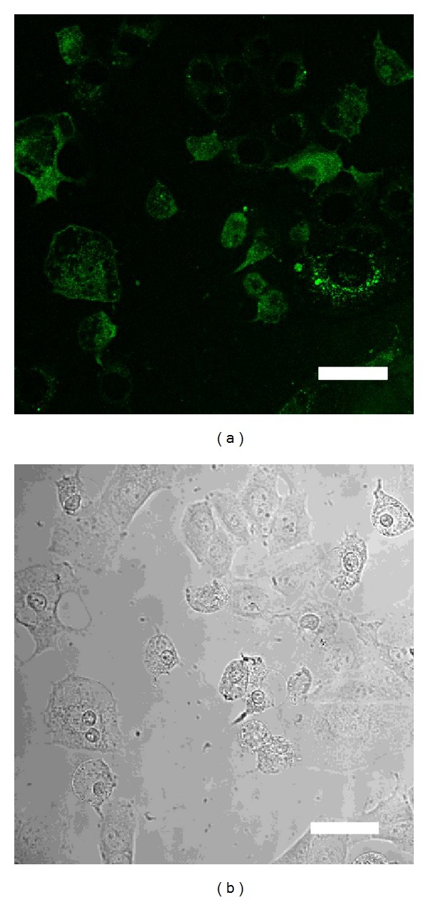
D-Glucosamine is a useful medicament in various fields of medicine and dentistry. With respect to stability of the cell membrane, it has been reported that bradykinin-induced nociceptive responses are significantly suppressed by the direct application of D-glucosamine. Electroporation is usually used to effectively introduce foreign genes into tissue culture cells. Buffers for electroporation with or without D-glucosamine are used in experiments of transfection vectors. This is the first study to indirectly observe the stability and protection of the osteoblast membrane against both electric stress and gene uptake (the proton sponge hypothesis: osmotic rupture during endosomes prior to fusion with lysosomes) in electroporation with D-glucosamine application. The transfection efficiency was evaluated as the fluorescence intensity of the transfected green fluorescent protein (GFP) in the cultured cells (osteoblasts; NOS-1 cells). The transfection efficiency increased over 30% in the electroporation samples treated with D-glucosamine-supplemented buffer after one day. The membrane absorption of D-glucosamine is the primary mechanism of membrane stress induced by electric stress. This new function of D-glucosamine is useful and meaningful for developing more effective transformation procedures.
Figures


Similar articles
-
Comparing chemical transfection, electroporation, and lentiviral vector transduction to achieve optimal transfection conditions in the Vero cell line.BMC Mol Cell Biol. 2024 May 13;25(1):15. doi: 10.1186/s12860-024-00511-x. BMC Mol Cell Biol. 2024. PMID: 38741034 Free PMC article.
-
Simulation and experimental demonstration of the electric field assisted electroporation microchip for in vitro gene delivery enhancement.Lab Chip. 2004 Apr;4(2):104-8. doi: 10.1039/b312804k. Epub 2004 Mar 10. Lab Chip. 2004. PMID: 15052348
-
Direct visualization of electroporation-assisted in vivo gene delivery to tumors using intravital microscopy - spatial and time dependent distribution.BMC Cancer. 2004 Nov 16;4:81. doi: 10.1186/1471-2407-4-81. BMC Cancer. 2004. PMID: 15546484 Free PMC article.
-
Electroporation of cells in microfluidic devices: a review.Anal Bioanal Chem. 2006 Jun;385(3):474-85. doi: 10.1007/s00216-006-0327-3. Epub 2006 Mar 14. Anal Bioanal Chem. 2006. PMID: 16534574 Review.
-
Transfection by electroporation.Curr Protoc Immunol. 2001 May;Chapter 10:10.15.1-10.15.3. doi: 10.1002/0471142735.im1015s03. Curr Protoc Immunol. 2001. PMID: 18432678 Review.
Cited by
-
Melatonin promotes the development of sheep transgenic cloned embryos by protecting donor and recipient cells.Cell Cycle. 2022 Jul;21(13):1360-1375. doi: 10.1080/15384101.2022.2051122. Epub 2022 Mar 20. Cell Cycle. 2022. PMID: 35311450 Free PMC article.
-
High-throughput mass spectrometry analysis revealed a role for glucosamine in potentiating recovery following desiccation stress in Chironomus.Sci Rep. 2017 Jun 16;7(1):3659. doi: 10.1038/s41598-017-03572-5. Sci Rep. 2017. PMID: 28623254 Free PMC article.
-
Enhancement effect of urea toward electroporation-mediated plasmid transfection efficiency in the HEK-293 cell line.Res Pharm Sci. 2024 Dec 15;19(6):766-773. doi: 10.4103/RPS.RPS_185_23. eCollection 2024 Dec. Res Pharm Sci. 2024. PMID: 39911892 Free PMC article.
References
-
- Shigemasa Y, Minami S. Applications of chitin and chitosan for biomaterials. Biotechnology and Genetic Engineering Reviews. 1996;13:413–420. - PubMed
-
- Muzzarelli RAA, Mattioli-Belmonte M, Pugnaloni A, Biagini G. Biochemistry, histology and clinical uses of chitins and chitosans in wound healing. In: Jolles P, Muzzarelli RAA, editors. Chitin and Chitinases. Basel, Switzerland: Birkhäuser; 1999. pp. 285–293. - PubMed
-
- Hua J, Sakamoto K, Nagaoka I. Inhibitory actions of glucosamine, a therapeutic agent for osteoarthritis, on the functions of neutrophils. Journal of Leukocyte Biology. 2002;71(4):632–640. - PubMed
-
- Crolle G, D’Este E. Glucosamine sulphate for the management of arthrosis: a controlled clinical investigation. Current Medical Research and Opinion. 1980;7(2):104–109. - PubMed
-
- Kaida K, Yamashita H, Toda K, Hayashi Y. Effects of glucosamine on tooth pulpal nociceptive responses in the rat. Journal of Dental Sciences. 2013;8:68–73.
Publication types
MeSH terms
Substances
LinkOut - more resources
Full Text Sources
Other Literature Sources
