. 2014 Mar 28:12:26.
doi: 10.1186/1741-7007-12-26.

Evolutionary origin of gastrulation: insights from sponge development

Affiliations

Evolutionary origin of gastrulation: insights from sponge development

Nagayasu Nakanishi et al. BMC Biol. .

Abstract

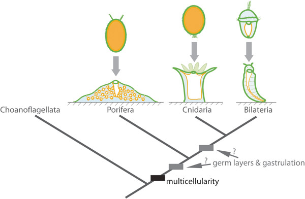
Background: The evolutionary origin of gastrulation--defined as a morphogenetic event that leads to the establishment of germ layers--remains a vexing question. Central to this debate is the evolutionary relationship between the cell layers of sponges (poriferans) and eumetazoan germ layers. Despite considerable attention, it remains unclear whether sponge cell layers undergo progressive fate determination akin to eumetazoan primary germ layer formation during gastrulation.

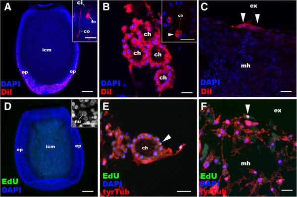
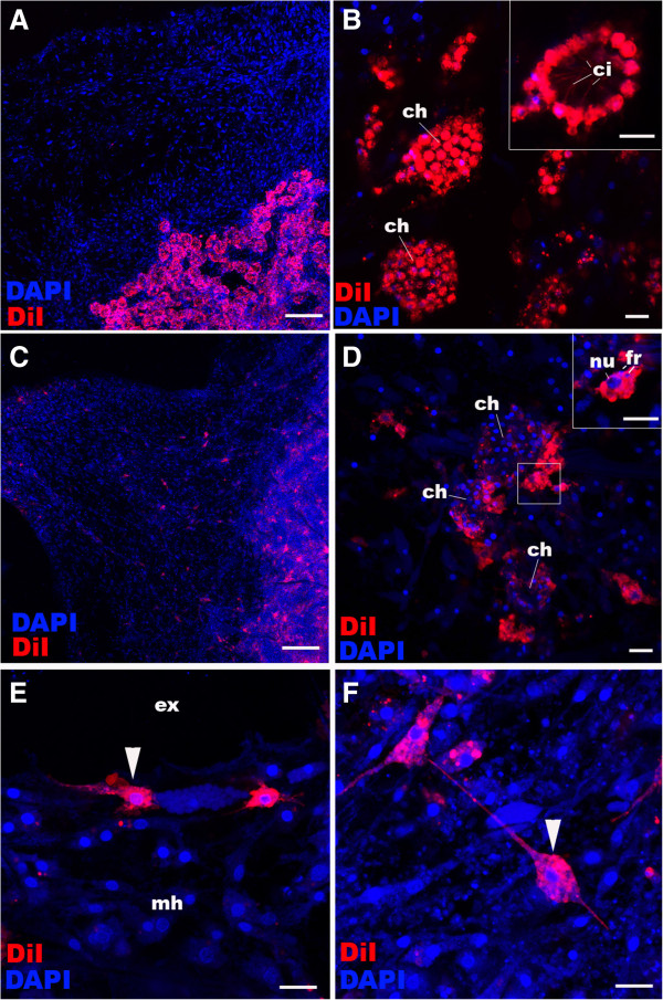
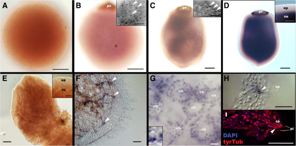
Results: Here we show by cell-labelling experiments in the demosponge Amphimedon queenslandica that the cell layers established during embryogenesis have no relationship to the cell layers of the juvenile. In addition, juvenile epithelial cells can transdifferentiate into a range of cell types and move between cell layers. Despite the apparent lack of cell layer and fate determination and stability in this sponge, the transcription factor GATA, a highly conserved eumetazoan endomesodermal marker, is expressed consistently in the inner layer of A. queenslandica larvae and juveniles.

Conclusions: Our results are compatible with sponge cell layers not undergoing progressive fate determination and thus not being homologous to eumetazoan germ layers. Nonetheless, the expression of GATA in the sponge inner cell layer suggests a shared ancestry with the eumetazoan endomesoderm, and that the ancestral role of GATA in specifying internalised cells may antedate the origin of germ layers. Together, these results support germ layers and gastrulation evolving early in eumetazoan evolution from pre-existing developmental programs used for the simple patterning of cells in the first multicellular animals.


Figures

Figure 1
Figure 1
Evolution of germ layers and gastrulation. Animal multicellularity, with the larval and juvenile/adult body plans of extant metazoans possessing multiple cell layers, is depicted above. The arrow represents metamorphosis from larval to juvenile/adult forms. Cell layers are coloured: orange, inner layer; green, outer layer; and blue, middle layer. In the case of cnidarians and bilaterians, these correspond to endoderm, ectoderm and mesoderm (bilaterians only), respectively. The phylogenetic relationship of these clades and the sister group to metazoans, the choanoflagellates, is shown below, along with the evolutionary origin of multicellularity. The origin of germ layers and gastrulation is debatable, occurring either before or after the divergence of sponges and eumetazoans.
Figure 2
Figure 2
Cell labelling and lineage-tracing through metamorphosis in Amphimedon queenslandica. A, D: Distributions of labelled cells in free-swimming larvae. Anterior (swimming axis) is to the bottom. B, C, E, F: Descendants of the labelled cells in juveniles. Nuclei are stained with DAPI. In E and F, the juveniles are labelled with an antibody against tyrosinated tubulin (tyrTub). A: Longitudinal confocal sections through the centre of a free-swimming larva incubated with CM-DiI, showing strong labelling in ciliated epidermal cell types, the columnar epithelial cell (co) and the flask cell (fc) (inset), with little labelling in inner cell mass (icm). B: Choanocytes in chambers (ch). In some cases, a subset of choanocytes is CM-DiI-labelled in a single choanocyte chamber (arrowhead in inset), suggesting that multiple precursor cells can be involved in development of a single chamber. C: Labelled exopinacocytes (arrowheads). D: A confocal longitudinal section through the centre of a free-swimming larva pulse-labelled with EdU. Note that the labelled cells localise in the inner cell mass (icm) and are likely to be proliferating archeocytes with characteristic large nucleoli (nu) [see Additional file 3: Figure S3]. E: An EdU-positive choanocyte in the chamber (arrowhead). F: An EdU-positive exopinacocyte (arrowhead). Other abbreviations: ep, outer layer epithelium; ci, cilium; ex external environment; mh, mesohyl. Scale bars: 100 μm (A, D), 10 μm (B, C, E, F, inset in D), 5 μm (inset in A). DAPI, 4',6-diamidino-2-phenylindole; EdU, 5-ethynyl-2′-deoxyuridine.
Figure 3
Figure 3
Choanocyte labelling and lineage-tracing in juvenile Amphimedon queenslandica. A, B: Choanocytes in chambers (ch) in juveniles are labelled with CM-DiI; note strong plasma membrane labelling in choanocytes (inset). C-F: Descendants of juveniles whose chambers were labelled with CM-DiI seven days earlier (as in A). Multiple other juvenile cell types are now labelled and spread through the sponge body. Note in (D) the limited number of CM-DiI-labelled choanocytes remaining in the choanocyte chambers (ch) seven days after labelling and the presence of labelled archeocytes with large nuclei (nu; inset), consistent with transdifferentiation of choanocytes into archeocytes. Note the presence of nuclear fragments (fr) presumably resulting from phagocytosis that has occurred during de-differentiation [see Additional file 5: Figure S5]. Arrowheads in E and F show a labelled exopinacocyte and a sclerocyte, respectively, indicating these cell types can also be produced from de-differentiating choanocytes. Nuclei are stained with DAPI. Scale bars: 100 μm (A, C), 10 μm (B, D-F). DAPI, 4',6-diamidino-2-phenylindole.
Figure 4
Figure 4
GATA mRNA expression during development in A. queenslandica. Late blastulae (A; sensu Leys and Degnan [10]), mid-stage embryos with a pigment spot (ps) (B), late-stage embryos with a pigment ring (pr) (C), a free-swimming larva (D), a settlement stage postlarva (E), and a juvenile (F-I) were labelled with the antisense riboprobe AqGATA. The juvenile in I is labelled with DAPI and an anti-tyrTub antibody. In B-D, the specimens are viewed from the lateral side with the posterior pigmented structures (ps and pr) placed up in each panel. In E and F, the specimens are viewed from the top of postlarva. Medial optical sections exposing the internally localised subepithelial cells are shown. Insets in B and C show GATA expression in individual subepithelial cells (arrowheads), and an inset in D shows the demarcation between the GATA-negative epidermal layer (ep) and the strongly GATA-positive subepithelial domain (su). An inset in E shows the downregulation of GATA expression in the subepithelial domain (su) relative to the outer layer epithelium at settlement. Arrowheads in F indicate the internal spicule-forming tent-pole-like structures in the juvenile that strongly express GATA. In G, an inset shows a GATA-expressing choanocyte with a cilium (ci) and a basal nucleus (nu). An arrowhead in H shows a cluster of GATA-expressing spicule (sp)-forming cells at the apex of the tent-pole-like structures shown in F; note in I that their cell bodies (arrowhead) are situated basal to the pinacoderm (pi). Scale bars: 100 μm (A-F), 50 μm (H, I, and insets in B-E), 10 μm (G). DAPI, 4',6-diamidino-2-phenylindole.

References

    1. Hyman LH. The Invertebrates: Protozoa Through Ctenophora, Volume 1. McGraw-Hill Book Company, Inc: New York and London; 1940.
    1. Hall BK. Germ layers and the germ-layer theory revisited - primary and secondary germ layers, neural crest as a fourth germ layer, homology, and demise of the germ-layer theory. Evol Biol. 1998;12:121–186.
    1. Martindale MQ. The evolution of metazoan axial properties. Nat Rev Genet. 2005;12:917–927. doi: 10.1038/nrg1725. - DOI - PubMed
    1. Martindale MQ, Pang K, Finnerty JR. Investigating the origins of triploblasty: ‘mesodermal’ gene expression in a diploblastic animal, the sea anemone Nematostella vectensis (phylum, Cnidaria; class, Anthozoa) Development. 2004;12:2463–2474. doi: 10.1242/dev.01119. - DOI - PubMed
    1. Nakanishi N, Renfer E, Technau U, Rentzsch F. Nervous systems of the sea anemone Nematostella vectensis are generated by ectoderm and endoderm and shaped by distinct mechanisms. Development. 2012;12:347–357. doi: 10.1242/dev.071902. - DOI - PubMed

Publication types

MeSH terms

LinkOut - more resources